R E S U M O
Os autores descrevem um caso de um doente com uma neoplasia da bexiga inoperável, internado por um quadro de síncope e hipotensão arterial. Na avaliação do doente é efectuado um ecocardiograma que mostra a existência de um trombo livre ao nível das cavidades direitas e dilatação das mesmas que levou à hipótese diagnóstica de embolia pulmonar. Perante a existência de uma neoplasia com hemorragia recente, algumas dúvidas terapêuticas surgiram, tendo sido iniciado heparina. Devido a agravamento da situação, com hipertensão pulmonar grave e presença de volumosos trombos ao nível de ambos os ramos da artéria pulmonar visualizados por ecocardiografia transesofágica, o doente acabou por ser submetido a trombólise, embora com algumas alterações ao esquema habitualmente realizado. O doente melhorou, sendo o ecocardiograma final normal.
C
ASO
C
LÍNICO
Trombo Livre Intra-Auricular Direito
e Embolia Pulmonar
[39]
ANAGALRINHO, TERESACRUZ, JOSÉLOUREIRO
Laboratório de Ecocardiografia da Unidade de Intervenção Vascular e Serviço 2 do Hospital Curry Cabral, Lisboa
531
A B S T R A C T
Free Floating Thrombus in Right Atrium and Pulmonary Thromboembolism
The authors describe a case of a patient with inoperable bladder cancer, who was admitted with syncope and arterial hypotension. After examination, an echocardiogram was requested, which showed a free-floating thrombus in the right atrium and dilatation of the right chambers. A diagnosis of
pulmonary embolism was made, and heparin was started. The patient’s clinical status worsened, with development of severe pulmonary hypertension due to the presence of large thrombi in the right and left
pulmonary arteries. Despite a recent hemorrhagic event related to the bladder cancer, thrombolytic therapy was begun with improvement of the patient. The final echocardiogram was almost normal, without pulmonary hypertension and no dilatation of the right chambers.
Recebido para publicação: Junho de 2002 • Aceite para publicação: Outubro de 2002 Received for publication: June 2002 • Accepted for publication: October 2002 INTRODUÇÃO
O
diagnóstico de trombos livres ao nível do coração direito, é uma situação pouco fre-quente, mas possivelmente subdiagnosticada se atendermos ao número de casos diagnostica-dos de embolia pulmonar(1). Este achado ocorrenormalmente em doentes com embolia pulmo-nar e resulta da migração de material trombó-tico do sistema venoso inferior ( membros infe-riores, pélvis) para a artéria pulmonar, no
INTRODUCTION
F
ree-floating thrombus in the right heart is diagnosed comparatively rarely, although it may be under-diagnosed considering the num-ber of patients diagnosed with pulmonary em-bolism(1). It is normally found in patients withpulmonary embolism as a result of thrombotic material migrating from the lower venous system (lower limbs or pelvis) to the pulmo-nary artery, although the material is rarely ob-Rev Port Cardiol 2003; 22 (4) : 531-540
Palavras-Chave Trombo livre na aurícula direita; Embolia pulmonar; Trombólise
Key words
Free floating thrombus in right atrium; Pulmonary thromboembolism; Thrombolysis
entanto este material em «trânsito» raramente se observa(2). Dado que o potencial embólico é
muito elevado(3), a taxa de mortalidade é
ele-vada, estimando-se em mais de 40 %(4). Os
au-tores descrevem um caso em que se encontrou um trombo de grandes dimensões ao nível da aurícula direita, originado a partir de um trombo ao nível da veia cava inferior num doente com uma neoplasia não operável e em início de quimioterapia. Esta situação levou a algumas considerações e esquemas terapêuti-cos diferentes do habitual.
CASO CLÍNICO
Doente de 74 anos de idade, sexo mascu-lino, com uma neoplasia da bexiga, submetido a cirurgia paliativa em Abril deste ano (urete-rostomia com interposição de conduto ileal e anastomose deste à pele), com episódios pré-vios de hemorragia vesical, e presentemente com uretrorragia. O doente iria iniciar quimio-terapia com esquema terapêutico apropriado e foi-lhe pedido um ecocardiograma prévio, dado se irem utilizar drogas cardiotóxicas. No dia em que o ecocardiograma estava marcado (14/05/02), o doente deu entrada na Urgência com um quadro de síncope, hipotensão arterial e escoriação da face.
Foi-lhe efectuado ECG, RX tórax, análises de rotina e um TAC craneo-encefálico devido ao traumatismo craneano resultante da síncope (escoriação da face).
Dos exames efectuados, o ECG mostrava al-terações da repolarização ao nível da parede anterior, o RX tórax era normal, o TAC craneo-encefálico não mostrava lesões, nomeadamente vasculares ou lesões ocupando espaço que jus-tificassem o quadro clínico, dado que o eco-cardiograma estava agendado e indicado no estudo deste doente, foi ao Laboratório de Ecocardiografia para realização do mesmo. No exame objectivo descrito na admissão existia referência a hipotensão arterial (89/53 mmHg), ligeira taquicardia sinusal (102/min) e uma li-geira hipoxémia (89 % de saturação periférica de O2), não havia referência a outros sinais,
nomeadamente dificuldade respiratória.
O ecocardiograma mostrava ausência de le-sões valvulares, boa função sistólica global do ventrículo esquerdo e movimento anómalo do septo interventricular, cavidades direitas ligei-ramente dilatadas (VD de 28 mm), ligeira hi-pertensão pulmonar sistólica – 42 mmHg. Em plano apical quatro câmaras e sobretudo em
532
served in transit(2). Given that the embolic
po-tential is very high(3), there is an elevated
mor-tality rate, estimated at more than 40 %(4). The
authors describe a case in which a large thrombus was found in the right atrium, origin-ating from a thrombus in the inferior vena cava of a patients with inoperable cancer who was about to begin chemotherapy. This situation led to the adoption of somewhat different ther-apy regimes from usual.
CASE REPORT
The patient was a 74-year-old male with bladder cancer who had undergone palliative surgery the previous April (ureterostomy with insertion of an ileal conduit and anastomosis to the skin) after episodes of vesicular haemor-rhage, and currently with urethral bleeding. The patient was due to begin chemotherapy with an appropriate therapeutic scheme and an echocardiogram had been requested as a pre-caution as cardiotoxic drugs were due to be administered. On the day on which the ECG was scheduled (14/5/2002) the patient was ad-mitted to the Emergency Department with a setting of syncope, hypertension and facial cuts.
The patient underwent ECG, chest X-ray, routine lab tests and a cranial CT scan, due to the head trauma resulting from syncope (facial cuts).
Although it was found that the ECG showed repolarization abnormalities in the anterior wall, the chest X-ray was normal, and the CT scan did not show signs of vascular damage or lesions in areas which could explain the clin-ical setting. As an echocardiogram had been arranged and would have been indicated in the study of this patient, it was decided to proceed with this. In the physical examination that had been carried out on admission, reference was made to hypertension (89/53 mmHg), slight si-nus tachycardia (102 bpm) and slight hypoxia (89 % of peripheral O2 saturation), but no other
signs such as respiratory difficulty.
The electrocardiogram showed no valvular lesions, good global left ventricular systolic function but anomalous motion of the left ven-tricular septum, right chambers slightly dilated (right ventricle=28 mm), and slight systolic pulmonary hypertension (42 mmHg). In both apical 4-chamber view and particularly sub-costal view, a highly mobile mass, serpentine in appearance, was observed moving freely, in
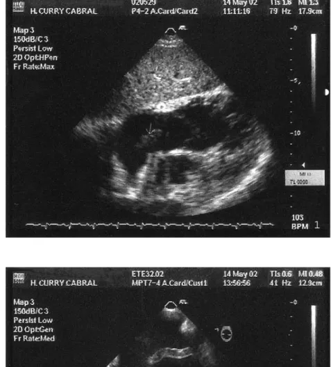
subcostal, observava-se uma massa, com as-pecto reptilíneo, muito móvel, com movimento anárquico, que se movia livremente dentro da aurícula direita (Fig. 1). Efectuou-se ecocar-diograma transesofágico que confirmou este diag-nóstico e melhorou a caracterização morfoló-gica da massa que se classificou como trom-bo. Este tinha um aspecto vermiforme, muito móvel e de formação recente (pouco ecogénico), e circulava livremente pela aurícula direita (Figs. 2 e 3). Em plano transgástrico, com rota-ção da sonda para as cavidades direitas, con-seguiu-se observar a veia cava inferior, que
es-533
an irregular trajectory, within the right atrium (Fig. 1). A transesophageal echocardiogram confirmed this and aided the morphological characterization of the thrombus, which was revealed to have a worm-like shape, highly mobile and of recent formation (being of low echogenicity), which circulated freely in the right atrium (Figs. 2 and 3). In transgastric view, with rotation of the probe towards the right chambers, it could be seen that the infe-rior vena cava was filled with thrombotic mate-rial, poorly echogenic, disorganized, typical of freshly coagulated matter (Fig. 4). This
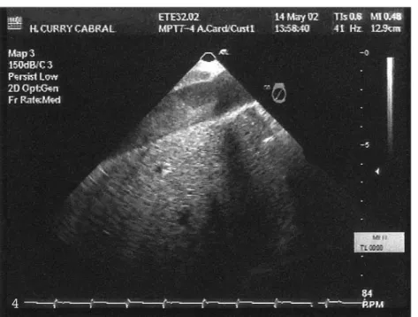
mate-Fig. 2 e 3 Ecocardiograma tran-sesofágico a 0º em que se vê a massa tubular ao nível da aurí-cula direita, com movimento anárquico, linear na Fig. 2 e como que enrolada sobre si pró-pria, em novelo, na Fig. 3. Fig. 2 and 3 Transesophageal echocardiogram at 0º, showing the free-floating tubular mass in the right atrium, linear in Fig. 2 and coiled round itself like a skein in Fig. 3.
Fig. 1 Plano subcostal no qual se vê a dilatação das cavidades direitas e uma massa de grandes dimensões ao nível da aurícula direita, com movimento anár-quico e aspecto tubular. Fig. 1 Subcostal view, showing dilatation of the right chambers and a large mass in the right atrium, with irregular motion and tubular appearance.
2 1
tava preenchida por material trombótico pouco ecogénico, não organizado, típico, de coágulo fresco (Fig. 4). Este material movia-se em pe-quenos movimentos de vaivém dentro da veia cava. Analisou-se a artéria pulmonar que não estava dilatada nem sugeria ter material trom-bótico.
Atendendo à situação clínica do doente, e não se conseguir mais informação do ponto de vista urológico, nomeadamente sobre fenóme-nos hemorrágicos graves relacionados com a neoplasia do doente (os tumores da bexiga são habitualmente muito sangrantes), optou-se por
534
rial was in motion, exhibiting small back-and-forth movements. Assessment of the pulmonary artery showed it to be non-dilated and free of thrombotic material.
Given the patient’s clinical situation, and as it was impossible to obtain further urological information on possible hemorrhages arising from the cancer (bladder tumors often bleed severely), it was decided to begin heparin in therapeutic dosages and to adjust dosage ac-cording to APTT. On the second day the pa-tient reported fatigue, with continuing sinus tachycardia and no hypoxemia (nasal O2 5 Fig. 3 Ecocardiograma
transeso-fágico a 0º em que se vê a massa tubular ao nível da aurícula di-reita, com movimento anárquico, linear na Fig. 2 e como que enro-lada sobre si própria, em novelo, na Fig. 3.
Fig. 3 Transesophageal
echocar-diogram at 0º, showing the free-floating tubular mass in the right atrium, linear in Fig. 2 and coi-led round itself like a skein in Fig. 3.
Fig. 4 Ecocardiograma transeso-fágico em plano transgástrico, com rotação para melhor visuliza-ção das cavidades direitas e em que se vê o fígado e a veia cava inferior, com grande trombo, pouco consistente e não organi-zado, com movimento de vaivém (movimento respiratório). Fig. 4 Transesophageal echocar-diogram in transgastric view, ro-tated for better visualization of the right chambers, showing the liver and the inferior vena cava with a large thrombus of low den-sity and disorganized, with to-and-fro motion (respiratory mo-tion).
3
se iniciar heparina em dose terapêutica e acer-tar a dose de acordo com aPTT. Ao 2.º dia, o doente referia cansaço, mantinha a taquicar-dia sinusal, ausência de hipoxémia com O2
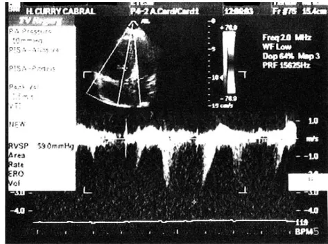
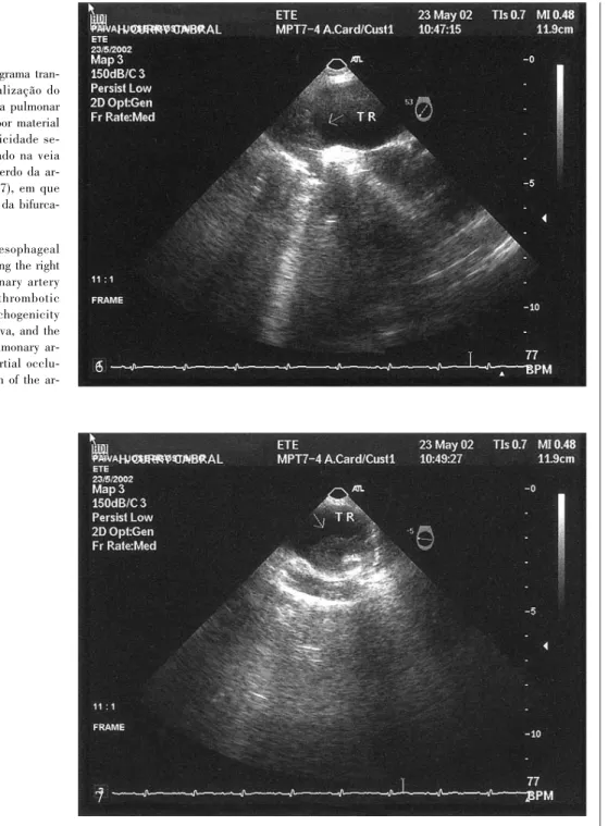
na-sal a 5 l/min, no entanto, surgiram grande al-terações das provas de função hepática, com transaminases muito elevadas (respectivamente TGO de 2200 U/l e TGP de 2380 U/l), LDH de 3100 mg/dl, troponina de 8,6 ng/ml e eleva-ção muito marcada dos d-dímeros (valores de 2420 ng/ml), compatíveis com embolia pulmo-nar significativa. Dado que o doente se encon-trava hemodinâmicamente estável, optou-se por se continuar a terapêutica com heparina e iniciar posteriormente warfarina. Entretanto, pediu-se uma opinião aos colegas de Urologia. O ecocardiograma é repetido seis dias depois (20/05/02), e verificou-se uma acentuada dila-tação das cavidades direitas (VD de 38 mm) com procidência do septo interauricular para a aurícula esquerda (sinal de sobrecarga de pressão), um ventrículo direito com parede li-vre imóvel (sinal de McConnell) e uma hiper-tensão pulmonar de ±60 mmHg, não se visua-lizava o trombo descrito inicialmente (Fig. 5). Efectuou-se novo ecocardiograma transesofá-gico que veio a confirmar a embolização do material trombótico descrito anteriormente, o qual ocupava quase totalmente os dois ramos da artéria pulmonar; estes trombos, dado a sua ecogenicidade, tinham aspecto de serem pouco consistentes (Figs. 6 e 7).
O caso foi discutido com os urologistas,
tendo-se optado por uma terapêutica trombolí- 535
l/min). There was, however, a considerable al-teration in hepatic function tests, with very high transaminases (GOT 2200 U/l and GPT 2380 U/l), 3100 mg/dl LDH, 8.6 ng/ml tropo-nin and a marked increase in d-dimers (2420 ng/ml), compatible with significant pulmonary embolism. As the patient was hemodynami-cally stable, it was decided to continue the he-parin treatment and then initiate warfarin. Meanwhile, specialists in the Urology Depart-ment were asked for an opinion. When echo-cardiography was repeated six days later (20/5/02) it showed accentuated dilatation of the right chambers (right ventricle 38 mm) with the interatrial septum bulging towards the left atrium (a sign of pressure overload), immo-bile right ventricular free wall (McConnell sign) and pulmonary hypertension of ±60 mmHg, but the thrombus described previously was not seen (Fig. 5). Another transesophageal echocardiogram confirmed the embolization of the thrombotic material, which now almost en-tirely filled the two branches of the pulmonary artery; on the basis of their echogenic char-acter these thrombi appeared to be of low dens-ity (Figs. 6 and 7).
The case was discussed with the urologists, and thrombolytic therapy was decided upon, with concomitant risk of surgery for ligation or sclerosing of bleeding vessels.
The warfarin treatment under way was sus-pended for 24 hours, enoxaparin was initiated but the initial heparin bolus was not administ-ered.
Fig. 5 Hipertensão pulmonar aos oito dias de heparina, com uma dilatação das cavidades direitas maior que no ecocardiograma de entrada.
Fig. 5 Pulmonary hypertension after eight days of heparin, with more dilatation of the right cham-bers than on admission echocar-diogram.
tica, correndo-se o risco de uma cirurgia para laqueação/esclerose de vasos sangrantes.
Dado que o doente já estava a fazer warfa-rina, suspendeu-se esta durante 24 h, iniciou-se enoxiparina, não iniciou-se tendo efectuado o bolus inicial de heparina.
O trombolítico utilizado foi a alteplase: bo-lus de 10 mg e infusão dos restantes 90 mg durante 2 horas.
Apesar de não haver complicações hemor-rágicas graves durante o procedimento, houve agravamento da uretrorragia, com eliminação
536
The thrombolytic used was alteplase in 10 mg bolus and infusion of the remaining 90 mg over 2 hours.
Although there were no severe hemorrhagic complications during the procedure, there was a worsening of the urethral bleeding, with the elimination of various clots, and decrease of hemoglobin from 9.4 to 8.5 g/dl. For this rea-son it was decided not to administer intrave-nous heparin and enoxaparin in therapeutic doses was recommenced.
On the echocardiogram 48 hours after thrombolysis there were no signs of dilatation
Fig. 6 e 7 Ecocardiograma tran-sesofágico com visualização do ramo direito da artéria pulmonar (Fig. 6), preenchida por material (trombo) com ecogenicidade se-melhante ao encontrado na veia cava, e no ramo esquerdo da ar-téria pulmonar (Fig. 7), em que existe parcial oclusão da bifurca-ção da artéria.
Fig. 6 and 7 Transesophageal echocardiogram showing the right branch of the pulmonary artery (Fig. 6) filled with thrombotic material of similar echogenicity to that in the vena cava, and the left branch of the pulmonary ar-tery (Fig. 7), with partial occlu-sion of the bifurcation of the
ar-Fig. 7 Ecocardiograma
transeso-fágico com visualização do ramo direito da artéria pulmonar (Fig. 6), preenchida por material (trombo) com ecogenicidade se-melhante ao encontrado na veia cava, e no ramo esquerdo da ar-téria pulmonar (Fig. 7), em que existe parcial oclusão da bifurca-ção da artéria.
Fig. 7 Transesophageal
echocar-diogram showing the right branch of the pulmonary artery (Fig. 6) filled with thrombotic material of similar echogenicity to that in the vena cava, and the left branch of the pulmonary artery (Fig. 7), with partial occlusion of the bi-furcation of the artery.
6
de vários coágulos e descida posterior dos va-lores de hemoglobina de 9,4 para 8,5 g/dl. Por esse motivo optou-se por não se efectuar hepa-rina endovenosa e recomeçou-se enoxipahepa-rina em dose terapêutica.
No ecocardiograma 48 horas após trombó-lise não existia dilatação das cavidades direi-tas (VD de 20 mm) e a pressão sistólica da ar-téria pulmonar era de 39 mmHg.
O doente efectuou eco_Doppler dos mem-bros inferiores após trombólise que nada reve-lou. Presentemente, encontra-se clinicamente estável, mantendo-se em anticoagulação oral e tendo dado início às sessões de quimioterapia previstas.
DISCUSSÃO
A observação de trombos livres nas cavida-des direitas é uma raridade(1, 2, 5) e um achado
nos doentes com suspeita de embolia pulmonar e que efectuam uma avaliação ecocardiográ-fica. O prognóstico é reservado com uma mor-talidade nas primeiras 24 horas que ronda os 21,1 %(4). A grande parte destes trombos tem
uma forma tubular, resultante da moldagem e formação do trombo numa veia(6, 7). Em todas as
séries, a existência deste tipo de trombos está sempre associada à ocorrência de embolia pul-monar, embora também se possam encontrar outros trombos ao nível das cavidades direitas, associados a situações de estase intra-cardíaca, baixo débito, doença reumática ou presença de material estranho intra-cardíaco, mas com me-nos potencial embólico(4, 7). A ecocardiografia é
o único meio de diagnóstico destas situações, com recurso nos últimos anos à ecocardiografia transesofágica, que nos pode dar uma informa-ção mais detalhada das cavidades direitas e da artéria pulmonar desde a sua porção proximal até à bifurcação dos ramos principais em arté-rias lobares. O ETE permite-nos diagnosticar a existência de trombos, e outras alterações por vezes concomitantes, como seja a existência de foramen oval patente, com alguns casos descri-tos de passagem de trombo através do mesmo(8).
Esta técnica é sobretudo útil nos doentes em situação crítica, sem diagnóstico de embolia pulmonar, mas cujo quadro clínico ou laborato-rial nos leva a essa suspeição(9). No nosso caso,
a realização de ecocardiograma foi efectuado um pouco por acaso, embora plenamente indi-cado. O quadro clínico de entrada no serviço de Urgência foi de síncope e hipotensão
arte-rial, e a ausência de dispneia, e a demora na 537
of the right chambers (right ventricle 20 mm) and pulmonary artery systolic pressure was 39 mmHg.
Doppler imaging of the patient’s lower limbs after thrombolysis showed no abnormal-ities. The patient is clinically stable at present, continues to take oral anticoagulants and has now begun the chemotherapy that had origin-ally been scheduled.
DISCUSSION
Observation of a free-floating thrombus in the right chambers is rare(1, 2, 3)and tends to be
found in patients with suspected pulmonary embolism who undergo echocardiography. Prognosis is poor, with a mortality of around 21.1 % in the first 24 hours(4). The great
major-ity of these thrombi are serpentine and tubular in form as a result of having been moulded and shaped when the thrombus was formed inside a vein(6, 7). In all series to date the existence of
this type of thrombus has been associated with pulmonary embolism; although other thrombi may also be encountered in the right chambers associated with intracardiac congestion, low output, rheumatic disease or the presence of foreign intracardiac matter, they have less em-bolic potential(4, 7). Echocardiography is the
only means of diagnosing these entities, with transesophageal echocardiography (TEE) being the method of choice in recent years as it can provide more detailed information on the heart chambers and valves, and additionally in these cases, on the pulmonary artery in its proximal section to where the principal branches bifur-cate into lobar arteries. TEE enables the exist-ence of thrombi to be diagnosed along with other concomitant abnormalities which may appear, such as patent foramen ovale through which the thrombus can pass, as has been des-cribed in some cases(8). This technique is
par-ticularly effective in the case of patients in a critical condition, who have not been diagnos-ed with pulmonary embolism, but whose clin-ical setting and test results raise this possibil-ity(9). In our own case, the echocardiogram was
run quite speculatively, although fully indicat-ed in the circumstances. The clinical setting on admission to the Emergency Department was one of syncope and hypertension without dyspnea, and the delay in obtaining laboratory tests (d-dimers) did not facilitate the diagnosis of pulmonary embolism.
obtenção dos exames analíticos (D-dímeros), dificultam o diagnóstico de embolia pulmonar. Nem sempre é fácil diagnosticar trombos com segurança, algumas situações podem levar a falsos positivos, como existência de rede de Chiari exuberante, válvulas de Eustáquio ou de Thebesius bem desenvolvidas, assim como tumores, cateteres ou vegetações(10).
Os outros métodos de diagnóstico em situa-ções de embolia pulmonar, nomeadamente a angiografia pulmonar(11), não tem significado na
presença de trombos livres a nível das cavida-des direitas e menos ainda no nosso caso, dado que a coexistência de um volumoso trombo ao nível da veia cava inferior, aumentaria o risco de embolia pulmonar maciça por manipulação dos catéteres.
A detecção de um trombo livre pode levar a modificações e orientações terapêuticas dife-rentes, sendo considerado uma terapêutica ur-gente(2, 4). Os melhores resultados, observando
as pequenas séries descritas, parecem facilitar a utilização dos trombolíticos(12-15), embora
al-guns centros, defendam o recurso à embolecto-mia cirúrgica sob circulação extra-corporal (CEC), com exploração das cavidades direitas e da artéria pulmonar(16). No entanto os
resulta-dos desta técnica estão muito relacionaresulta-dos com a experiência de cada centro, sendo na maioria destes uma terapêutica de recurso, em casos de insucesso da terapêutica trombolítica. Os dois centros cirúrgicos, para onde habitual-mente referenciamos os doentes, possuem um pequeno número de casos de embolectomia pulmonar cirúrgica, além de que o estado geral do doente, não era o melhor para ser subme-tido a este tipo de cirurgia. Outras técnicas, nomeadamente de extracção de êmbolos por via percutânea(17), utilizadas em situações de
contra-indicação para trombólise e em doentes clinicamente graves, dependem muito da expe-riência do operador, e no nosso centro nunca foi efectuada. A trombólise tem várias vanta-gens, pode levar à lise do coágulo e caso já te-nha havido embolização para a artéria pulmo-nar leva à melhoria da perfusão pulmopulmo-nar, diminuição da hipertensão pulmonar, melhoria da função do ventrículo direito, e devido à in-terdependência dos dois ventrículos, melhoria da função do ventrículo esquerdo e do débito cardíaco. Os trombolíticos, tem ainda acção extra cardíaca, com lise de eventuais coágulos a nível sistémico, diminuindo a possibilidade de uma recorrência(12-15). Neste nosso caso, o
atraso na terapêutica trombolítica, deveu-se à
538
It is not always possible to diagnose thrombi with a high degree of confidence as some pathologies, such as the existence of a pronounced Chiari net, or well-developed eus-tachian or thebesian valves, or the presence of tumors, catheters or vegetations, may lead to false positives(10).
Other diagnostic methods employed in cases of pulmonary embolism, notably pulmo-nary angiography(11), are not relevant in the
de-tection of free thrombi in the right chambers and would be particularly inappropriate in this case as the coexistence of a bulky thrombus near the inferior vena cava would increase the risk of massive pulmonary embolism arising from manipulation of the catheters.
Detection of a free-floating thrombus can lead to modifications to the therapeutic ap-proach, as it is considered to require urgent treatment(2, 4). According to the relatively
limi-ted number of cases described in the litera-ture, best results would seem to be obtained from the use of thrombolytics(12-15), although
some centers favor surgical embolectomy un-der extracorporeal circulation (ECC), with ex-ploration of the right chambers and of the pul-monary artery(16). It would appear that the
results of this technique vary from center to center and it is generally resorted to in cases where thrombolytic treatment has not been successful. The two surgical centers to which we normally refer our patients deal with a small number of cases of surgical pulmonary embolec- tomy and in this case the overall condition of the patient suggested that under-going ECC would not be appropriate. Other techniques, for example percutaneous embolism extraction(17) which are employed in situations
where thrombolysis is contraindicated or for patients whose clinical status is serious, are heavily dependent on the experience of the operator and have never been performed at our centre. On the other hand, thrombolysis has a number of advantages in that it may lead to a rapid recovery through lysis of the clot and im-provement of pulmonary perfusion while at the same time reducing pulmonary hypertension, improving right ventricular function, and due to the interdependence of the two ventricles, improving left ventricular function and cardiac output and aiding recovery from shock. Throm-bolytics also have an extracardiac action in lysing clots which may be present at the syste-mic level, thus reducing the risk of recurrence
throm-existência de uma neoplasia com fenómeno he-morrágico escasso, mas visível – uretrorragia, que nos levou a tentar primeiro apenas hepa-rina e a consultar os especialista de Urologia para uma opinião acerca do assunto.
A decisão de efectuar trombólise tardia neste doente, deveu-se por um lado, ao desen-volvimento de hipertensão pulmonar grave e visualização de vários trombos com aspecto re-cente ao nível de ambos os ramos da artéria pulmonar e por outro lado, ao facto de alguns autores referirem o benefício da trombólise em situações de embolismo maciço mesmo sem significativo compromisso hemodinâmico(18).
Neste doente, dado a ineficácia da heparina durante cerca de oito dias e hipótese elevada de recorrência, decidimos optar por esta forma terapêutica. O benefício da trombólise no caso do tromboembolismo pulmonar está descrito até às duas semanas, o que se verificou no nosso caso.
O esquema terapêutico habitual na embolia pulmonar, com utilização de rTPA(19), envolve o
uso concomitante de heparina (bolus e perfu-são contínua), no entanto, dado que o doente já tinha iniciado warfarina, optámos por esperar a normalização do INR, recomeçar enoxiparina nesse intervalo e avaliar a situação de modo a minimizar os riscos hemorrágicos. Não efectuá-mos heparina endovenosa após o trombolítico por agravamento da uretrorragia, com emissão de vários coágulos, traduzindo-se por queda posterior dos valores da hemoglobina.
Houve uma melhoria franca de perfusão pulmonar com descida da pressão sistólica na artéria pulmonar de 61 para 39 mmHg, desa-parecimento da dilatação das cavidades direi-tas e do movimento paradoxal do septo. Não se repetiu o ecocardiograma transesofágico, mas os bons resultados obtidos, levaram-nos a con-siderar que terá ocorrido a lise dos coágulos visualizados bilateralmente.
Presentemente, o doente encontra-se assin-tomático, sob anticoagulação oral e em vias de iniciar o esquema de quimioterapia previsto. Não foi considerado a hipótese de colocação de um filtro ao nível da veia cava inferior, porque não se conseguiu esclarecer a razão daquele trombo gigante ao nível da mesma, e sabe-se que os filtros por si só podem ser trombogéni-cos(20). Neste caso, não se pode excluir que
es-tas manifestações sejam parte de um síndrome para-neoplásico.
Em conclusão; os autores apresentam um
caso de um doente com um trombo livre intra- 539
bolytic therapy was due to the existence of cancer with a slight but visible hemorrhagic phenomenon – urethral bleeding, which led us first to try heparin alone and consult urology specialists.
The decision to apply thrombolysis in the patient at this stage was due on the one hand to the existence of severe pulmonary hyperten-sion and the appearance of several thrombi, apparently recent, in both branches of the pul-monary artery, and on the other to the fact that some authors refer to the benefits of throm-bolysis in situations of massive embolism even without significant hemodynamic compromise(18).
For this patient, due to the heparin not proving effective over an eight-day period and consider-ing there was a high probability of recurrence, we decided to opt for the therapy, given that he was still within the therapeutic band, as the benefits of thrombolysis in pulmonary embo-lism have been described as extending up to two weeks.
The customary therapeutic scheme for ca-ses of pulmonary embolism, using rTPA(19),
in-volves concomitant heparin treatment (bolus and continuous infusion), but as this patient had already begun warfarin, we opted to await normalization of INR, restart enoxaparin dur-ing this period and assess the situation so as to minimize risk of hemorrhage. Intravenous he-parin was not administered after the throm-bolysis because of worsening urethral bleeding, with several clots being discharged, resulting in a fall in hemoglobin values.
There was a clear improvement in pulmo-nary perfusion with a fall in pulmopulmo-nary artery systolic pressure from 61 to 39 mmHg; this was accompanied by the disappearance of dila-tation of the right chambers and paradoxical septal motion. Transesophageal echocardio-graphy was not repeated but the good results obtained lead us to believe that lysis of the clots which were seen bilaterally had indeed taken place.
The patient is presently asymptomatic, tak-ing oral anticoagulants and beginntak-ing the che-motherapy which had originally been schedul-ed. The possibility of placing a filter in the inferior vena cava was not considered appro-priate because it had not been possible to clar-ify the reasons for the giant thrombus at this site and it is known that filters can themselves be thrombogenic(20). In this case, we cannot
ex-clude the possibility of these manifestations being part of a paraneoplastic syndrome.
1. Goldhaber SZ. Optimal strategy for diagnosis and treat-ment of pulmonary embolism due to right atrial thrombus. Mayo Clin Proc 1988;63:1261-4.
2. Chapoutot L, Nazeyrollas P, Metz D, Maes D, Maillier B, Jennesseaux C, Elaerts J. Floating right heart thrombi and pulmonary embolism: diagnosis, outcome and therapeutic management. Cardiology 1996;87:169-74.
3. Farfel Z, Shechter M, Vered Z, Rath S, Goor D, Gafni J. Review of echocardiographically diagnosed right heart en-trapment of pulmonary emboli-in-transit with emphasis on management. Am Heart J 1987;113:171-8.
4. European Working Group on Echocardiography. The Eu-ropean Cooperative Study on the clinical significance of right heart thrombi. Eur Heart J 1989;118:569-73.
5. Casazza F, Bongarzoni A, Centonze F, Morpurgo M. Pre-valence and prognostic significance of right-sided cardiac mobile thrombi in acute massive pulmonary embolism. Am J Cardiol 1997;79:1433-5.
6. Chartier L, Béra J, Delomez M, Asseman P, Beregi J, Bauchart J, Warembourg H, Théry C. Free-floating thrombi in the right heart. Diagnosis, management and prognostic indexes in 38 consecutive patients. Circulation 1999;99: 2779-83.
7. Panidis IP, Kotler MN, Mintz GS, Ross J. Clinical and echocardiographic features of right atrial masses. Am Heart J 1984;107:745-58.
8. Wittlich N, Erbel R, Eichler A, Schuster S, et al. Detec-tion of central pulmonary artery thromboemboli by transeso-phageal echocardiography in patients with severe pulmonary embolism. J Am Soc Echocardiogr 1992;5:515-24.
9. Pruszczyk P, Torbicki A, Pacho R, Chlebus M, Kuch-Wo-cial A, Pruszynski B, Gurba H. Noninvasive diagnosis of suspected severe pulmonary embolism - transesophageal echocardiography vs. spiral CT. Chest 1997;112:722-8. 10. Pruszczyk P, Torbicki A, Kuch-Wocial A, et al. Trans-esophageal echocardiography for definitive diagnosis of
he-modynamically significant pulmonary embolism. Eur Heart J 1995;16:534-8.
11. Stein PD, Athanasoulis C, Alavi A, et al. Complications and validity of pulmonary angiography in acute pulmonary embolism. Circulation 1992;85:462-8.
12. Golhaber SZ, Morpurgo M, for the WHO/ISFC task force on pulmonary embolism. Diagnosis, treatment and prevention of pulmonary embolism. JAMA 1992;268:1727-33.
13. Cracowski J, Tremel F, Baguet J, Mallion J. Thromboly-sis of mobile right atrial thrombi following severe pulmonary embolism. Clin Cardiol 1999;22:151-4.
14. Arcasoy SM, Kreit JW. Thrombolytic therapy of pulmo-nary embolism: A comprehensive review of current evidence. Chest 1999;115:1695-707.
15. Goldhaber SZ, Visani L, De Rosa M, for ICOPER. Acute pulmonary embolism: clinical outcomes in the International Cooperative Pulmonary Embolism Registry (ICOPER). Lan-cet 1999;353:1386-9.
16. Gray HH, Morgan JM, Paneth M, Miller GAH. Pulmo-nary embolectomy for acute massive pulmoPulmo-nary embolism: an analysis of 71 cases. Br Heart J 1988;60:196-200. 17. Sharafuddin MJA, Hicks ME. Current status of percutan-eous mechanical thrombectomy. Part II. Devices and mecha-nisms of action. J Vasc Interv Radiol 1998;9:15-31. 18. Konstantinides S, Geibel A, Olschewski M et al. Associa-tion between thrombolytic treatment and the prognosis of he-modynamically stable patients with major pulmonary embo-lism. Circulation 1997;96:882-8.
19. Goldhaber SZ, Agnelli G, Levine MN. Reduced dose bo-lus alteplase vs. conventional alteplase infusion for pulmo-nary embolism thrombolysis. An international multicenter randomized trial. Chest 1994;106:718-24.
20. Decousus H, Leizorovicz A, Parent F, et al. A clinical trial of vena caval filters in the prevention of pulmonary em-bolism in patients with proximal deep-vein thrombosis. N Engl J Med 1998;338:409-15.
auricular direito associado a outro coágulo de grandes dimensões ao nível da veia cava infe-rior, em que a ecocardiografia (transtorácica e transesofágica) foi um bom método diagnóstico. A existência de neoplasia e fenómenos hemor-rágicos recentes condicionaram a abordagem terapêutica inicial do doente, que levou a um esquema de trombólise com alteplase e hepa-rina de baixo peso molecular, efectuada tardia-mente
540
CONCLUSION
The authors describe the case of a patient with a free-floating right interatrial thrombus associated with another large clot at the infe-rior vena cava, for which transthoracic and transesophageal echocardiography proved to be effective diagnostic methods, and also indicat-ed certain therapeutic options. The existence of cancer and recent hemorrhages hampered initial approaches to treating the patient and a therapeutic scheme with alteplase and low mo-lecular weight heparin was settled upon.
Pedido de separatas para: Address for reprints: ANA GALRINHO
UIV – Hospital Curry Cabral 1069-166 LISBOA