Compensatory orthodontic treatment of skeletal
Class III malocclusion with anterior crossbite
José Valladares Neto1
How to cite this article: Valladares Neto J. Compensatory orthodontic treat-ment of skeletal Class III malocclusion with anterior crossbite. Dental Press J Orthod. 2014 Jan-Feb;19(1):113-22. doi: http://dx.doi.org/10.1590/2176-9451.19.1.113-122.bbo
Submitted: November 08, 2013 - Revised and accepted: November 10, 2013
Contact address: José Valladares Neto
Rua 132, número 113, Setor Sul - Goiânia-GO / Brazil CEP: 74093-210
E-mail: [email protected]
» The patient displayed in this article previously approved the use of her facial and intraoral photographs.
1 Adjunct Professor, Department of Orthodontics, Federal University of Goiás.
Certified by the Brazilian Board of Orthodontics and Facial Orthopedics.
» The author reports no commercial, proprietary or financial interest in the products or companies described in this article.
Introduction:This case report describes the orthodontic treatment of an adult patient with skeletal Class III maloc-clusion and anterior crossbite. A short cranial base led to difficulties in establishing a cephalometric diagnosis. The pa-tient’s main complaint comprised esthetics of his smile and difficulties in mastication. Methods: The patient did not have the maxillary first premolars and refused orthognathic surgery. Therefore, the treatment chosen was orthodontic camouflage and extraction of mandibular first premolars. For maxillary retraction, the vertical dimension was tempo-rarily increased to avoid obstacles to orthodontic movement. Results: At the end of the treatment, ideal overjet and overbite were achieved. Conclusion: Examination eight years after orthodontic treatment revealed adequate clinical stability. This case report was submitted to the Brazilian Board of Orthodontics and Facial Orthopedics (BBO) as part of the requirements to become a BBO diplomate.
Keywords:Crossbite. Tooth extraction. Corrective orthodontics.
Introdução:o presente relato de caso clínico versa sobre o tratamento ortodôntico em um paciente adulto com má oclusão de Classe III esquelética e mordida cruzada anterior. Dificuldades de diagnóstico cefalométrico foram geradas pela base craniana encurtada. A queixa principal se direcionou à estética do sorriso e a problemas relacionados com a função mastigatória. Métodos: o tratamento ortodôntico de escolha foi a compensação dentoalveolar por meio da extração de primeiros pré-molares inferiores, uma vez que o paciente apresentava ausência dos primeiros pré-molares superiores e recusou-se à realização da cirurgia ortognática. A retração inferior foi auxiliada pelo levantamento provi-sório da dimensão vertical da oclusão para que a movimentação ortodôntica ocorresse sem entraves. Resultados: ao final do tratamento, sobressaliência e sobremordida ideais foram obtidas. Conclusão: a reavaliação oito anos após o tratamento ortodôntico revelou adequada estabilidade clínica. O presente caso foi apresentado ao Board Brasileiro de Ortodontia e Ortopedia Facial (BBO), como parte dos requisitos para se tornar diplomado pelo BBO.
INTRODUCTION
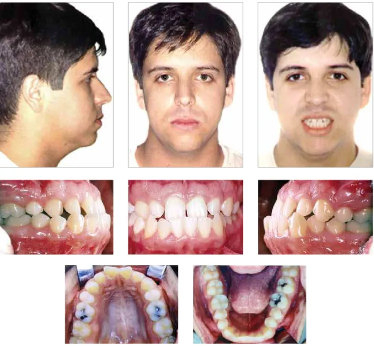
A 22-year and 10-month-old male patient ar-rived for his initial examination in good general health, complaining about his smile, particularly an anterior crossbite and maxillary diastemas, as well as difficulties associated with mastication. His den-tal history included the extraction of maxillary first premolars at the age of 12, carried out by a clinical dentist due to lack of adequate space for eruption of maxillary canines.
Figure 1 - Initial facial and intraoral photographs.
DIAGNOSIS
The examination of temporomandibular joints re-vealed bilateral clicking at mandibular opening and closing, maximal mouth opening of 43 mm and an ir-regular path, but no pain.
Intraoral clinical examination revealed adequate oral hygiene. Malocclusion was classii ed as Angle Class I with anterior crossbite, absence of maxillary i rst
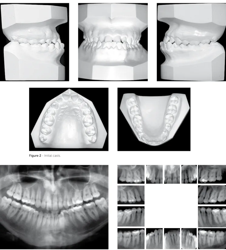
premolars, canines in full Class III relationship, anterior mandibular crowding, rotated maxillary central incisors and anterior diastemas (Figs 1 and 2). There were no dif erences between usual maximal intercuspation and centric relation. Radiographs showed that the patient had good dental and periodontal health and no end-odontic problem or bone loss (Figs 3 and 4).
Figure 2 - Initial casts.
Figure 4 - Initial periapical radiographs.
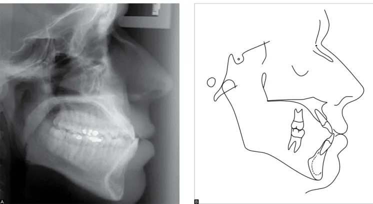
Cephalometry revealed that the maxillomandibu-lar relationship was apparently normal (ANB = 2°) and that a few angles were slightly greater than normal (Conv. = 5.5°; SNA = 86°; SNB = 84°) (Fig 5). However, the ANB angle is known to be markedly af ected by geo-metrical factors.1 When the cranial base is short, maxil-lomandibular discrepancies cannot be evaluated on the sagittal plane using the ANB angle (Fig 6). Other ceph-alometric parameters (Wits = -8 mm; S-N = 71.5 mm) and particularly facial analysis should be used to eluci-date this confounding factor.2,3
When evaluated by cephalometry and having the cranial base as reference, maxillary and mandibular incisors showed buccal inclination and marked pro-trusion (1-NA = 25°, 1-NA = 7 mm) in mandibu-lar teeth (1-NB = 34°, 1-NB = 11 mm). In contrast, the inclination of mandibular incisors in relation to the mandibular plane was good and met the Brazilian standards (IMPA = 94°).2
TREATMENT PLAN
The i rst treatment plan presented to the patient was the orthodontic combined with orthognathic surgery,
which the patient promptly refused. For this reason, an alternative plan was suggested. It included orthodontic camoul age with orthodontic appliances in both arches and extraction of mandibular i rst premolars. The pa-tient had undergone extraction of maxillary i rst premo-lars and, therefore, our aim was to achieve normal molar and canine occlusion. Mandibular extractions followed by retraction of anterior teeth should be supported by adequate anchorage control.
The dentist and the patient agreed on the follow-ing objectives for the treatment selected: preservation of maxillary and mandibular bones position; align-ment and reduction in maxillary diastemas; alignalign-ment of mandibular teeth; normal occlusion, correction of negative overjet and functional occlusion; esthetic im-provement at er lower lip retraction; and achievement of a pleasant smile.
Treatment plan was divided into the follow-ing phases: modii ed Nance lingual arch (away from mandibular incisors); i xed orthodontic appli-ances in both arches using the straight-wire system and 0.022 x 0.028-in slots; extraction of mandibu-lar i rst premomandibu-lars; tooth leveling and alignment with
Figure 5 - A) Initial cephalometric profile radiograph and B) cephalometric tracing.
0.012-in, 0.014-in and 0.016-in nickel-titanium wires and 0.018-in, 0.020-in and 0.017 x 0.025-in stainless steel wires; retraction of mandibular anterior teeth us-ing slidus-ing mechanics and 0.019 x 0.025-in stainless steel wire; removal of lingual arch; orthodontic treat-ment inishing; retention.
TREATMENT PROGRESSION
Treatment progression was in accordance with the plan. Mandibular second molars were included in initial
leveling to aggregate an anchorage unit for the retrac-tion of incisors. Maxillary second molars were bonded and included in leveling during orthodontic inishing.
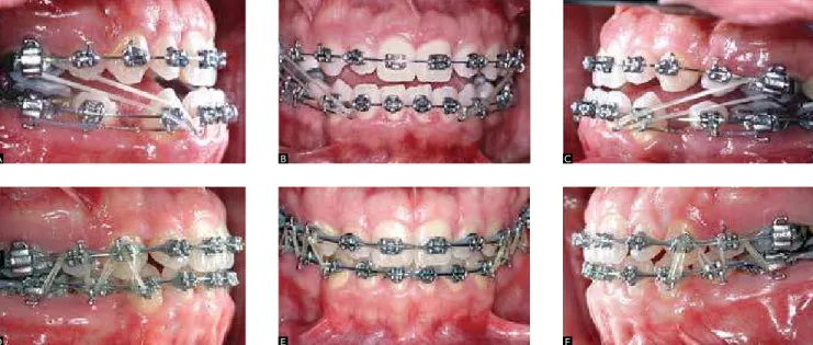
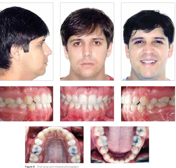
The vertical dimension had to be temporarily in-creased with glass-ionomer cement built-up on pos-terior teeth. This procedure was used for retraction of mandibular anterior teeth because anterior crossbite and marked overjet were obstacles to movement (Figs 7A, B and C). Spaces were closed by means of sliding me-chanics (0.019 x 0.025-in wire) and hooks were soldered between canines and lateral incisors. Class III intermax-illary elastics (¼-in, medium force) were used to con-trol anchorage together with the lingual arch which was removed ater retraction of anterior teeth and closing of extraction spaces. No skeletal anchorage was used. Treatment was completed with 0.018-in archwires, elastic chains in both arches to retain interproximal con-tacts, and Class II intermaxillary elastics (5/6-in, me-dium force) to retain the movement achieved (Figs 7D, E and F). Ater orthodontic completion, intercuspation was good, and canine and molar occlusion relationships, as well as overjet, were normal (Fig 8, 9). Maxillary (2 x 2) and mandibular (4 x 4) V-looped braided bonded lingual archwires were placed for retention.
Figure 6 - Diagram illustrating Class III skeletal relationship with short (N’) and normal (N) anterior cranial bases.
RESULTS
The inal radiograph showed that root parallelism was good ater space closure and that root size was pre-served (Figs 10 and 11).
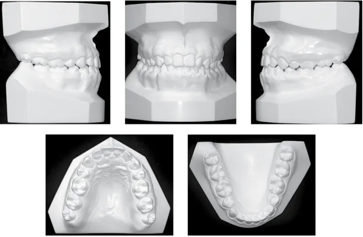
In the maxillary arch, diastemas were reduced, molars were slightly extruded, intercanine distance (35.5 mm) was preserved and intermolar distance was shortened
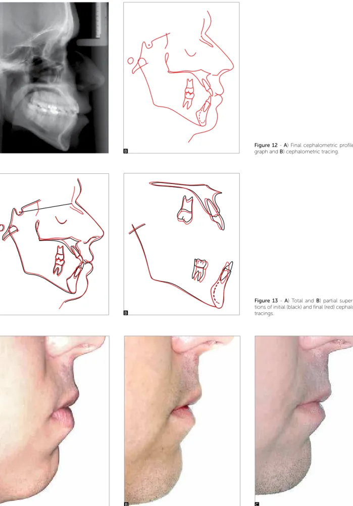
(from 43.5 mm to 42.0 mm). A marked cephalometric ef-fect was found in the mandibular arch with anterior retrac-tion, intrusion and mesial movement of mandibular mo-lars (Fig 12 and Table 1). However, intercanine (21.5 mm) and intermolar (33.0 mm) distances did not change.
There were no signiicant changes in the position of the maxilla or the mandible (Fig 13). Facial
Figure 9 - Final casts.
Figure 10 - Final panoramic radiograph. Figure 11 - Final periapical radiographs of maxillary and mandibular incisors.
ics improved due to less marked lower lip protrusion, coni rmed by reduction of 2.5 mm in the cephalo-metric variable that describes the lower lip (S line) (Fig 14 and Table 1).
The relationship between the maxilla and the man-dible showed good intercuspation and coordination, although sagittal skeletal discrepancy was camoul aged
Figure 12 - A) Final cephalometric profile radio-graph and B) cephalometric tracing.
Figure 13 - A) Total and B) partial superimposi-tions of initial (black) and final (red) cephalometric tracings.
Figure 14 - Comparison of facial profile close-up: A) initial, B) final and C) control eight years later.
A
A A
B
B B
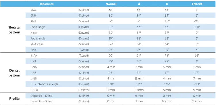
Table 1 - Initial (A) and final (B) cephalometric values.
Measures Normal A B A/B dif.
Skeletal pattern
SNA (Steiner) 82° 86° 85° 1°
SNB (Steiner) 80° 84° 83° 1°
ANB (Steiner) 2° 2° 2.5° -0.5°
Facial angle (Downs) 0° 5,5° 6.5° -1.0°
Y axis (Downs) 59° 57° 57° 0°
Facial angle (Downs) 87° 93° 92° 1°
SN-GoGn (Steiner) 32° 34° 34° 0°
FMA (Tweed) 25° 26° 23° 3°
Dental pattern
IMPA (Tweed) 90° 94° 78° 16°
1.NA (Steiner) 22° 26° 25° 1°
1-NA (Steiner) 4 mm 7 mm 6 mm 1 mm
1.NB (Steiner) 25° 34° 17° 17°
1-NB (Steiner) 4 mm 11 mm 4 mm 7 mm
1.1 – Interincisal angle (Downs) 130° 115° 135° -20°
1-APo (Ricketts) 1 mm 10 mm 5 mm 5 mm
Profile Upper lip – S line (Steiner) 0 mm 0 mm 0 mm 0 mm
Lower lip – S line (Steiner) 0 mm 3 mm 0.5 mm 2.5 mm
1. Hussels W, Nanda RS. Analysis of factors afecting angle ANB. Am J
Orthod. 1984;85(5):411-23.
2. Martins DR, Janson GRP, Almeida RR, Pinzan A, Henriques JFC, Freitas
MR. Atlas de crescimento craniofacial. 1a ed. São Paulo: Ed. Santos; 1998.
3. Arnett GW, Gunson MJ. Facial planning for orthodontistis and oral
surgeons. Am J Orthod Dentofacial Orthop. 2004;126(3):290-5.
4. Benyahia H, Azaroual MF, Garcia C, Hamou E, Abouqal, R, Zaoui F.
Treatment of skeletal Class III malocclusions: orthognathic surgery or orthodontic camoulage? How to decide. Int Orthod. 2011;9(2):196-209.
5. Burns NR, Musich DR, Martin C, Razmus T, Gunel E, Ngan P. Class III
camoulage treatment: what are the limits? Am J Orthod Dentofacial Orthop. 2010;137(1):9.e1-13; discussion 9-11.
REFERENCES
FINAL CONSIDERATIONS
Cranial base abnormalities strongly afect the inter-pretation of cephalometric variables in this region, par-ticularly SNA, SNB, ANB and convexity angle. Other cephalometric parameters, correction factors and, above all, facial analysis indings contributed to making the diagnosis and developing a treatment plan. In adults, Class III skeletal patterns may oten be treated with