Abstract
Tendon rupture is a common injury. Inadequate endogenous repair often leaves patients symptomatic, with tendons susceptible to re-rupture. Administration of certain growth factors improves tendon healing in animal models, but their delivery remains a challenge. Here we evaluated the delivery of TGF-β1 to tendon defects by the implantation of genetically modifi ed muscle grafts. Rat muscle biopsies were transduced with recombinant adenovirus encoding TGF-β1 and grafted onto surgically transected Achilles tendons in recipient animals. Tissue regenerates were compared to those of controls by biomechanical testing as well as histochemical and immunohistochemical analyses. Healing was greatly accelerated when genetically modifi ed grafts were implanted into tendon defects, with the resulting repair tissue gaining nearly normal histological appearance as early as 2 weeks postoperatively. This was associated with decreased deposition of type III collagen in favour of large fi bre bundles indicative of type I collagen. These differences in tendon composition coincided with accelerated restoration of mechanical strength. Tendon thickness increased in gene-treated animals at weeks 1 and 2, but by week 8 became signifi cantly lower than that of controls suggesting accelerated remodelling. Thus localised TGF-β1 delivery via adenovirus-modifi ed muscle grafts improved tendon healing in this rat model and holds promise for clinical application.
Keywords: Transforming growth factor-β; tendon healing; gene transfer; muscle graft; adenovirus
*Address for Correspondence: Dr. med. Martin Majewski
Orthopädische Universitätsklinik Basel Spitalstrasse 21
CH-4031 Basel, Switzerland
Telephone number: 0041 61 328 78 13 Fax number: 0041 61 328 78 09 E-mail: [email protected]
Introduction
Tendon ruptures are common sporting injuries (Schepsis et al., 2002). Such injuries have a certain ability to heal spontaneously via endogenous biological processes that occur during three overlapping phases: infl ammation, regeneration, and remodelling coupled to maturation. During the fi rst two phases, infl ammatory and then mesenchymal cells enter the lesion where they proliferate, differentiate into tenocytes and lay down a collagenous matrix (Kajikawa et al., 2007). Initially, this matrix contains a high proportion of type III collagen, characterised by small, poorly cross-linked fi brils that provide limited mechanical strength. During remodelling and maturation, these are replaced by larger, highly cross-linked fi bres of type I collagen that provide considerable tensile strength. Although a functional tendon regenerate forms within 6-8 weeks of rupture, complete regeneration of the tendon is never achieved, leading to re-rupture in up to 10 % of cases (Longo et al., 2009; Majewski et al., 2002). Advances in the treatment of tendon rupture, such as percutaneous suturing (Ma and Griffi th, 1977) or functional treatment (Thermann et al., 1995) have improved outcomes, but it has become increasingly clear that further improvements in the healing of tendon ruptures will require the implementation of novel, biological approaches (DeFranco et al., 2004; James et al., 2008; Rees et al., 2009). It is particularly important to accelerate the initial stages of healing, as re-rupture of repaired tendons normally occurs early in the healing process.
A number of different growth factors have been shown to promote one or more of the biological processes involved in tendon healing (Evans, 1999; Molloy et al., 2003). These include fi broblast growth factor-2 (FGF-2) (Chan et al., 2000), platelet-derived growth factor (PDGF) (Thomopoulos et al., 2007), bone morphogenetic protein (BMP)-12 (growth and differentiation factor (GDF)-7; cartilage-derived growth factor (CDGF)-3) (Lou et al., 2001), BMP-13 (GDF-6; CDMP-2) (Forslund and Aspenberg, 2001), BMP-14 (GDF-5; CDMP-1) (Rickert et al., 2001), and transforming growth factor-β (TGF-β) (Kashiwagi et al., 2004). Problems in delivering proteins to sites of tendon injury in a sustained fashion restrain clinical application of such growth factors. Gene transfer provides the basis of a strategy for obviating this limitation (Evans et al., 2004; Hildebrand et al., 2004). In principle, the transfer to the injury site of cDNA encoding appropriate factors could lead to their sustained, local, endogenous production, thereby eliminating the problems associated with the exogenous delivery of proteins.
IMPROVEMENT OF TENDON REPAIR USING MUSCLE GRAFTS TRANSDUCED
WITH TGF-
β
1 cDNA
Martin Majewski1*, Ryan M. Porter2,4, Oliver B. Betz2, Volker M. Betz2, Harald Clahsen3, Rudolf Flückiger2
and Christopher H. Evans2,4
1 Orthopädische Klinik, Universität Basel, Basel, Switzerland
2 Center for Molecular Orthopaedics, Brigham and Women’s Hospital, Harvard Medical School, Boston, USA 3 Anatomisches Institut, Universität Düsseldorf, Düsseldorf, Germany
4 Center for Advanced Orthopaedic Studies, Beth Israel Deaconess Medical Center, Harvard Medical School, Boston,
Data from several previous experimental studies confi rm the merit of this approach for delivering potentially reparative cDNAs to injured tendons. Relevant genes include those encoding BMP-12 (Lou et al., 2001; Majewski et al., 2008), BMP-14/GDF-5 (Basile et al., 2008; Bolt et al., 2007; Rickert et al., 2005), FGF-2 (Tang et al., 2008), interleukin-10 (Ricchetti et al., 2008), TGF-β1 (Hou et al., 2009b), insulin-like growth factor-1 (Schnabel et al., 2009), vascular endothelial growth factor (Hou et al., 2009a), and PDGF (Suwalski et al., 2010). Most such studies have used viral vectors for gene transfer because of their high effi ciency. These vectors can be used by direct, in vivo application or in an indirect, ex vivo fashion. Although promising data have been reported following in vivo delivery (Bolt et al., 2007; Lou et al., 2001; Ricchetti et al., 2008; Rickert et al., 2005; Suwalski et al., 2010; Tang et al., 2008) tendons have low cellularity, a circumstance that limits opportunities for in vivo gene transfer. Moreover, in vivo gene transfer raises greater safety concerns when clinical application is considered.
Ex vivo delivery strategies often use mesenchymal stem cells (MSCs) as vehicles (Hou et al., 2009a; Hou et al., 2009b; Schnabel et al., 2009), but in a clinical setting such manipulations are cumbersome, expensive and invasive. In response to these considerations, we have been interested in developing expedited ex vivo gene delivery technologies that can be accomplished conveniently in a single operative session (Evans et al., 2007). One such approach involves the genetic modifi cation of autologous muscle biopsies (Evans et al., 2009; Evans et al., 2007). As well as providing a convenient tissue for expedited gene transfer, muscle contains progenitor cells with the potential to contribute to tendon healing (Pelinkovic et al., 2003).
In previous experiments we have used a recombinant adenovirus vector to deliver BMP-12 cDNA to muscle biopsies that were then sutured to sites of Achilles tendon rupture in a rat model (Majewski et al., 2008).BMP-12 was selected as a morphogen that promotes the differentiation of progenitor cells into tenocytes (Violini et al., 2009; Wang et al., 2005). In the present experiments, we have used adenovirus to deliver TGF-β1 cDNA as a growth factor that promotes a different component of the healing process, the deposition of a collagenous matrix.
Materials and Methods
Study design
We used 133 adult male Sprague-Dawley rats, each weighing 400-425 g. Each experimental group consisted of 40 rats. The fi rst group of 40 animals (surgical control) received no muscle graft after transection and suturing of the tendon. The second group (muscle control) received a muscle graft without modification by recombinant adenovirus. The third group (Ad.TGF-β1 group) received a muscle graft transduced with a recombinant adenovirus carrying TGF-β1 cDNA (Ad.TGF-β1). Ten rats per timepoint were sacrifi ced after 1, 2, 4 and 8 weeks. For each time-point, 7 animals were used for biomechanical testing and 3 animals for histological examination. Four
additional rats were used for muscle harvesting and 9 rats were used to test for transduction after 1, 3 and 7 d in vitro. Approval for all procedures was obtained beforehand from the local Institutional Animal Care and Use Committee (Boston, MA, USA).
Vector preparation
A fi rst generation (E1/E3-deleted), serotype 5, recombinant adenoviral vector carrying the cDNA for human TGF-β1 (Ad.TGF-β1) was constructed by Cre-lox recombination as described earlier (Hardy et al., 1997). The transgene, under transcriptional control of the human cytomegalovirus immediate early promoter, was inserted into the E1 domain. Recombinant adenoviruses were propagated in 293/Cre8 cells. High-titer preparations were generated by amplifi cations in 293 cells, purifi cation on CsCl gradients, and dialysis against 4 % sucrose with 10 mM Tris-HCl (pH 7.8), 150 mM NaCl, and 10 mM MgCl2.
Transduction of muscle graft
Skeletal muscle tissue was harvested with a round punch of 4 mm diameter from a donor rat 1 d before Achilles tendon surgery. To transduce the muscle discs, 5 x 10¹⁰
Ad.TGF-β1 particles were dropped onto the surface of the muscle, and the grafts were incubated for 30 min at 37 °C and 5 % CO2 without further dilution to enhance muscle cell modifi cation. After this incubation, the muscle grafts were cultured in 1 mL DMEM medium containing 5 % foetal bovine serum in 24-well plates for 1 d in an incubator with 37 °C and 5 % CO2. To confi rm transgene expression, additional muscle grafts were transduced as described above, and conditioned media samples were tested after 1, 3 and 7 d for TGF-β1 secretion by ELISA (R&D Systems, Minneapolis, MN, USA).
Surgical procedure
Rats were placed under general anaesthesia by the administration of isofl urane using a precision vapouriser. The animals then received intramuscular injections of 20 mg/kg cefazolin (antibiotic) and 0.06 mg/kg buprenorphine (analgesic) into the left thigh. Each animal’s right hind leg was shaved and disinfected with Betadine®-Scrub 3x using aseptic techniques and rinsed with 70 % alcohol. Subsequently, it was transferred to a heated surgery table and covered with a sterile surgical drape such that only the prepared limb was exposed.
Post-operative treatment
The animals woke up on a heating pad to aid the return to normal body temperature. During the next three days, animals received 20 mg/kg i.m. injections of cefazolin and 0.06 mg/kg i.m. injections of buprenorphine, each twice per day. For the duration of the experiment, all animals were monitored for signs of pain and infection. After the operation, no cast or dressing was applied. The animals were allowed unrestricted cage movement and provided rat chow and water ad libitum.
Euthanasia
Groups of animals were euthanised after 7, 14, 28 and 56 d by CO2 asphyxiation under general anaesthesia and subsequent bilateral thoracotomy. Muscle–Achilles tendon–bone units were harvested by transection of the middle part of the gastrocnemius muscle and of the calcaneus.
Mechanical testing
The muscle-tendon-bone units were wrapped in cotton gauze soaked in lactated Ringer’s solution and stored at -20 °C before testing. On the day of testing, the specimens were thawed in Ringer’s solution for 4 h. Before testing, tendon thickness was measured with a precision calliper at the former site of tendon repair. The muscle-tendon-bone units were fastened in the clamping device by freezing the muscular segment between the cryo-jaws and by fi xing the bony segment between the copper clamp (Wieloch et al., 2004). The clamping device was attached to an electro-hydraulic materials testing machine (Fa. Zwick, Einsingen-Ulm, Germany). The tendons were not preconditioned or cyclically stretched prior to tensile testing. The room temperature was kept constant at 25 °C. To prevent the specimens from dehydrating they were kept moist with Ringer’s solution. Both reservoirs of the cryo-jaws were
fi lled with liquid nitrogen. The experiment was started as soon as the expansion of the freezing-zone reached the border of the metal clamp but did not extend into the tendon substance or into the repair site (registered manually with a metal-needle). The displacement rate was set at 1000 mm/min. Force-displacement curves were recorded and transferred to an IBM-compatible computer for subsequent data analysis. Load to failure (N, peak of the curve) and stiffness (N/mm) were measured. Load values were not corrected for area of tendon.
Histology
For each group 3 specimens were examined per time point per group, with 5 sections for each specimen, giving a total of 15 sections per time point per group. The tendons were immediately fi xed in 4 % buffered formalin (pH 7.4) for 24 h, dehydrated and embedded in paraffi n wax. Longitudinal sections (5 μm) at the mid-substance of the tendon were stained with haematoxylin and eosin. Collagen III was specifi cally detected using the primary rabbit anti-rat collagen type III polyclonal antibody (1:300, AbD Serotec, Dusseldorf, Germany) and visualised with biotinylated goat anti-rabbit IgG antibodies (1:50, B6648, E8386, Sigma, Hamburg, Germany) and extavidin peroxidase.
analysing software (Image-Pro Plus, Media Cybernetics, Silver Spring, MD, USA). The results are expressed as percentage of the total area staining for type III collagen. Additional immunohistochemical staining for TGF-β1 (rabbit polyclonal; Santa Cruz, CA, USA) was performed.
Cross polarisation microscopy
For cross polarisation microscopy, the same number of sections was used as for histology (15 per time point per group). 5 μm sections were stained for 1 h in picrosirius solution (0.1 % solution of Sirius Red F3BA in saturated aqueous picric acid, pH 2) according to the method of Junqueira (Junqueira et al., 1979). The sections were washed for 2 min in 0.01 N HCl, dehydrated, cleared and mounted in synthetic resin. To analyse collagen ratios, tissue samples were observed under polarised light microscopy. Thicker mature collagen fi bres, including type I collagen fi bres, appeared red-orange, whereas thinner collagen fi bres, including type III collagen appeared pale-green (Junqueira et al., 1978).
For each sample 5 regions (centre of tendon, left border, right border, 2 mm above tendon centre, 2 mm below tendon centre) within the interface (400x, area 100 μm x 100 μm) were captured by a digital camera (Olympus C-3030, Hamburg, Germany). Collagen ratios were obtained by measuring the area of thicker mature red-orange versus thinner pale-green using a digital image analysing software (Image-Pro Plus, Media Cybernetics, Silver Spring, MD, USA).
Semiquantitative histological analysis
For additional objective information, histological sections were analysed with a scoring system based on the Bonar score, validated by Maffulli et al. (Maffulli et al., 2008). Sections were evaluated for tenocyte appearance (0 = normal to 3 = large round nucleus with abundant cytoplasm), collagen ordering (0 = normal to 3 = separation of fi bres with complete loss of architecture) and number of cells. Two blinded investigators performed the evaluation. Mean tenocyte appearance and collagen ordering, as well as mean cell count divided by ten were averaged to give a score for each time point.
Statistics
The Mann Whitney test was used to test for statistically signifi cant differences between the cohorts. The level of signifi cance was set to p < 0.05.
Results
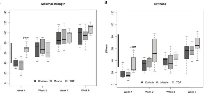
time point, the strength of defects treated with TGF-β cDNA was already well within the normal range of 60-80 N (Fig. 1A). Defects treated with TGF-β cDNA also had much greater stiffness at weeks 1 and 2 (Fig. 1B) and were within the 30-85 N/mm range of normal tendon at 2 weeks. Genetic delivery of TGF-β also increased tendon thickness at weeks 1 and 2, after which time thickness declined. By 4 weeks, there was no signifi cant difference from controls. At 8 weeks, the regenerate formed in response to the TGF-β cDNA was less thick than the healing controls, although it was still thicker than normal tendon (Fig. 2).
Histological examination of the repair tendon tissue showed that by 2 weeks delivery of TGF-β cDNA led to the formation of well-organised, tendinous tissue with large collagen fibres. Control specimens, in contrast, showed more tissue with a disorganised matrix and greater cellularity (Fig. 3). Scoring of the histological slides displayed better scores for the TGF group from 2 weeks onwards (Fig. 4). Quantitative immunohistochemistry (Fig. 5) confi rmed that delivery of TGF-β cDNA suppressed
the expression of type III collagen that is associated with repair tissue. The collagenous components of the matrix were further investigated by polarised light microscopy in conjunction with sirius red staining (Fig. 3). Two weeks post-operatively the majority of the collagen fi bres in the defects treated with TGF-β cDNA stained orange, indicating the presence of large fi bres indicative of type I collagen. The fibres of control defects, in contrast, contained a considerable proportion of smaller fi bres staining green, indicative of type III collagen (Fig. 6).
Fig. 1. Effect of TGF-β1 cDNA delivery on the mechanical properties of the rat Achilles tendon at progressive
times after transection. (A) Maximum load to failure and (B) stiffness were determined using explanted muscle-tendon-bone units. For each plot, the thick horizontal bars within each box indicate the median, the top and bottom of each box indicate the upper quartile and the lower quartile, and the outer bars show the highest and lowest values that were measured. Statistical signifi cance is indicated by an asterisk. Range of normal tendon is indicated by the black bar on the y-axis.
A B
Fig. 2. Effect of TGF-β1 cDNA delivery on the thickness
Fig. 3. Effect of TGF-β1 cDNA delivery on the histological appearance of the repair site in the rat Achilles tendon.
H&E sections: Compared to untreated lesions (Control) and lesions receiving untransduced muscle (Muscle), the TGF-β1 group (TGF-β1) showed larger collagen bundles in a parallel orientation, generally reduced number of cells between the collagen bundles and less scar tissue. Sirius Red sections under polarised light: large collagen
fi bres stain orange-red and small fi brils stain green. Remodelling of the matrix was much slower in untreated lesions (Control) and lesions receiving untransduced muscle (Muscle) than in the TGF-β1 group (TGF-β1), as indicated by the greater fraction of thinner, green-staining fi brils relative to red-orange fi bres. Immunohistochemical staining for TGF-β1: the TGF-β1 group displays the greatest amount of stained TGF-β1 compared to untreated lesions (Control) or lesions receiving untransducted muscle (Muscle). Black and striped bars in the H&E images represent 200 μm, whereas black and striped bars in the sirius red and immunohistochemical images represent 100 μm.
●
●
●
●
1 2 3 4 5 6 7 8
1.0
1.5
2.0
2.5
3.0
3.5
Histological Score
Week
Score
● Control
Muscle TGF
●
●
●
●
1 2 3 4 5 6 7 8
0
1
02
03
04
05
0
Collagen III
Week
T
ype III collagen (in % of total)
● Controls Muscle TGF
Fig. 4. Semiquantitative scoring of histological slides. Semiquantitative scoring for tenocyte appearance, collagen organisation and cell count was performed for all histological slides. Mean values are displayed for each time point and group.
Fig. 5. Effect of TGF-β1 cDNA delivery on the
Discussion
These data suggest that ex vivo delivery of TGF-β1 cDNA to sites of tendon rupture using muscle biopsies accelerates the early repair response of rat Achilles tendon. This was seen as an early recovery of mechanical strength which, in turn, is likely to refl ect the precocious deposition of type I collagen and the attenuated deposition of type III collagen. Histologically, the matrix showed greater organisation, lower cellularity and an emerging crimp pattern. A rapid increase in mechanical strength is important clinically, because re-rupture of a tendon commonly occurs early in the repair process.
Although we did not measure the duration of TGF-β1 transgene expression in vivo, genetically modifi ed muscle continued to express high levels of TGF-β1 for at least one week in vitro. This suggests that TGF-β1 expression persists locally within the lesion during the critical early stages of healing, which is consistent with the early gains in mechanical strength and thickness noted here. Under in vivo conditions, fi rst generation adenovirus vectors typically express transgenes at high levels for about 2 weeks. In this context, it is worth noting that no adhesions were observed at the time of explantation in the treatment group. Adhesions form as a result of prolonged exposure to TGF-β1. If exposure to TGF is short, as is likely to be the case here, it may improve biomechanical stability without increasing adhesion formation. Exposure to TGF-β1 during this time-frame would also explain the rapid increase in thickness of the genetically-treated regenerate, accompanied by increases in stiffness and strength. By 8
weeks these differences have largely disappeared, probably due to tapering of TGF-β1 expression and catching up by the innate healing process. The important point, however, is the dramatic early increase in these properties, during the time when the tendon is most liable to re-rupture.
These data are in qualitative agreement with those of Hou et al. (Hou et al., 2009a), who employed adenovirus to deliver TGF-β cDNA in an ex vivo fashion to injured rabbit Achilles tendon using MSCs. Like us, they found that TGF-β cDNA accelerated healing, based upon histology, collagen analysis and mechanical strength. In a subsequent study, the same group (Hou et al., 2009b) confi rmed that delivery of TGF-β cDNA promoted the synthesis of type I collagen and improved the ratio of type I to type III collagen. In these studies, TGF-β expression was detected up to 56 d. In the present study, TGF-β expression in transfected muscle was measured for a period of 7 d and might be increased beyond this time point. However, immunohistochemical staining for TGF-β confi rmed its elevated presence only in the fi rst week.
Compared to the genetic delivery of BMP-12 in this model, which we examined in an earlier study (Majewski et al., 2008), TGF-β gives a larger increase in mechanical strength during the first week, but in other respects the results are strikingly similar. Given that BMP-12 and TGF-β are likely to act on different components of the healing process, they have the potential to act synergistically when co-administered. This possibility has not yet been examined experimentally. Alternative isoforms of TGF-β also remain to be evaluated. Transfer of cDNAs encoding BMP-14/GDF-5 (Bolt et al., 2007; Rickert et al., 2005), PDGF (Suwalski et al., 2010), and FGF-2 (Tang et al., 2008) have also shown promise in treating animal models of tendon damage, but differences in the models, methods of gene delivery, and other variables preclude direct comparison with the present work.
The precise mechanisms through which TGF-β expression accelerates healing in our model are unknown. From what is known about the properties of TGF-β we can suggest that it enhances collagen synthesis and the differentiation of mesenchymal progenitor cells. Muscle is a rich source of such cells, and could thus provide progenitors to the healing tendon. Histology did not provide evidence for this, but it should be a subject of future research.
A limitation of the present study is the use of transected rat Achilles tendon, which does not accurately refl ect human Achilles tendon tears that arise as a result of tendinopathy (Lui et al., 2011). There is a struggle to fi nd an appropriate model for the human condition, which has not yet produced satisfying results (Longo et al., 2011). However, it is crucial to understand this vastly complex system of healing on a simplifi ed level, for which precisely this simplifi ed injury model has been chosen.
A further limitation was that sutures had not dissolved completely by the time of biomechanical testing. However, since all tendons were sutured in the exact same way, differences in load bearing capacity were only due to the different tissue properties at the site of repair.
●
●
●
●
1 2 3 4 5 6 7 8
02468
1
0
Ratio of Collagen I to Collagen III
Week
col I / col III
● Controls Muscle TGF
Regardless of the transgene that is used, successful translation of these concepts into the clinic will require careful attention to additional matters such as cost, safety and practicality (Evans et al., 2007). As noted, ex vivo delivery using expanded, autologous cells and the in vivo delivery of most viral vectors are likely to be problematic. In response to these constraints, Basile et al. (Basile et al., 2008) have developed a technology whereby recombinant adeno-associated virus (AAV) vectors are freeze-dried onto allograft tendon, thus forming a construct that can be implanted into a defect. Because AAV is relatively stable, such constructs have the potential to provide an off the shelf product. Proof of principle was demonstrated in a murine, fl exor digitorum longus tendon model with GDF-5 as the transgene. The muscle biopsy method we used in the present work provides an additional strategy. In a clinical setting, the autologous muscle biopsy could be harvested, genetically modifi ed and implanted in a single operative session, thereby reducing cost and complexity. Before contemplating human application, it is necessary to consider the limitations of rodent models such as the one used here, and the need for large animal studies (Evans, 2011). Moreover, the use of gene therapy always raises safety concerns (Evans et al., 2011).
Acknowledgments
We are grateful to Mrs H. Schaller and Mrs C. Pilapil for histology preparation, Mrs E. Krott for assistance during histology examination. Dr. L. Dürselen for help during mechanical testing, and Mr A. Todorov for proofreading and help with graphics. This work was supported by NIH grant number R01 AR052809 to CHE from NIAMS.
References
Basile P, Dadali T, Jacobson J, Hasslund S, Ulrich-Vinther M, Soballe K, Nishio Y, Drissi MH, Langstein HN, Mitten DJ, O’Keefe RJ, Schwarz EM, Awad HA (2008) Freeze-dried tendon allografts as tissue-engineering scaffolds for Gdf5 gene delivery. Mol Ther 16: 466-473.
Bolt P, Clerk AN, Lu u HH, Kang Q, Kummer JL, Deng Z-L, Olson K, Primus F, Montag AG, He T-C, Haydon RC, Toolan BC (2007) BMP-14 gene therapy increases tendon tensile strength in a rat model of Achilles tendon injury. J Bone Joint Surg Am 89: 1315-1320.
Chan BP, Fu S, Qin L , Lee K, Rolf CG, Chan K (2000) Effects of basic fi broblast growth factor (bFGF) on early stages of tendon healing: a rat patellar tendon model. Acta Orthop Scand 71: 513-518.
DeFranco MJ, Derwin K, Iannotti JP (2004) New therapies in tendon reconstruction. J Am Acad Orthop Surg
12: 298-304.
Evans CH (1999) Cyto kines and the role they play in the healing of ligaments and tendons. Sports Med 28: 71-76.
Evans CH (2011) Barr iers to the clinical translation of orthopedic tissue engineering. Tissue Eng Part B Rev
Evans CH, Ghivizzani SC, Robbins PD (2004) The 2003 Nicolas Andry Award. Orthopaedic gene therapy. Clin Orthop Relat Res 429: 316-329.
Evans CH, Palmer GD, Pascher A, Porter R, Kwong FN, Gouze E, Gouze JN, Liu F, Steinert A, Betz O, Betz V, Vrahas M, Ghivizzani SC (2007) Facilitated endogenous repair: making tissue engineering simple, practical, and economical. Tissue Eng 13: 1987-1993.
Evans CH, Liu FJ, Gl att V, Hoyland JA, Kirker-Head C, Walsh A, Betz O, Wells JW, Betz V, Porter RM, Saad FA, Gerstenfeld LC, Einhorn TA, Harris MB, Vrahas MS (2009) Use of genetically modifi ed muscle and fat grafts to repair defects in bone and cartilage. Eur Cell Mater 18: 96-9111.
Evans CH, Ghivizzani SC, Robbins PD (2011) Orthopaedic gene therapy – lost in translation? J Cell Physiol 227:416-420.
Forslund C, Aspenberg P (2001) Tendon healing stimulated by injected CDMP-2. Med Sci Sports Exerc
33: 685-687.
Hardy S, Kitamura M, Harris-Stansil T, Dai Y, Phipps ML (1997) Construction of adenovirus vectors through Cre-lox recombination. J Virol 71: 1842-1849.
Hildebrand KA, Frank CB, Hart DA (2004) Gene intervention in ligament and tendon: current status, challenges, future directions. Gene Ther 11: 368-378.
Hou Y, Mao Z, Wei X, Lin L, Chen L, Wang H, Fu X, Zhang J, Yu C (2009a) Effects of transforming growth factor-beta1 and vascular endothelial growth factor 165 gene transfer on Achilles tendon healing. Matrix Biol 28: 324-335.
Hou Y, Mao Z, Wei X, Lin L, Chen L, Wang H, Fu X, Zhang J, Yu C (2009b) The roles of TGF-beta1 gene transfer on collagen formation during Achilles tendon healing. Biochem Biophys Res Commun 383: 235-239.
James R, Kesturu G, Balian G, Chhabra AB (2008) Tendon: biology, biomechanics, repair, growth factors, and evolving treatment options. J Hand Surg Am 33: 102-112.
Junqueira LC, Cosser melli W, Brentani R (1978) Differential staining of collagens type I, II and III by Sirius Red and polarization microscopy. Arch Histol Jpn 41: 267-274.
Junqueira LC, Bignolas G, Brentani RR (1979) Picrosirius staining plus polarization microscopy, a specifi c method for collagen detection in tissue sections. Histochem J 11: 447-455.
Kajikawa Y, Morihara T, Watanabe N, Sakamoto H, Matsuda K, Kobayashi M, Oshima Y, Yoshida A, Kawata M, Kubo T (2007) GFP chimeric models exhibited a biphasic pattern of mesenchymal cell invasion in tendon healing. J Cell Physiol 210: 684-691.
Kashiwagi K, Mochizu ki Y, Yasunaga Y, Ishida O, Deie M, Ochi M (2004) Effects of transforming growth factor-beta 1 on the early stages of healing of the Achilles tendon in a rat model. Scand J Plast Reconstr Surg Hand Surg 38: 193-197.
Longo UG, Ronga M, M affulli N (2009) Acute ruptures of the achilles tendon. Sports Med Arthrosc 17: 127-138.
Shoulder Pathologies: From Bench to Bedside. Sports Medicine and Arthroscopy Review 19: 184-193
Lou J, Tu Y, Burns M , Silva MJ, Manske P (2001) BMP-12 gene transfer augmentation of lacerated tendon repair. J Orthop Res 19: 1199-1202.
Lui PPY, Maffulli N, Rolf C, Smith RKW (2011) What are the validated animal models for tendinopathy? Scand J Med Sci Sports 21: 3-17.
Ma GW, Griffi th TG ( 1977) Percutaneous repair of acute closed ruptured achilles tendon: a new technique. Clin Orthop Relat Res 128: 247-255.
Maffulli N, Longo UG , Franceschi F, Rabitti C, Denaro V (2008) Movin and Bonar scores assess the same characteristics of tendon histology. Clin Orthop Relat Res
466: 1605-1611.
Majewski M, Widmer K H, Steinbruck K (2002) [Achilles tendon ruptures: 25 year’s experience in sport-orthopedic treatment]. Sportverletz Sportschaden 16: 167-173.
Majewski M, Betz O, Ochsner PE, Liu F, Porter RM, Evans CH (2008) Ex vivo adenoviral transfer of bone morphogenetic protein 12 (BMP-12) cDNA improves Achilles tendon healing in a rat model. Gene Ther 15: 1139-1146.
Molloy T, Wang Y, Mu rrell G (2003) The roles of growth factors in tendon and ligament healing. Sports Med
33: 381-394.
Pelinkovic D, Lee JY , Engelhardt M, Rodosky M, Cummins J, Fu FH, Huard J (2003) Muscle cell-mediated gene delivery to the rotator cuff. Tissue Eng 9: 143-151.
Rees JD, Maffulli N, Cook J (2009) Management of tendinopathy. Am J Sports Med 37: 1855-1867.
Ricchetti ET, Reddy SC, Ansorge HL, Zgonis MH, Van Kleunen JP, Liechty KW, Soslowsky LJ, Beredjiklian PK (2008) Effect of interleukin-10 overexpression on the properties of healing tendon in a murine patellar tendon model. J Hand Surg Am 33: 1843-1852.
Rickert M, Jung M, A diyaman M, Richter W, Simank HG (2001) A growth and differentiation factor-5 (GDF-5)-coated suture stimulates tendon healing in an Achilles tendon model in rats. Growth Factors 19: 115-126.
Rickert M, Wang H, W ieloch P, Lorenz H, Steck E, Sabo D, Richter W (2005) Adenovirus-mediated gene transfer of growth and differentiation factor-5 into tenocytes and the healing rat Achilles tendon. Connect Tissue Res 46: 175-183.
Schepsis AA, Jones H , Haas AL (2002) Achilles tendon disorders in athletes. Am J Sports Med 30: 287-305.
Schnabel LV, Lynch M E, van der Meulen MCH, Yeager AE, Kornatowski MA, Nixon AJ (2009) Mesenchymal stem cells and insulin-like growth factor-I gene-enhanced mesenchymal stem cells improve structural aspects of healing in equine fl exor digitorum superfi cialis tendons. J Orthop Res 27: 1392-1398.
Suwalski A, Dabboue H, Delalande A, Bensamoun SF, Canon F, Midoux P, Saillant G, Klatzmann D, Salvetat J-P, Pichon C (2010) Accelerated Achilles tendon healing by PDGF gene delivery with mesoporous silica nanoparticles. Biomaterials 31: 5237-5245.
Tang JB, Cao Y, Zhu B, Xin K-Q, Wang XT, Liu PY (2008) Adeno-associated virus-2-mediated bFGF gene
transfer to digital fl exor tendons signifi cantly increases healing strength. an in vivo study. J Bone Joint Surg Am
90: 1078-1089.
Thermann H, Frerichs O, Biewener A, Krettek C, Schandelmeier P (1995) [Functional treatment of acute rupture of the Achilles tendon. An experimental biomechanical study]. Unfallchirurg 98: 507-513.
Thomopoulos S, Zaege l M, Das R, Harwood FL, Silva MJ, Amiel D, Sakiyama-Elbert S, Gelberman RH (2007) PDGF-BB released in tendon repair using a novel delivery system promotes cell proliferation and collagen remodeling. J Orthop Res 25: 1358-1368.
Violini S, Ramelli P , Pisani LF, Gorni C, Mariani P (2009) Horse bone marrow mesenchymal stem cells express embryo stem cell markers and show the ability for tenogenic differentiation by in vitro exposure to BMP-12. BMC Cell Biol 10: 29-29.
Wang Q-W, Chen Z-L, Piao Y-J (2005) Mesenchymal stem cells differentiate into tenocytes by bone morphogenetic protein (BMP) 12 gene transfer. J Biosci Bioeng 100: 418-422.
Wieloch P, Buchmann G, Roth W, Rickert M (2004) A cryo-jaw designed for in vitro tensile testing of the healing Achilles tendons in rats. J Biomech 37: 1719-1722.
Discussion with Reviewers
Reviewer I: The thickness of the TGF-β1 treated tendon
never returns close to normal but is similar to untreated tendon by week 8, suggesting an inadequate heal. Why does TGF-β1 not continue to “improve’ the thickness? This has not been discussed at all. Is TGF-β1 the “wrong” isoform, and where does TGF-β3 come into play in the healing process? Do these need to be applied sequentially to improve the heal further?
Authors: The precise mechanisms through which TGF-β
expression accelerates healing in our model are unknown. From what is known about the properties of TGF-β, we can suggest that it enhances collagen synthesis and the differentiation of mesenchymal progenitor cells. Muscle is a rich source of such cells, and could thus provide progenitors to the healing tendon. Histology did not provide evidence for this, but it should be a subject of future research. The literature suggests that all isoforms of TGF-β stimulate the synthesis of types I and III collagen by tendon fi broblasts to a similar degree. There are a few in vivo studies in which the ability of the different isoforms to promote tendon healing has been directly compared. Such data do not suggest large quantitative or qualitative differences between the effects of the different isoforms. However, this matter deserves further experimental investigation. The sequential application of different growth factors as healing progresses is an attractive general concept. However, much more research is needed to determine which factors to add, how much to add, when to add them, and how to add them.
there any quantitiative differences in the fi bril diameter and organisation at 8 weeks, between the controls and the TGF-β transfected biopsies?
Authors: When maintained in tissue culture, muscle discs secreted nanogramme quantities of TGF-β1 into the medium for at least one week. The immunohistochemistry shown in the present article confirms that TGF-β1