SOCIEDADE BRASILEIRA DE ORTOPEDIA E TRAUMATOLOGIA
w w w . r b o . o r g . b r
Technical
Note
Reconstruction
of
the
distal
biceps
tendon
using
triceps
graft:
a
technical
note
夽
Thiago
Medeiros
Storti
∗,
Alexandre
Firmino
Paniago,
Rafael
Salomon
Silva
Faria
HospitalOrtopédicoeMedicinaEspecializada(Home),Servic¸odeCirurgiadeOmbroeCotovelo,Brasília,DF,Brazil
a
r
t
i
c
l
e
i
n
f
o
Articlehistory:
Received26October2015
Accepted29March2016
Availableonline13May2017
Keywords:
Elbow
Tendoninjuries
Reconstructivesurgicalprocedures
Transplantationautologous
Reconstruction
a
b
s
t
r
a
c
t
Ruptureofthedistalbicepsbrachiitendontypicallyoccurinacontractionagainstresistance
withtheelbowin90◦offlexion.Chronicrupturesareuncommonandarecomplicatedby
tendonandmuscleretractionandpoorquality.Somereconstructiontechniqueshavebeen
describedintheliterature,withvariationsonthesurgicalexposures,typeofgraft(alloor
autograft),graftdonorsite,andtypeofattachmenttotheradialtuberosity.Theauthors
reportthecaseofapatientpresentedaruptureofthedistalbicepsbrachiitendonthattook
placefiveweeksearlierand,therefore,underwentreconstructionusingautograftfromthe
centralstripoftricepstendonthroughdoubleincisionandfixationwithanchorstotheradial
tuberosity.Theuseofthetricepsbrachiiasautograftforreconstructionofchronicruptures
ofthedistalbicepshadnotyetbeendescribedintheliterature.Theauthorshavechosento
useitduetoitsbiomechanicalcharacteristicsthatqualifyitassuitableforthisprocedure
andbecausethisiseasierforcollection,usingthesameoperatingfieldatthesamejoint,
minimizingthenegativeeffectsofthedonorarea.Aftersixmonthspostoperatively,the
patienthasfullmovementarcandrestorationof96%oftheflexionstrengthand90%ofthe
supinationstrengthwhencomparedwiththecontralaterallimb.Thisprocedureappears
tobeagoodoptionforcasesofchronicdistalbicepsruptureinolderpatientswhohave
functionaldemandofsupination.
©2017PublishedbyElsevierEditoraLtda.onbehalfofSociedadeBrasileiradeOrtopedia
eTraumatologia.ThisisanopenaccessarticleundertheCCBY-NC-NDlicense(http://
creativecommons.org/licenses/by-nc-nd/4.0/).
Reconstruc¸ão
do
tendão
distal
do
bíceps
com
enxerto
do
tríceps:
nota
técnica
Palavras-chave:
Cotovelo
Traumatismosdostendões
r
e
s
u
m
o
Rupturasdotendão distaldobícepsbraquial ocorremtipicamentecomumacontrac¸ão
contrarresistênciacomocotoveloem90◦ deflexão.Rupturascrônicassãolesões
inco-munsesãocomplicadaspelaretrac¸ãoepobrequalidadetendíneaemuscular.Algumas
夽
StudyconductedattheHospitalOrtopédicoeMedicinaEspecializada(Home),Servic¸odeCirurgiadeOmbroeCotovelo,Brasília,DF,
Brazil.
∗ Correspondingauthor.
E-mail:[email protected](T.M.Storti).
http://dx.doi.org/10.1016/j.rboe.2016.03.010
2255-4971/©2017PublishedbyElsevierEditoraLtda.onbehalfofSociedadeBrasileiradeOrtopediaeTraumatologia.Thisisanopen
Procedimentoscirúrgicos reconstrutivos
Transplanteautólogo
Reconstruc¸ão
técnicasdereconstruc¸ãotêmsidodescritasnaliteratura,comvariac¸õesnaviadeacesso,
notipodeenxerto(aloouautoenxertos),naáreadoadoradoenxertoenotipodefixac¸ão
àtuberosidaderadial.Descrevemosocasodeumpacientequeapresentavarupturado
tendãodistaldobícepsbraquialhaviacincosemanas,foisubmetidoàreconstruc¸ãocom
autoenxertodatiracentraldotendãotricipitalatravésdeduplaincisãoefixac¸ãocom
ânco-rasàtuberosidaderadial.Ousodotrícepsbraquialcomoautoenxertoparareconstruc¸ãode
rupturascrônicasdobícepsdistalaindanãohaviasidodescritonaliteratura.Osautores
optaramporeledevidoàscaracterísticasbiomecânicasqueocredenciamcomoadequado
paraesseprocedimentoeàfacilidadedecoletacomomesmocampocirúrgiconamesma
articulac¸ão,queminimizamosefeitosnegativosdaáreadoadora.Apósseismesesde
pós-operatório,opacienteapresentaarcodemovimentocompletoerestaurac¸ãode96%daforc¸a
deflexãoe90%daforc¸adesupinac¸ãoquandocomparadocomomembrocontralateral.A
técnicadescritapareceserumaboaopc¸ãoparacasosderupturacrônicadobícepsdistal
parapacientesmaisvelhosequeapresentamdemandafuncionaldesupinac¸ão.
©2017PublicadoporElsevierEditoraLtda.emnomedeSociedadeBrasileirade
OrtopediaeTraumatologia.Este ´eumartigoOpenAccesssobumalicenc¸aCCBY-NC-ND
(http://creativecommons.org/licenses/by-nc-nd/4.0/).
Introduction
Thebicepsbrachii isthe primarysupinatorand secondary
flexoroftheforearm.1 Rupturesofthedistaltendonofthe
bicepsarerareinjuriesthatusuallyaffectthedominantarmof
middle-agedmen.Theinjurytypicallyoccursduringresisted
contraction,withtheelbowat90◦offlexion.2Significantloss
offlexionstrengthandmorepronouncedlossofsupination
strengthareoftenassociatedwithchronicruptures.2Ruptures
areconsideredchronic4–6weeksaftertheinjury.1 Inthese
cases,themuscle-tendonunitretractsandthereisformation
offibrosis,whichhinderstheradialtuberosityrepair.3–5
Sev-eralprocedureshavebeendescribedtotreatchronicruptures
ofthedistalbicepstendon,includingtenodesisinthebrachial
tendonandtheuseoftendongraft.3
The authors describe the surgical technique used in a
patientwhopresentedchronicretractedruptureofthedistal
tendonofthebicepsbrachii,whichwasreconstructedusing
doubleincisionwithgraftingfrom the distaltendonofthe
brachialtriceps.
Case
report
Patient,51years,male,taxidriver,right-handed,attendedto
thisservicewithhistoryofsuddenpainanddeformityonthe
anterioraspectoftheleftarmwhenattendingtoliftweights
athomefiveweeksbefore.Hereportedhavingpainand
diffi-cultieswhiledriving,whichimpairedhisprofessionalactivity.
Hehadnosignificanthistoryofdiseasesorpreviouselbow
pain.Hedidnotpracticeanyphysicalactivities.
Upon physical examination, evident deformity was
observedontheanterioraspectoftheleftarm,withbulging
contourofthebicepsmusclebelly.Hehadpainatpalpation
andabsenceofthebicepstendonontheanterioraspectofthe
elbow,inadditiontoagreatstrengthreductionduring
supina-tionandpainduringflexion.Neurologicalandvascularstatus
waspreserved.
Magneticresonanceimagingdisclosedsignsofcomplete
ruptureofthedistalbicepstendons,with4.4cmretraction.
Surgical
technique
The surgicaltreatment was selected due tothe functional
demand of the patient’s professional activity (taxi driver),
whichreliesheavilyonthemovementsoftheupperlimbs.
Theauthorsoptedforareconstructionofthedistalbiceps
tendonthroughthe doubleincision techniquedescribedby
BoydandAnderson6 andmodifiedbyMorreyetal.5 Tendon
graftfromthedistaltricepswasused;thistechniquehasnot
been described inthe literature,but theauthors’literature
research7,8 indicatedthatthisprocedurewouldbeusefulin
thepresentcaseofamiddle-agedpatientwithhighfunctional
demandoftheaffectedlimbforhisworkactivitiesand no
sportsdemand.
Thepatientwasplacedontheoperatingtableinthesupine
position,withouttourniquet.Atransverseincisionof
approx-imately 3cm was made in the anterior cubital fold. The
bicepstendoniseasilycapturedwhenthe skinisretracted
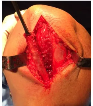
proximally,separatedfromthedeeptissues.Themostdistal
portionofthedegeneratedtendonwasresected;thetendon
wasrepairedwithBunnellsuturesusingnonabsorbableNo.5
thread(Fig.1).
Then, the radial tuberosity was palpated and a curved
Kellyforcepswaspassedthroughthebicepstendontunnel,
betweentheulnaandtheradius,anditwasadvanceduntil
itstipcouldbepalpatedonthedorsalaspectoftheproximal
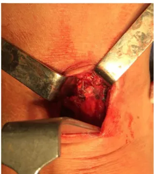
forearm.Asecondincisionwasmadeovertheforceps.The
tuberosity was exposed throughmuscle divulsionwith the
forearminmaximalpronation(Fig.2).Theradialtuberosity
wasscarifieduntilbleedingwasobserved.Twobioabsorbable,
double-loaded2.9-mmanchorswerepositioned.
Then, the brachial triceps tendon graft was collected,
withoutolecranonbonefragments,throughaposterior
longi-tudinalincisionandsubcutaneousdissectionuntilthetendon
wasexposed.Theauthorschosetoremoveastripfrom its
Fig.1–Intraoperativeimageshowingtherepairofthe
rupturedtendon.
Fig.2–Intraoperativeimageshowingtheexposedradial
tuberosity.
no need to explore the ulnar nerve (Fig. 3). Subsequently,
themedialandlateralbordersoftheremovedportionwere
approximatedandtheintervalwasclosed.
The most distal end of the graft was attached to the
tuberositybyfourU-shapedsutureswithanchorwires(Fig.4).
Theotherendofthetendonwasthenpassedtotheregion
oftheantecubitalfossaincisionthroughnonabsorbableNo.5
sutures(Krackow)topullthetendonthroughthetunnel
previ-ouslyoccupiedbythebicepstendon.Thebicepswasmobilized
andthenpulledwiththeuseofAllisclamps.Theelbowwas
positionedat40–60◦offlexion,withtheforearminfull
supina-tion.Moderate traction wasapplied tothe graft,whilethe
tendonstumpwasdistallytractioned.Thetwostructureswere
initiallystabilizedwithnon-absorbableNo.5U-shapedsuture;
thenseveralsinglesuturesweremadeatthe edges(Fig.5).
Once the reconstructionwas completed, the woundswere
Fig.3–Intraoperativeimageshowingremovalofthe
tricepstendongraft.
Fig.4–Intraoperativeimageshowingthefixationofthe
graftintheradialtuberosity.
closed;compressivedressingswereapplied,andthelimbwas
immobilizedwithabrachialsplint,maintainingtheelbowat
90◦offlexionandtheforearminmildsupination.
Immobilizationwithaslingwasmaintainedfortwoweeks;
thereafter,physicaltherapywasinitiated.Initially,exercisesof
passiveflexionandlimitedactiveextensionwiththeforearm
insupinationwereperformed,aswellaspassivesupination
andactivepronationto50◦.Thelimbwasimmobilizedwitha
Fig.5–Intraoperativeimageshowingthefixationofthe
grafttotherupturedbicepstendon.
theendofthethirdweek,whenexercisestoincrease
flex-ionandactivesupinationwithoutloadwereinitiated;atthis
phase,thepatientwasinstructedtointerrupttheuseofslings.
Musclestrengtheningexerciseswereinitiatedafterthesixth
weekwithlightloads,whichwereprogressivelyincreased.
Results
Three months after surgery, the patient had full range of
motionwithout pain, but stillpresented decreasedmuscle
strength.Afterthefourthmonth,hewasallowedtoreturn
to his work activities. At five months post-operative, the
patienthadrecoveredfullmusclestrengthandhadcompletely
returnedtodailyactivities.
Inhislastfollow-upassessment,sixmonthsaftersurgery,
thepatienthadfullrangeofmotion:0◦extension,135◦flexion,
85◦ supination,and 85◦ pronation. Atthatmoment,a
digi-taldynamometerwas used;theobservedflexion forcewas
17.35kgf(19.29kgfinthecontralateralelbow)andthe
supina-tionforce,7.14kgf(7.40kgfinthecontralateral).Furthermore,
theextensionforcewas 16.25kgfintheoperatedelbowvs.
15.45kgfinthecontralateral.
Thepatient’sresultisencouraging,withrecoveryof90%of
theflexionstrengthand96%ofthesupinationstrength,and
maintenanceofextensionforce,evenaftergraftremoval.
Discussion
Theprimaryrepairofachronicruptureofthedistalbrachial
bicepsistechnicallychallenging. Non-anatomicaltenodesis
inthebrachialismusclehasbeen proposedasatreatment
option. However, despite the high satisfaction rate of the
patientswhounderwentthisprocedure,Klonzetal.9observed
that half oftheir patients lost over 50% of the supination
strength.Theriskofweaknessinsupinationafterthis
tech-niquemaybeunacceptableforpatientswithhighfunctional
demand.
Several techniques for the reconstruction of the distal
biceps tendon have been described; they differ in their
approach,thegraft choice,andthetypeoffixation.1–4 Both
auto-andallograftshavebeenusedforthispurpose.Several
allograftshavebeenreportedintheliterature,1,10,11including
theAchillestendon,semitendinosus,anteriortibial,and
gra-cilis.Regardingautografts,1–4somestudiesindicatedtheuse
ofthefascialata,semitendinosus,andpalmarislongus.
Nodescriptionsoftheuseofthedistalbrachialtriceps
ten-donforthispurposewereretrievedintheliterature.Theuse
ofthistendonasanautograftforchronicrupturesofthedistal
brachialbicepswasdevisedbytheauthorstoavoidthe
disad-vantagesobservedintherecoveryperiodwhenthedonorarea
isnotlocatedinthesamejointastherecipientarea.
More-over,otheradvantagesincludeitspresenceineveryindividual,
theabsenceofneurovascularrisksduringharvesting,andthe
possibilityofvariablesizesandlengths,accordingtotheneed.
Martinetal.7 assessedthebiomechanicalcharacteristics
ofgraftsfromthecentralportionofthetricepsbrachii,
com-paringthemtothoseofthelongpalmar,andconcludedthat
thetricepsgraftiscomparableinultimateload-to-failureand
stiffness withthe palmarislongus tendon graft.They also
observedthatthetricepstendonpresentsgreaterdeformation
thanthepalmarislongus,butwithoutclinicalsignificance.In
anotherbiomechanicalstudy,Baumfeldetal.8evaluatedthe
propertiesofthemedial,central,andlateralstripsofthedistal
tricepsandconcludedthatthelateralportionissignificantly
thinnerandlessrigidthanthecentralandmedialportions,
andthatthecentralportionofthetricepsbrachiipresented
anultimateloadtofailureof704N,vs.357Nforthepalmaris
longus.
Wileyetal.2comparedtwogroupsofpatientswithchronic
ruptures ofthe distalbiceps;onegroupwasconservatively
treatedandtheotherunderwentreconstructionwith
semi-tendinosusautograftthroughdoubleincision.Theyconcluded
thatthepatientswhounderwentreconstructionobtainedan
improvementinflexionandsupinationstrengthwhen
com-paredtothosetreatedconservatively.
Although there is stilldebate on the best approachfor
fixationofdistalbicepstendonruptures,whetherdoubleor
singleincision,recentstudiesshowanegligibledifferencein
resultsand complicationsbetweenthetwo techniques.12,13
Thechoiceofthebestapproachforthesepathologiesshould
beguidedbysurgeonexperienceandconfidence.
Conflicts
of
interest
Theauthorsdeclarenoconflictsofinterest.
r
e
f
e
r
e
n
c
e
s
1.DarlisNA,SotereanosDG.Distalbicepstendonreconstruction inchronicruptures.JShoulderElbowSurg.2006;15(5):614–9.
asemitendinosusautografttechnique.JShoulderElbowSurg. 2006;15(4):440–4.
3. LevyHJ,MashoofAA,MorganD.Repairofchronicrupturesof thedistalbicepstendonusingflexorcarpiradialistendon graft.AmJSportsMed.2000;28(4):538–40.
4. HangDW,BachBRJr,BojchukJ.Repairofchronicdistalbiceps brachiitendonruptureusingfreeautogenoussemitendinosus tendon.ClinOrthopRelatRes.1996;(323):188–91.
5. MorreyBF,AskewLJ,AnKN,DobynsJH.Ruptureofthedistal tendonofthebicepsbrachii.Abiomechanicalstudy.JBone JointSurgAm.1985;67(3):418–21.
6. BoydHB,AndersonMD.Amethodforreinsertionofthedistal bicepsbrachiitendon.JBoneJointSurgAm.1961;43(7):1041–3.
7. MartinCR,HildebrandKA,BaergenJ,BittingS.Tricepstendon fasciaforcollateralligamentreconstructionabouttheelbow: aclinicalandbiomechanicalevaluation.AmJOrthop(Belle MeadNJ).2011;40(9):E163–9.
8. BaumfeldJA,vanRietRP,ZobitzME,EygendaalD,AnKN, SteinmannSP.Tricepstendonpropertiesanditspotentialas anautograft.JShoulderElbowSurg.2010;19(5):697–9.
9. KlonzA,LoitzD,WöhlerP,ReilmannH.Ruptureofthedistal bicepsbrachiitendon:isokineticpoweranalysisand
complicationsafteranatomicreinsertioncomparedwith fixationtothebrachialismuscle.JShoulderElbowSurg. 2003;12(6):607–11.
10.Sanchez-SoteloJ,MorreyBF,AdamsRA,O’DriscollSW. Reconstructionofchronicrupturesofthedistalbiceps tendonwithuseofanAchillestendonallograft.JBoneJoint SurgAm.2002;84(6):999–1005.
11.PattersonRW,SharmaJ,LawtonJN,EvansPJ.Distalbiceps tendonreconstructionwithtendoachillesallograft:a modificationoftheEndobuttontechniqueutilizinganACL reconstructionsystem.JHandSurgAm.2009;34(3): 545–52.
12.GrewalR,AthwalGS,MacDermidJC,FaberKJ,Drosdowech DS,El-HawaryR,etal.Singleversusdouble-incision techniquefortherepairofacutedistalbicepstendon ruptures:arandomizedclinicaltrial.JBoneJointSurgAm. 2012;94(13):1166–74.