Abstract
Submitted: February 3, 2017
0RGL¿FDWLRQ0D\
Accepted: May 21, 2017
Topical application of bFGF on
acid-conditioned and non-acid-conditioned
dentin: effect on cell proliferation and
gene expression in cells relevant for
periodontal regeneration
Periodontal regeneration is still a challenge in terms of predictability and magnitude of effect. In this study we assess the biological effects of combining chemical root conditioning and biological mediators on three relevant cell types for periodontal regeneration. Material and Methods: Bovine dentin slices were conditioned with 25% citric acid followed by topical
ELISA to assess the dynamics of bFGF release from the dentin surface and
stromal cells (BMSC) grown onto these dentin slices. We also assessed the
increased cell proliferation, except for BMSC grown on non-conditioned dentin slices. Dentin substrate discretely increased expression of Col1a1 in all cell types. Expression of Runx2, Col1a1 and Fn was either unaffected or inhibited by bFGF application in all cell types. We could not detect expression of the target genes on BMSC grown onto conditioned dentin. Conclusion: Acid conditioning of dentin improves the release of topically-applied bFGF. Topical application of bFGF had a stimulatory effect on proliferation of PDL
Ke y w or d s: Intercellular signaling peptides and proteins. Periodontal ligament. Dental cementum. Bone marrow. Cells.
Fernanda Regina Godoy ROCHA1
João Antônio Chaves de SOUZA2
Morgana Rodrigues
GUIMARÃES-STABILI3
José Eduardo Cezar SAMPAIO4
Carlos ROSSA JUNIOR4
http://dx.doi.org/10.1590/1678-7757-2017-0051
1University of Florida at Gainesville, Department of Oral Biology and Periodontology, Gainesville, USA. 2Universidade Federal de Goiás, Faculdade de Odontologia, Departmento de Periodontia, Goiania,
GO, Brasil.
3Universidade de São Paulo, Faculdade de Odontologia, Departmento de Estomatologia, São Paulo,
SP, Brasil.
4Univ Estadual Paulista – UNESP, Faculdade de Odontologia de Araraquara, Departmento de
Diagnóstico e Cirurgia, Araraquara, SP, Brasil.
Corresponding address: Carlos Rossa Jr Faculdade de Odontologia de Araraquara - UNESP.
Introduction
Evidence indicates that current cause-related
periodontal therapy is effective in stopping disease progression and reducing local inflammation,
but regeneration of periodontal tissues lost as a
consequence of disease still poses a challenge both in
terms of predictability and magnitude of effect5. Several studies2,17,24 have evaluated the potential of a host of
biological and chemical mediators, particularly growth
and differentiation factors, to improve the results of
regenerative therapy. Acid root conditioning is one of
intent4,7. There is great controversy in regard to the
conditioning agents and methods of application. In fact, there are products currently available
commercially for applications in periodontal
regeneration based on the biological properties of
growth factors. One such product includes in its surgical procedure protocol acid conditioning of the root surface
to remove smear layer from the dentin4 and there is
evidence indicating that chemical conditioning with
EDTA improved PDGF-induced adhesion of periodontal
PDGF without previous EDTA conditioning3.
Chemical conditioning of dentin has also been
extensively studied and may favor the repair/
on dentin surface induced by chemical conditioning,
including removal of smear layer, greater exposure of
8,16 may favor the adsorption of topically
applied growth factors and also turn the dentin surface
more conductive to cell attachment to promote blood
clot attachment.
Our research group has systematically evaluated
the most commonly used conditioning agents
(citric acid, tetracycline hydrochloride and EDTA),
optimizing conditions for their topical use based on the effectiveness of smear layer removal, exposure of
cells6,11,16,23. These in vit r o studies indicated that citric acid is the best candidate for a chemical conditioning
agent to improve removal of smear layer, exposure
conditioning include: 1) the exposure of endogenous
growth factors and biological mediators stored in the
dentin or cementum matrix; and 2) the increased
adsorption of exogenous growth factors topically
applied to the root surfaces due to the exposed
There are reports of promising results for
periodontal regeneration with the use of root
conditioning in pre-clinical in v iv o models, but the
results of clinical studies using root conditioning as a sole technique or even combined with other techniques
are, in general, disappointing1,18,28. However, very few
studies have assessed the combination of chemical root/dentin conditioning with topical application of
biologically-active mediators. Polypeptide growth
factors may act locally or systemically and modulate
several cell functions, including proliferation, migration, chemotaxis, differentiation, and gene expression12.
factor (bFGF) is a growth factor with biological effects
that may have important implications for periodontal regeneration, namely induction of angiogenesis, cell
proliferation and differentiation, resulting in increased
extracellular matrix production15. bFGF may be useful
for periodontal regeneration, because of its angiogenic properties and also the chemotactic and proliferative
effects on PDL cells, which may improve healing and
regeneration processes. Both pre-clinical studies19,21,25
and one clinical study14 assessed the use of bFGF in various concentrations and modes of application,
suggesting that bFGF may enhance periodontal
regeneration. This study addresses the hypothesis that
root conditioning improves the biological effect of bFGF topically applied onto the dentin surface.
Material and methods
Preparation of dentin slices
Rectangular dentin slices of 1.0x0.5x0.3 cm
(length x width x heigth) were obtained manually
from the roots of bovine incisors, by sectioning 2-3 mm apically to the cementum enamel junction with
a diamond disc mounted on a slow speed dental
handpiece. The portion of the dentin facing the pulp
used at low speed. The external surface (i.e., facing
the periodontal ligament and bone) of the dentin
slices was submitted to 5 scaling strokes by a single operator using a 5-6 Gracey curette. Cells were always
the manual scaling) of the dentin slices with an area
of 50 mm2. After preparation, the dentin slices were
then sterilized by ethylene oxide gas to avoid changes to the microstructure of the dentin or accumulation
of chemical residues on the dentin surface that could
occur by other means of sterilization.
Cell lines
line (PDL) and cementoblast cell line (OCCM) were
kindly provided by Dr. Martha J. Somerman, National
Institutes of Health/National Institute of Dental and
Craniofacial Research (NIH/NIDCR), Bethesda, MD9.
The primary murine bone marrow stromal cells (BMSCs)
bones (tibia and femur) of C57BL/6 mice. These cells
Waltham, MA, USA), supplemented with 10% volume
to volume (v:v) heat-inactivated fetal bovine serum
Waltham, MA, USA), and penicillin (100 IU/mL)/
streptomycin (100 μg/mL) (Life Technologies, Thermo
atmosphere containing 5% CO2. For the experiments,
preliminary experiments- data not shown) to achieve
Waltham, MA, USA) and resuspended in low-FBS
(2% v:v) culture medium (DMEM) at 1x10⁶ cells/
mL. Fifty μL of this cell suspension (5x10⁴ cells) were seeded onto the dentin slices or on the control wells
volume of culture medium (which varied according
procedure aimed at obtaining similar cell densities in
dentin slices and cell culture plastic, as cell density
the stimulus with the growth factor.
Experimental procedures
The experimental conditions were the following:
1) Control (cells on dentin slices without any growth
factor); 2) 10 ng of bFGF (R&D Systems, Minneapolis, MN, USA) applied topically; 3) 50 ng of bFGF applied
topically. For each of these conditions we had 12 dentin
slices. Of these 12 dentin slices for each experimental
condition, 6 were submitted to 25% citric acid conditioning for 3 minutes, followed by a wash with 10
mL of sterile phosphate buffered saline (PBS, pH 7.2,
MA, USA) before the seeding of cells (experimental condition #1) or before topical application of bFGF
and seeding of cells (experimental conditions #2 and
#3). Having 12 dentin slices (6 without citric acid
conditioning, 6 with citric acid conditioning) for each experimental condition allows for two replicates with
each cell type (PDL, OCCM or BMSC). All experiments
were repeated independently (using different cell passages of PDL and OCCM cells, and BMSC obtained
from different animals) three times.
Dentin slices were placed into 12-well cell culture
plates (1 slice/well). Topical application of the growth factors was carried out by preparing a suspension
of 50 μL of DMEM (without FBS and antibiotics)
containing the desired concentration of the growth
factor(s). The 50 μL volume yielded optimal conditions to completely cover the dentin slices. This suspension
of DMEM containing the growth factors was removed
by aspiration after 2 minutes. Immediately, 50 μL of a
1x10⁶ cells/mL suspension of each cell line was pipetted onto the dentin slices and placed in a CO2 incubator
at 37°C for 4 hours for initial cell adhesion. After the
initial 4-hour period, 1.5 mL of DMEM supplemented
with 2% FBS were added to each well, and they were incubated for a further 20-hour period. In the negative
control wells (without dentin slices), the same quantity
of cells was plated and the growth factors added
directly to the culture medium. Cells were collected mechanically with sterile plastic cell scrapers. The
exact same procedure was used to plate the cells for
the tissue culture-treated plastic substrate controls in
which no dentin slice was used as substrate.
Release of bFGF from the dentin substrate
analysis
We prepared dentin slices using the exact same
procedure described under “experimental procedures”,
except for the seeding of the cells. After topical application of bFGF (both with and without citric acid
conditioning), the dentin slices were placed in 12-well
tissue culture plates and 2 mL of non-supplemented
DMEM was added to each well. Controls included the addition of bFGF directly to the non-supplemented
200 uL of the culture medium were collected from wells
after 10, 30, 60, 120, 240 e 480 minutes of incubation, and the concentration of bFGF was assessed by
independent experiments were performed, and all
samples were analyzed in duplicate.
Cell proliferation analysis
Cells were plated onto 12-well plates directly on cell culture-treated plastic or on dentin slices
according to each experimental group, as described
before. Genomic DNA was quantitated from the cells
harvested by mechanically scraping the dentin slices/ bottom of tissue culture wells in all experimental
groups. These samples were collected at 24, 48 and
72 h after plating. Genomic DNA was isolated using the
Waltham, MA, USA), according to the manufacturer’s
instructions. The concentration of DNA in all samples
was measured using a nanoscale spectrophotometer (Nanovue Plus, GE Healthcare, Marlborough, MA, USA)
and all experiments were performed independently
three times.
Gene expression analysis
and treated with DNase (RNAqueous kit-4-PCR,
USA). Concentration and purity of total RNA were
determined by A260 and A260/A280, respectively, in a UV spectrophotometer for microvolumes. cDNA was
synthesized from 100 ng of total RNA using random
hexamers primers and moloney leukemia virus reverse
transcriptase, according to the instructions of the supplier (High Capacity cDNA synthesis kit, Applied
Biosystems, Thermo Fisher Scientific, Waltham,
MA, USA). The qPCR reaction was performed using
TaqMan chemistry (TaqMan Fast Universal PCR Master
Waltham, MA, USA) and pre-designed and optimized
sets of primers and probes (TaqMan Gene Expression
Waltham, MA, USA) for detection of target genes
(GAPDH) on a StepOne Plus thermocycler (Applied
USA). All samples were analyzed in duplicate and the
cycle threshold (Ct) value of each well was determined.
using the thermocycler’s software. Results are
expressed as fold change compared to negative control
(cells grown on tissue culture-treated plastic in
12-well plates without stimulation with growth factors).
Statistical analysis
The statistical analysis was done using GraphPad 5.0 (GraphPad Software Inc., San Diego, CA, USA). Central
tendency and dispersion measures were calculated
from different experiments. Pairwise comparisons between experimental conditions were performed
using non-paired Student’s t-test, considering each
experimental condition as an independent event.
Results
Effect of acid conditioning on the release of
bFGF from the dentin substrate
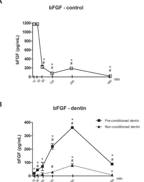
Acid conditioning previously to the topical
application of the growth factor increased the
concentration of bFGF released into the culture
medium at 10 and 30 minutes. Concentration of bFGF released into the culture medium from dentin samples
was lower than that measured in the positive control
samples, in which bFGF was added directly to the
culture medium, to account for
of the culture medium, tissue culture plastic or of the
spontaneous degradation of bFGF in its detection by
ELISA.
detected over the experimental periods indicates a quick degradation of the bFGF in culture medium (at
1 h, the concentration in the culture medium dropped
by 6-fold - Figure 1A). The results suggest that some
of the growth factor applied onto the dentin substrate was either strongly bound to the substrate and not
released from it over 30 minutes; or that it was
inactivated (Figure 1).
Effect of bFGF and culture substrate on
and BMSC
Next, we assessed the effects of bFGF on the
proliferation of the different cell types. This assessment
was performed indirectly, by quantifying genomic DNA.
In these experiments, we also determined the effect of the culture substrate by comparing the quantity
of genomic DNA of cells cultured on cell
culture-treated plastic or onto dentin slices. bFGF treatment
proliferation in comparison to non-treated dentin
substrate, but interestingly, there was no
dose-acid conditioned-dentin was increased at 24 and 48
h and then reduced at 72 h in comparison with
non-conditioned dentin (Figure 2A).
Proliferation of cementoblasts on plastic substrate was increased by bFGF at 48 h and subsequently
reduced in comparison to vehicle control at 72 h.
Similarly to the PDL fibroblasts, proliferation of cementoblasts grown on dentin slices was enhanced
by topical application of bFGF, with the lower
concentration producing greater increases in cell
proliferation (Figure 2B). Proliferation of BMSC cells
by bFGF at 48 and 72 h. However, when plated onto
dentin substrates, only the higher concentration
of bFGF increased proliferation of BMSC on
acid-conditioned dentin, whereas in non-acid-conditioned dentin
topical application of bFGF inhibited proliferation of BMSCs (Figure 2C).
Effect of bFGF and culture substrate on gene
expression of Runx2, Col1a1, Bglap and Fn
Since bFGF has a range of biological effects beyond
cell proliferation, we then assessed its effects on the expression of selected target genes related with the
repair/regeneration of periodontal tissues, taking into
consideration the nature of the culture substrate. All
the gene expression experiments were carried out using 50 ng/mL of bFGF. Cells plated directly onto
plastic culture-treated plates served as a positive
control to determine the biological effect of this
growth factor on target gene expression in each cell
Figure 1- Concentration of bFGF in acqueous-based cell culture medium, after 10 to 480 min of incubation. 50 ug/mL of bFGF was applied
topically to dentin slices with and without previous acid conditioning and the quantity of bFGF in the medium was determined in aliquots of the culture medium by ELISA at the indicated periods. In positive control experiments (without dentin slices), the same concentration of bFGF was added directly to the culture medium. Bars and vertical lines indicate means and standard deviations of three independent
type. To account for the effect of the dentin substrate,
the negative control was represented by the data on
normalized gene expression of cells plated onto non-conditioned dentin slices that were not treated with the
growth factor (0 ng/mL of bFGF, control). Results of
the experiments in which cells were plated onto dentin
slices treated with bFGF are presented according to the cell type and analyzed in terms of fold change
of normalized target gene expression in comparison
with the expression in the negative control cells. Target gene expression for positive control samples
was calculated as a fold change of the expression in
untreated cells (0 ng/mL of bFGF) also plated onto
tissue culture-treated plastic.
Cementoblasts (OCCM cells)
bFGF increased expression of all target genes, but
more markedly of Bglap and Col1a1. When these cells
difference observed was a higher expression of Col1a1
in cells plated onto non-conditioned dentin treated
with 50 ng/mL of bFGF in comparison with cells plated onto conditioned dentin and treated with the same
concentration of bFGF. Overall, the acid conditioning
and concentration of bFGF had no marked effect on
target gene expression in these cells (Figure 3A).
Expression of all genes studied, except Bglap, was
higher when cells were grown onto acid-conditioned
dentin slices in comparison with cells grown on non-conditioned dentin. Moreover, there was no
dose-dependent effect of bFGF on target gene expression,
independently of the acid conditioning of dentin
substrate. bFGF treatment of cells plated onto tissue culture-treated plastic increased expression of Col1a1
and Bglap, but not of Fn and Runx2 (Figure 3B).
Figure 2-*HQRPLF'1$TXDQWLWDWLRQLQGLFDWLQJWKHSUROLIHUDWLRQRIPXULQHSHULRGRQWDOOLJDPHQW¿EUREODVWVP3'/SDQHO³$´FHPHQWREODVWV
2&&0SDQHO³%´DQGERQHPDUURZVWURPDOFHOOV%06&SDQHO³&´DFFRUGLQJWRWKHH[SHULPHQWDOFRQGLWLRQV&HOOVZHUHSODWHGRQWR
dentin slices on which bFGF (10 and 50 ng) was applied topically both with and without previous acid conditioning. Cells were grown in complete culture medium for 24, 48 and 72 h. In positive control samples, cells were plated on tissue culture plastic (no dentin slices) and stimulated with 50 ng/mL of bFGF and genomic DNA collected at the same periods. The different symbols indicate mean and the vertical lines indicate the standard deviation of genomic DNA quantity from three independent experiments, measured in triplicate at each time
SRLQWLQGLFDWHVSFRQWUROvsQJP/E)*)LQGLFDWHVSFRQWUROvsQJP/E)*)DQGLQGLFDWHVSQJP/E)*)
Figure 3- 5HODWLYH FKDQJHV LQ JHQH H[SUHVVLRQ P51$ RI 5XQ[ &ROD )Q DQG %JODS E\ FHPHQWREODVWV 2&&0 SDQHO ³$´
SHULRGRQWDOOLJDPHQW¿EUREODVWVP3'/SDQHO³%´DQGERQHPDUURZVWURPDOFHOOV%06&SDQHO&´FXOWXUHGRQGLIIHUHQWVXEVWUDWHV
Bone Marrow Stromal Cells (BMSCs)
increased the expression of collagen, but not of the
other target genes. We did not detect target gene
expression in BMSCs cultured onto acid-conditioned
dentin slices, in spite of comparable cycle thresholds (Cts) for the expression of the housekeeping gene
(Gapdh). In non-conditioned dentin slices, the lower
concentration of bFGF (10 ng/mL) significantly inhibited expression of Bglap by BMSCs; but
whereas the higher concentration of bFGF (50 ng/mL)
only increased the expression of Col1a1. There were opposite effects of the dose of bFGF on the expression
of Col1a1 (higher for 50 ng/mL of bFGF) and Fn (higher
for 10 ng/mL of bFGF) (Figure 3C).
Discussion
In this study we evaluated, i n v i t r o, topical application of bFGF on dentin surface with and
without previous acid conditioning, assessing the
release of bFGF from dentin and its effects on cell
proliferation and on the expression of selected genes associated with the formation/regeneration of soft and
mineralized periodontal tissues. These assessments
were carried out in three different cell types that may
have access to the root surface in the clinical situation and which are also shown to be important for the
repair/regeneration of periodontal tissues: periodontal
stromal cells.
We used bovine dentin as a substrate to simulate
the clinical situation of treating an osseous defect
associated with periodontal disease progression. Bovine teeth were used because they may be easily
obtained in large quantities, with good condition and
have a more uniform composition26,27. Yassen, et
al.27 (2011), discussed the issue of inconsistent data reported with the use of bovine teeth in studies focusing
on caries and dental erosion/abrasion; however the
same authors comment on the comparison between
micro-morphological structure, chemical composition and physical properties of human and bovine dentin;
which indicated very similar characteristics. It is
on the biological effects and bioavailability of topically applied growth factors, since the substrate may bind
to the growth factor reversibly or irreversibly and it
may also alter the chemical structure of the topically
applied polypeptide, affecting its biological activity. In the clinical situation associated with surgical
periodontal treatment, the tooth surface is very likely
to be completely devoid of cementum due to the
mechanical treatment performed to remove soft and mineralized bacterial deposits.
Acid conditioning of the root surface previously
the release of bFGF into aqueous-based culture medium. This may suggest that chemical conditioning
may prolong the release of topically applied growth
factors to the surrounding microenvironment, which could favor their bioactivity considering the short
half-life of most growth factors10,22. On the other
hand, it is also possible that the bFGF adsorbed and
not released into the culture medium remains on the dentin substrate where it may still exert its biological
functions. Comparison between concentration of
bFGF added directly to the culture medium in control
samples with the concentration of bFGF present in the culture medium of the test samples (in which the
growth factor was applied topically onto the dentin
slices) indicates that most of the growth factor applied
topically is either retained on the dentin substrate or lost/degraded, regardless of chemical conditioning
previous to the topical application. The rationale
for chemical conditioning includes exposure of the
organic matrix, which may favor the adsorption of the topically applied growth factors. This is
supported by the fact that after 8 h the concentration
of bFGF in the culture medium incubated with
pre-conditioned dentin samples was greater than that in control samples. Also, it is important to consider the
possibility of dentin matrix substrate-derived factors
released by the chemical conditioning on the cells
in the microenvironment; which is supported by the report demonstrating increased mineralization and
expression of mineralization-associated genes (bone
sialoprotein and alkaline phosphatase, for example) by odontoblasts stimulated with proteins in demineralized
dentin extracts13.
Clinically, immediately after completion of
treatment the exposed root surface is covered by a blood clot and cells derived primarily from the
adjacent bone marrow, particularly when some type of
mechanical barrier has been interposed between the
regeneration technique. However, other cell types in
the microenvironment, including periodontal ligament
the area, and there is evidence of an important role
for these cell types in periodontal regeneration5,20.
In fact, probably the main reason for the low
predictability and high variability of results in periodontal regeneration is the fact that the attachment
unit is composed of various tissues, which are produced
attached to cementum on the surface of the root via
a functional periodontal ligament. We have previously
dentin, and particularly that dentin conditioning has
a positive effect on these parameters22. Our present
results indicate that chemical conditioning of dentin
comparison with non-conditioned dentin only at 24 and
48 h, with a reversal in this effect at 72 h. We also
increased cell number, 72 h after bFGF stimulation,
with no changes detected at 24 or 48 h. In a previous
study using primary human periodontal ligament
23 we also did not observe changes in cell
proliferation after a 24 h-stimulation with 1 and
10 ng/mL of bFGF. In this report we expand these
proliferation of cementoblasts at 48 h and bone
marrow stromal cells at 48 and 72 h.
Increased proliferation of these relevant cell
types is important to support the repopulation of the substrate/wound area and favor the repair/
regeneration; however it is also critical to consider
other biological effects of bFGF, particularly on cell differentiation and matrix production. In a previous
study23, we report that 10 ng/mL of bFGF inhibited
expression of Col1a1 by human periodontal ligament
a higher concentration of bFGF (50 ng/mL) we only
observed a discrete increase in Col1a1 expression.
Interestingly, 50 ng/mL of bFGF strongly induced Col1a1 gene expression by cementoblasts and also
in BMSC cells, albeit less potently.
When bFGF was applied on the dentin substrate,
grown on acid-conditioned dentin, but not in
non-conditioned dentin; whereas in BMSC cells the
dose-dependent, but only in non-conditioned dentin.
However, cementoblasts were less responsive to bFGF
in terms of increase in Col1a1 expression, particularly
when grown on acid-conditioned dentin.
bFGF stimulation of all three cell types had little
effect on the expression of Fn and Runx2. The most
expression by BMSC cells grown on non-conditioned dentin exposed to the lower concentration of bFGF
increase in the expression of both Fn (without dose-dependent effect) and Runx2 (inverse dose-dose-dependent
effect) only when grown in acid-conditioned dentin.
Cementoblasts were the least responsive cell type,
as the expression of Fn, Runx2 and Bglap by OCCM cells were not markedly regulated by bFGF in any of
the experimental conditions. Notably, BMSCs grown
on conditioned dentin had barely detectable levels
of mRNA for all target genes (data not shown). This suggests that chemical conditioning may render the
substrate unfavorable for cell metabolism of BMSCs.
In fact, we cannot rule out a cytotoxic effect in
acid-conditioned dentin, as the proliferation was assessed
DNA integrity or of cell viability by other methods.
It is important to note that in the positive control
samples (cells grown directly on tissue culture plastic, without dentin slices) the concentration of bFGF was
much greater than in the experimental samples. This
of bFGF for the topical application, whereas in the positive control samples (cells grown on tissue culture
the same growth factor. We note that it is not possible to “topically apply” or “condition” tissue culture plastic
with the growth factor, similarly to what was done with
the dentin slices. Also, even though we seeded the
cells using the same protocol in both cell culture plastic and dentin slices to assure the exact same n=initial
numbers of cells and also a similar cell density, it is
possible that the cells grown in tissue culture plastic
proliferated faster than those plated onto dentin slices, resulting in different cell numbers at the end of the
experimental period. We speculate that this possible
the initial cell numbers and cell density. Anyway, these
considerations preclude direct comparison between the
results from positive control and experimental groups (cells grown on dentin slices), but the information
provided by the negative control samples (treated
with the vehicle used to resuspend the bFGF) indicates
that bFGF used was biologically active and the information provided by the positive control samples
(cells grown on tissue culture plastic and treated with
bFGF) informs the general trend for the effects on the expression of the target genes in the different cell
types. Nevertheless, we have not determined how
much of the initial quantity of the topically applied
bFGF was retained/adsorbed on the dentin substrate,
eventually present on the dentin surface retained its
full biological activity, but we consider the modulation
of gene expression and cell proliferation as indirect evidence of bFGF bioactivity.
In summary, we show that previous acid conditioning
of dentin improves the release of topically-applied
bFGF in aqueous medium. We have also shown that different cell types that are relevant for periodontal
regeneration respond differently to bFGF; however
there was an overall increase in cell proliferation and
in the expression of bone and connective tissue matrix gene expression by cells grown on dentin with bFGF.
Cementoblasts were the most responsive cell type in
terms of regulation of gene expression by bFGF in the
absence of a dentin substrate; whereas PDL cells were most responsive to bFGF in terms of gene expression
when grown on acid-conditioned dentin. Although no
clear-cut conclusion may be drawn from our results,
the data indicate that topical application of bFGF has biological effects on relevant cell types for periodontal
regeneration and warrants in v iv o studies to fully
assess the potential of this approach on periodontal
regeneration.
Acknowledgements
This study was supported by a research grant
awarded by the São Paulo State Foundation for
Research Support (FAPESP, grant# 2010/11749-4 to JECS) and also by the CAPES Foundation (Brazilian
Ministry of Education). We thank ACECIL (Campinas,
SP, Brazil), which kindly provided the sterilization by
ethylene oxide gas to the dentin slices used in this study.
References
1- Al-Hamdan K, Eber R, Sarment D, Kowalski C, Wang HL. Guided tissue regeneration-based root coverage: meta-analysis. J Periodontol. 2003;74:1520-33.
2- Basan T, Welly D, Kriebel K, Scholz M, Brosemann A, Liese J, et al. Enhanced periodontal regeneration using collagen, stem cells or growth factors. Front Biosci (Schol Ed). 2017;9:180-93.
3- Belal MH, Watanabe H, Ichinose S, Ishikawa I. Effect of PDGF-BB combined with EDTA gel on adhesion and proliferation to the root surface. Odontology. 2012;100:206-14.
4- Blomlöf L, Jonsson B, Blomlöf J, Lindskog S. A clinical study of root surface conditioning with an EDTA gel. II. Surgical periodontal treatment. Int J Periodontics Restorative Dent. 2000;20:566-73. 5- Bosshardt DD, Sculean A. Does periodontal tissue regeneration really work? Periodontol 2000. 2009;51:208-19.
of EDTA-T gel for smear layer removal at root surfaces. Quintessence Int. 2005;36:551-8.
7- Chaves E, Cox CF, Morrison E, Caffesse R. The effect of citric acid application on periodontally involved root surfaces. II. An in vitro scanning electron microscopic study. Int J Periodontics Restorative Dent. 1993;13:188-96.
8- Dantas AA, Fontanari LA, Ishi EP, Leite FR, Zandim DL, Rached RS, et al. Blood cells attachment after root conditioning and PRP application: an in vit r o study. J Contemp Dent Pract. 2012;13:332-8.
9- D'Errico JA, Ouyang H, Berry JE, MacNeil RL, Strayhorn C, Imperiale MJ, et al. Immortalized cementoblasts and periodontal ligament cells in culture. Bone. 1999;25:39-47.
10- Gamal AY, Mailhot JM, Garnick JJ, Newhouse R, Sharawy MM.
IGF-1 application on tetracycline HCI conditioned root surfaces. J Clin Periodontol. 1998;25:404-12.
11- Ishi EP, Dantas AA, Batista LH, Onofre MA, Sampaio JE. Smear layer
conditioning. J Contemp Dent Pract. 2008;9:25-33.
12- Kao RT, Murakami S, Beirne OR. The use of biologic mediators and tissue engineering in dentistry. Periodontol 2000. 2009;50:127-53. 13- Kim HS, Lee DS, Lee JH, Kang MS, Lee NR, Kim HJ, et al. The effect of odontoblast conditioned media and dentin non-collagenous proteins on the differentiation and mineralization of cementoblasts in
vit r o. Arch Oral Biol. 2009;54:71-9.
14- Kitamura M, Nakashima K, Kowashi Y, Fujii T, Shimauchi H, Sasano
randomized controlled phase II clinical trial. PLoS One. 2008;3:e2611. 15- Lee J, Stavropoulos A, Susin C, Wikesjö UM. Periodontal regeneration: focus on growth and differentiation factors. Dent Clin North Am. 2010;54:93-111.
16- Leite FR, Sampaio JE, Zandim DL, Dantas AA, Leite ER, Leite AA.
on clot stabilization. Quintessence Int. 2010;41:341-9.
17- Li F, Yu F, Xu X, Li C, Huang D, Zhou X, et al. Evaluation of recombinant human FGF-2 and PDGF-BB in periodontal regeneration: a systematic review and meta-analysis. Sci Rep. 2017;7:65.
treatment of periodontal disease. A systematic review. Ann Periodontol. 2003;8:205-26.
19- Murakami S, Takayama S, Kitamura M, Shimabukuro Y, Yanagi K,
21- Rossa C Jr, Marcantonio E Jr, Cirelli JA, Marcantonio RA, Spolidorio LC, Fogo JC. Regeneration of Class III furcation defects with basic
histometric study in dogs. J Periodontol. 2000;71:775-84.
surfaces with or without conditioning with tetracycline or EDTA. J Oral Sci. 2007;49:213-20.
23- Silverio-Ruiz KG, Martinez AE, Garlet GP, Barbosa CF, Silva JS, Cicarelli RM, et al. Opposite effects of bFGF and TGF-beta on collagen
2007;39:130-7.
24- Suárez-López Del Amo F, Monje A, Padial-Molina M, Tang Z, Wang HL. Biologic agents for periodontal regeneration and implant site development. Biomed Res Int. 2015;2015:957518.
25- Takayama S, Murakami S, Shimabukuro Y, Kitamura M, Okada H. Periodontal regeneration by FGF-2 (bFGF) in primate models. J Dent Res. 2001;80:2075-9.
26- Wegehaupt FJ, Widmer R, Attin T. Is bovine dentine an appropriate substitute in abrasion studies? Clin Oral Investig. 2010;14:201-5. 27- Yassen GH, Platt JA, Hara AT. Bovine teeth as substitute for human teeth in dental research: a review of literature. J Oral Sci. 2011;53:273-82.