Key words:
Prostatitis; Cells; Receptors, Purinergic P2X7; Tumor Necrosis Factor-alpha; Interleukin-1
Int Braz J Urol. 2013; 39: 276-85
__________________ Submitted for publication: June 04, 2012
__________________ Accepted after revision: January 17, 2013 Purpose: Evidence shows that adenosine triphosphate (ATP) is involved in the
transmis-sion of multiple chronic pain via P2X7 receptor. This study was to investigate the P2X7 and microglial cells in the chronic prostatitis pain.
Materials and Methods: Rats were divided into control group and chronic prostatitis group (n = 24 per group). A chronic prostatitis animal model was established by injecting complete Freund’s adjuvant (CFA) to the prostate of rats, and the thermal withdrawal la-tency (TWL) was detected on days 0, 4, 12 and 24 (n = 6 at each time point in each group). Animals were sacrificed and the pathological examination of the prostate, detection of mRNA expression of P2X7 and ionized calcium binding adaptor molecule 1 (IBA-1) and measurement of content of tumor necrosis factor-α (TNF-α) and interleukin-1β (IL-1β) in the dorsal horn of L5-S2 spinal cord were performed on days 0, 4, 12 and 24. In addition, the content of TNF-α and IL-1β in the dorsal horn of L5-S2 spinal cord was measured after intrathecal injection of inhibitors of microglial cells and/or P2X7 for 5 days.
Results: The chronic prostatitis was confirmed by pathological examination. The expres-sion of P
2X7 and IBA-1 and the content of TNF-α and IL-1β in rats with chronic
prosta-titis were significantly higher than those in the control group. On day 4, the expressions of pro-inflammatory cytokines became to increase, reaching a maximal level on day 12 and started to reduce on day 24, but remained higher than that in the control group. Following suppression of microglial cells and P2X7 receptor, the secretion of TNF-α and IL-1β was markedly reduced.
Conclusion: In chronic prostatitis pain, the microglial cells and P2X7 receptor are activated resulting in the increased expression of TNF-α and IL-1β in the L5-S2 spinal cord, which might attribute to the maintenance and intensification of pain in chronic prostatitis.
INTRODUCTION
Chronic prostatitis, a common urologi-cal condition in young and middle-age men, is caused by multiple etiological factors. Pain is a major presentation of chronic prostatitis (1). Pre-vious studies focused on the pathological chan-ges in the prostate, while the pathways related
to neurotransmission and the regulatory mecha-nisms of chronic prostatitis pain have not been studied. Recent studies have identified the chro-nic prostatitis pain as a visceral referred pain, which is usually accompanied by the dysfunction of pelvic floor muscles. The prostate is innerva-ted largely by the pelvic nerves arising from the L5-S2 spinal cord (2,3).
P2X7 receptor mediates activation of microglial cells in
prostate of chemically irritated rats
_______________________________________________
Heng Zhang, Limei Liu, Zhong Yang, Jinhong Pan, Zhiwen Chen, Qiang Fang, Weibin Li, Longkun Li,
Gengsheng Lu, Zhansong Zhou
Department of Urology (HZ, JP, ZC, QF, WL, LL, GL, ZZ); Department of Pathology (LL) and Department of Neurobiology (ZY), Southwest Hospital, Third Military Medical University, Chongqing 400038, China
ABSTRACT
ARTICLE
INFO
It has also been shown that the transmis-sion and regulation of pain are associated with not only the neurons but the microglia and astrocytes (4,5). Studies also demonstrated that astrocytes and microglias may secrete pro-inflammatory cytokines such as tumor necrosis factors (TNF), in-terleukin-1 (IL-1), nerve growth factor (NGF), and nitric oxide (NO), which may lead to the neuronal injury and chronic pain (6,7). Especially, the mi-croglias are widely distributed in the central ner-vous system (CNS). The detrimental stimulation of CNS (such as trauma, ischemia and infection) may activate microglias. Under this condition, their morphology, the receptor expression on these cells and their function alter; these cells are ameboid; the markers for activation increase on these cells (8). There is evidence showing that the microglias in the posterior horn of spinal cord are significan-tly activated after damage to peripheral nerves (9). This suggests that the activation of microglias in the spinal cord is related to the occurrence and transmission of neuropathic pain. However, the role of microglias in chronic prostatitis pain is still poorly understood, and molecules activated after injury on these cells and the exact mechanisms are unclear.
There are a lot of P2X7 receptors of ade-nosine triphosphate (ATP). ATP is a type of pain--causing neurotransmitter, and its receptors can be classified as P2X receptors and P2Y receptors. P2X7 receptor is a special subtype of purinergic re-ceptor P2X family and an ATP-gated non-selective cation channel. P2X7 receptor contains 595 amino acids and three P2X7 receptors form homologous polymers generally. P2X7 receptor is a dual func-tional receptor. Under pathological conditions, P2X7 receptor is involved in the transmission of pain. It was reported that microglias may be acti-vated by the P2X7 receptor, which is up-regulated in various types of chronic pain (10). Chessell et al. found P2X7 knockout mice failed to present with hyperalgesia to heat and mechanical stimulation after nerve injury (11). However, whether the mi-croglias and P2X7 receptor in the posterior horn of L5-S2 spinal cord are activated to regulate the chronic prostatitis pain remains unclear. Hence, the present study was to investigate the role of microglias and PX receptor in the chronic
pros-tatitis pain and the possible therapeutic strategies for chronic pelvic pain syndrome.
MATERIALS AND METHODS
Animals
The specific pathogen free (SPF) rats wei-ghing 200 ± 25 g were purchased from the Experi-mental Animal Center of the Third Military Medical University and randomized into experiment group and control group. All rats were intraperitoneally anesthetized with 1% pentobarbital and then the prostate was exposed through a ventral midline inci-sion (1 cm). For rats in the experiment group, injec-tion with complete Freund’s adjuvant (CFA; Sigma--Aldrich, Sigma) was done once at bilateral ventral lobes (10µL for each). For rats in the control group, 20µL of normal saline was injected (Nackley et al. (12), Butler et al. (13) and Zhou et al. (14)). Then, the wound was closed. Rats were sacrificed on days 0, 4, 12 and 24 d after injection (n = 6 at each time point in each group) and pathological examination and detection of the mRNA expression of P2X7 and ioni-zed calcium binding adaptor molecule 1 (IBA-1) and the content of TNF-α and IL-1β in spinal cord were performed. All procedures were performed in accor-dance with the guidelines for animal care and use of National Institute of Health, and this study was approved by the Ethics Committee of our Hospital.
Prostatic Inflammation Model Identification
Rats were intraperitoneally anesthetized with 10% chloral hydrate (0.3 mL/100 g) on days 0, 4, 12 and 24 after CFA injection, and transcardially perfused with 200 mL of 0.9% saline and then with 0.01 mol/L phosphate buffer (about 300 mL) contai-ning 4% paraformaldehyde at 48º C Subsequently, the left and right prostatic tissues were collected, fi-xed in 4% paraformaldehyde at 4º C overnight, em-bedded in paraffin, cut into sections, stained, and finally examined under a microscope (15).
Detection of heat pain threshold
a light spot (5 mm in diameter; 50 W, 12 V) was pro-duced through a radiant heat stimulator and used to stimulate the paw. The time to paw withdraw was re-corded as the TWL. The stimulation was done for no longer than 30 s and measurement was performed 5 times in each rat with an interval of 10 min. between two detections. The maximal or minimal TWL was removed, and the TWL in remaining 3 measurements was employed for the calculation of average (16).
Detection of mRNA expression of P2X7 and IBA-1 in L5-S2 posterior horn
Animals were sacrificed by decapitation. The posterior horn of L5-S2 spinal cord was carefully col-lected on ice under a microscope, and stored at -70º C for use. Total RNA was extracted from the L5-S2 spinal cord using the RNAgents Total RNA Isolation System (Promega, Madison, WI) according to the manufacturer’s instructions. The concentration and purity of total RNA were determined by spectropho-tometric analysis at A260 and A280 (1.8-2.0). The qua-lity of RNA was determined by methanal agarose gel electrophoresis following ethidium bromide staining. Total RNA (2µg) was subjected to reverse transcrip-tion using the Reverse Transcriptranscrip-tion System (Jikang, Shanghai, China) with random primer oligo(DT)18 (0.5µg). The reaction conditions were as follows: 70º C for 5 min, 37º C for 60 min. and 70º C for 10 min., and products were then stored at -70º C.
The resulting cDNA (20 ng) was used as templates for real-time fluorescence quantitative (FQ) PCR using a SYBR green PCR core reagent kit (Applied Biosystems, Foster City, CA) in DNA En-gine OPTIONTm2 (MJ RESEARCH, USA). The
pri-mers were designed using the Geneworks software package as follows: P2X7: 5’-GACAAACAAAGT-CACCCGGAT-3’ (forward) and 5’-CGCTCACCAAAG-CAAAGCTAAT-3’ (reverse); IBA-1: 5’-TTGATCTGA
ATGGCAATGGA-3’ (forward) and 5’-CCTCC AAT-TAGGGCAACTCA-3’ (reverse). The PCR conditions were as follows: reverse transcription at 50º C for 30 min., Hot Start Taq (1.25 unit/sample) activation for 15 min at 95º C, 40 cycles of denaturation at 94º C for 15 s, annealing at 56º C for 30 s, and extension at 72º C for 30 s. The SYBR Green fluorescence was acquired by a final extension at 79º C. The melting curve analysis was performed after each reaction. GAPDH (5’-TTTAACTCTGGTAAAGTGGATATTG-TTG-3’ [forward] and 5’-ATTTCCATTGATGACAA GCTTCC-3’) served as an internal control (Table-1). The expression of target genes could be calculated according to the amplification standard curve and regression equation of GAPDH automatically by the DNA Engine OPTIONTm2. The expression of target genes was normalized to that of GAPDH as the re-lative expression. Average was obtained from 6 ani-mals in each group.
Contents of TNF-α and IL-1β in L5-S2 posterior horn
L5-S2 posterior horn was homogenized in 0.5 mL of ice-cold lysis buffer containing 50 mM Tris, 150 mM NaCl, 1% TritonX 100, 0.5% sodium deoxycholate, l mM phenylmethanesulfonyl fluoride (PMSF), 0.1% sodium dodecyl sulfate (SDS), 10 mM NaF and 1 mM vitriolu acid sodium. Homogenates were centrifuged at 1800 rpm for 10 min. and the supernatant was collected and stored at -70º C. The contents of IL-1β and TNF-α were detected using the commercially available ELISA kits according to the manufacturer’s instructions. Briefly, 50µg) was subjected to reverse transcription using the L of bio-tinylated antibody was added to 100µg) was subjec-ted to reverse transcription using the L of samples in an anti-rat TNF-α or IL-1β pre-coated plate (Santa Cruz Biotechnology, Inc., Santa Cruz) followed by incubation for 3 h at room temperature. After
wa-Table 1 - Primers and conditions for RT-PCR.
Forward Reverse
P2X7 5’-GACAAACAAAGTCACCCGGAT-3’ 5’-CGCTCACCAAAGCAAAGCTAAT-3’
IBA-1 5’-TTGATCTGA ATGGCAATGGA-3’ 5’-CCTCC AATTAGGGCAACTCA-3’
shing three times, a prepared 100µg) was subjected to reverse transcription using the L of strepta vidin horseradish peroxidase solution and 3, 3′, 5, 5′ -te-tramethylbenzidin substrate solution were added followed by incubation. Absorbance was measured at 492 nm in a microplate reader (Salzburger La-bortechnik, Salzburg, Austria). A standard curve was delineated to determine the content of TNF-α and IL-1β. The sensitivity of this assay is > 10 pg/mL. Average was obtained from 6 animals in each group.
Intrathecal injection of agonist or antagonist of
P2X7 and microglial cells
The second part of the experiment in-volved intrathecal cannulation according to the procedures described by Yaksh and Rudy (17). In brief, rats were intraperitoneally anesthetized with pentobarbital, a cannula (PE-10 tubing) was in-serted through the cisterna magna at 6 cm to the L1 spinal cord via the spinal subarachnoid space. A recovery period of 7 d was allowed, and rats showing no motor impairment following surgery were used for further experiments. The prostati-tis was induced as mentioned above. From day 7, intrathecal injection of drug was done for con-secutive 5 days. Rats were divided into 5 groups (n = 6 per group) and treated as follows: 1) P2X7 receptor agonist: 2’-3’-O-(4-Benzoylbenzoyl)--adenosine 5’-triphosphate (BzATP; 100µmol/L); 2) P2X7 receptor antagonist: oxidized ATP (oATP; 100µmol/L); 3) inhibitor of microglial cells: mi-nocycline (50µg); 4) P2X7 receptor agonist + inhi-bitor of microglial cells; 5) in the control group, injection was done with artificial cerebrospinal fluid (ACSF; pH5.5) of equal volume. Six rats were included in each group. Drugs were injected at a
volume of 15µL and flushing was done with 5µL of ACSF. Injections were finished within 5 min. The content of TNF-α and IL-1β in the spinal cord was determined.
Statistical analysis
All data were expressed as means ± stan-dard error (SEM) and statistical analysis was per-formed with SPSS version 13.0 for Windows. When F-test showed significance, means were compared with the LSD test of post hoc analysis (Dunnett’s t test). Analysis of variance (ANOVA) was conducted for comparisons of data among di-fferent groups. A value of P < 0.05 was considered statistically significant.
RESULTS
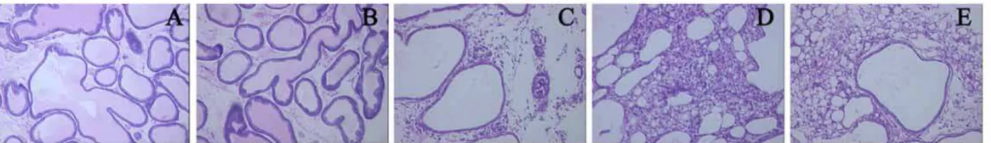
Pathological changes in prostate
Pathological examination showed thAT CFA treated prostates presented with degenera-tion, necrosis and exfoliation of mucosal cells in the prostate gland. Infiltration of large amounts of lymphocytes and monocytes was noted in the interstitium, and some lymphocytes aggregated in cluster. In the control group, the mucosal epithe-lial cells were regularly arranged, and the infiltra-tion of leukocytes was not observed in the inters-titium (Figure-1).
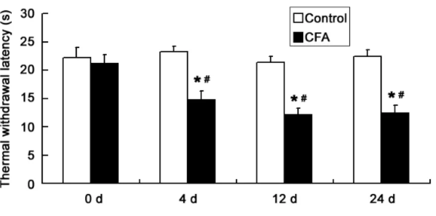
Detection of pain sensation
The TWL of CFA treated rats was 14.73 ± 0.93s, 12.15 ± 0.99s and 12.45 ± 1.19 respectively at 4 d, 12 d and 24 d after injection, respectively, which were significantly shorter than that in the
control group (P < 0.01). This suggests that hype-ralgia was induced following CFA injection at the prostate and the rat chronic prostatitis model was successfully established (Figure-2).
mRNA expression of P2X7 and IBA-1 in poste-rior horn
The mRNA expression of P2X7 and IBA-1 was significantly increased in the posterior horn of L5-S2 spinal cord in the experiment group on days 4, 12 and 24 as compared to the control group at the corresponding time points and to the experi-ment group at baseline (P < 0.01). The P2X7 expres-sion reached a maximal level on day 12 (P < 0.05) (Figures 3A and 3B).
Content of TNF-α and IL-1β in L5-S2 posterior horn
The content of TNF-α and IL-1β in the pos-terior horn of L5-S2 spinal cord was significantly increased in the experiment group on days 4, 12 and 24, as compared to the control group at cor-responding time points (P < 0.01) and to the ex-periment group at baseline (P < 0.01). The content of TNF-α and IL-1β reached a peak on day 12 (P < 0.05) (Figures 3C and 3D).
Content of TNF-α and IL-1β following inhibition of microglial cells and/or P2X7
In the experiment group, following injection of minocycline and oATP, the content of TNF-α and IL-1β was markedly reduced (P < 0.01). However, the
Figure 2 - Thermal withdrawal latency (s) in two groups.
*P < 0.01 vs. control group; #P < 0.01 vs. 0 d.
P2X7 agonist (BzATP) could promote the secretion of TNF-α and IL-1β (P < 0.01 and < 0.05, respectively), and minocycline inhibited the bioeffects of BzATP (P < 0.05). In the control group, intrathecal injection of ASCF had no influence on the contents of TNF-α and IL-1β in the spinal cord (Figure-4).
DISCUSSION
Figure 4 - Contents of IL-1β and TNF-α in dorsal horn after injection of different agonist or antagonist (ng/mL).
*P < 0.01 vs. control group; #P <0.01 vs. 0 d; &P <0.05 vs. 4 d and 24 d.
Figure 3 - mRNA expression of P2X7 (A) and IBA-1 (B) in dorsal horn and contents of TNF-α (C) and IL-1β (D) (ng/mL) in dorsal
and treatment of chronic prostatitis have been a challenge in urology. In addition, the long-lasting pain may result in physical and psychological di-sorders. Thus, the investigation of the etiology of chronic prostatitis and the pathogenesis of pain in chronic prostatitis are crucial for the accurate diagnosis and development of effective strategies for the treatment of chronic prostatitis.
Pain appears to be a most prominent ma-nifestation of chronic prostatitis. However, the diagnosis and treatment of pain in chronic pros-tatitis are still challenging because of the com-plicated pathogenesis of chronic prostatitis pain (21). Patients with chronic prostatitis often expe-rience pains not only at prostate, but at the sites adjacent to or tissues outside the prostate which are found to be also controlled by the L5-S2 spi-nal cord. Moreover, some patients feel pain even after prostatitis disappears. Hence, the pain in the chronic prostatitis is often characteristic of “extra--territorial” and “mirror” image pain. Increasing evidence demonstrates that there are abnormali-ties in the cell-mediated neurological regulation and the transmitters in the L5-S2 spinal cord in chronic prostatitis (22,23).
Accumulating studies have revealed that the pathological pain is due to not only neuronal dysfunction, but the activation of astrocytes and microglias (24), especially in the chronic exaggera-ted and continuing pain. Microglias and astrocytes are regarded as “immune cells” in the nervous system, and can secrete some pro-inflammatory cytokines such as IL-1β, TNF-α, NGF, NO, pros-taglandin and bradykinin following activation, le-ading to the exaggeration and persistence of pain by acting on other glial cells and neurons (25,26). Therefore, microglial cells play important roles in the pathogenesis of pathological pain (27,28). In rats with sciatic inflammation, intrathecal injec-tion of minocycline, an inhibitor of microglial cell activation, was found to inhibit the abnormal me-chanical pain with low threshold (29).
In the pathogenesis of pathological pain, P2X7 plays an important role in the secretion of pro-inflamamtory cytokines mediated by micro-glial cell activation (27). P2X7 is an ATP recep-tor, a transmitter and modulator in the nervous system. P2X7 is a special subtype of purinergic
receptor P2X family. In rats with inflammatory pain, visceral pain and neuropathic pain, focal or intraperitoneal injection of antagonist of P2X7 (oATP or A-740003) was found to inhibit the me-chanical hyperalgesia, allodynia and hypersensiti-vity (30,31). In the P2X7 receptor deficiency mice, the neuropathic hypersensitivity to mechanical or heat stimulation was absent following nerve injury (11). However, under physiological condi-tions, P2X7 receptor is not activated. Under the pathological conditions, P2X7 receptor is activated and involved in the pain transduction. Our findin-gs also revealed that P2X7 receptor activation sig-nificantly increased the secretion of pro-inflam-matory cytokines in animal inflammation model. Following activation, P2X7 receptor involves in the pain transduction, which is associated with the calcium related signal transduction (32,33).
inflam-mation. However, the focal drug concentration is at a low level leading to unfavorable efficacy. In Traditional Chinese Medicine, the surface projec-tion of L5-S2 spinal cord is also known as Shenshu point and acupuncture of Shenshu point has been used in the treatment of chronic prostatitis pain (35). To date, we have applied “water-needle the-rapy” for chronic prostatitis pain on the basis of our previous findings, in which the acupuncture of acupoint at L5-S2 spinal cord was performed followed by focal injection of B12, B1, hydrocor-tisone and Chinese herbs. This treatment achieves favorable efficacy, but is still in its infancy stage.
Of note, our findings can not explain the whole molecular mechanisms underlying the ac-tivation of microglial cells in chronic prostatitis because there are other receptors (such as P2X4, P2Y12 and Toll like receptor) related to the neu-ropathic pain (36,37). Studies have shown that the P2X7 receptor is related to the P2X4 receptor in structure and function, and there is interac-tion between P2X7 and P2X4 in the microglial cell mediated pain (38). In addition, the activation of Toll-like receptor 4 in the dorsal horn and the release of IL-1β are dependent on the activation of P2X7 receptor, and inhibitors of P2X7 recep-tor (oxidized ATP, A-438079) may suppress the hyperalgesia to heat and mechanical stimulation following intrathecal injection of LPS (a agonist of Toll-like 4 receptor) (39). These findings de-monstrate that the P2X7 receptor on the microglial cells can interact with the above molecules, whi-ch then aggregates the neuropathic pain Howe-ver, the specific mechanisms of their interactions require further studies.
CONCLUSIONS
The chronic prostatitis is related to the ac-tivation of P2X7 and microglial cells and the high expression of TNF-α and IL-1β in the dorsal horn of L5-S2 spinal cord. Moreover, TNF-α and IL-1β expression in the L5-S2 spinal cord can be inhibi-ted by inhibitors of P2X7 receptor and microglial cells. These findings indicate that chronic pelvic pain syndrome may cause secondary inflamma-tion in the L5-S2 spinal cord by activating the microglial cells via PX receptor, a phenomenon
probably associated with the persistence and in-tensification of chronic prostatitis pain.
ABBREVIATIONS
ACSF = artificial cerebrospinal fluid
ANOVA = analysis of variance
ATP = adenosine triphosphate
BzATP = 2’-3’-O-(4-Benzoylbenzoyl)-adenosine 5’-triphosphate
CFA = complete Freund’s adjuvant
CNS = central nervous system
IBA-1 = ionized calcium binding adaptor mole-cule 1
IL-1 = interleukin-1
NGF = nerve growth factor
NO = nitric oxide
oATP = oxidized ATP
PMSF = phenylmethanesulfonyl fluoride
SDS = sodium dodecyl sulfate
SEM = standard error
SPF = specific pathogen free TNF-α = tumor necrosis factor-α
TWL = thermal withdrawal latency
ACKNOWLEDGMENT
This study was supported by the National Natural Science Foundation (NO.81100537).
CONFLICT OF INTEREST
None declared.
REFERENCES
1. Luzzi GA: Chronic prostatitis and chronic pelvic pain in men: aetiology, diagnosis and management. J Eur Acad Dermatol Venereol. 2002; 16: 253-6.
2. Ishigooka M, Nakada T, Hashimoto T, Iijima Y, Yaguchi H: Spi-nal substance P immunoreactivity is enhanced by acute chem-ical stimulation of the rat prostate. Urology. 2002; 59: 139-4. 3. Chen Y, Song B, Jin XY, Xiong EQ, Zhang JH: Possible
4. Beggs S, Salter MW: Microglia-neuronal signalling in neuropathic pain hypersensitivity 2.0. Curr Opin Neuro-biol. 2010; 20: 474-80.
5. Watkins LR, Milligan ED, Maier SF: Glial activation: a driv-ing force for pathological pain. Trends Neurosci. 2001; 24: 450-5.
6. Madiai F, Hussain SR, Goettl VM, Burry RW, Stephens RL Jr, Hackshaw KV: Upregulation of FGF-2 in reactive spinal cord astrocytes following unilateral lumbar spinal nerve ligation. Exp Brain Res. 2003; 148: 366-76.
7. Kucher BM, Neary JT: Bi-functional effects of ATP/P2 re-ceptor activation on tumor necrosis factor-alpha release in lipopolysaccharide-stimulated astrocytes. J Neuro-chem. 2005; 92: 525-35.
8. DeLeo JA, Tanga FY, Tawfik VL: Neuroimmune activation and neuroinflammation in chronic pain and opioid toler-ance/hyperalgesia. Neuroscientist. 2004; 10: 40-52. 9. Beggs S, Salter MW: Stereological and somatotopic
anal-ysis of the spinal microglial response to peripheral nerve injury. Brain Behav Immun. 2007; 21: 624-33.
10. Suzuki T, Hide I, Ido K, Kohsaka S, Inoue K, Nakata Y: Production and release of neuroprotective tumor necrosis factor by P2X7 receptor-activated microglia. J Neurosci. 2004; 24: 1-7.
11. Chessell IP, Hatcher JP, Bountra C, Michel AD, Hughes JP, Green P, et al.: Disruption of the P2X7 purinoceptor gene abolishes chronic inflammatory and neuropathic pain. Pain. 2005; 114: 386-96.
12. Nackley AG, Suplita RL 2nd, Hohmann AG: A peripheral cannabinoid mechanism suppresses spinal fos protein expression and pain behavior in a rat model of inflamma-tion. Neuroscience. 2003; 117: 659-70.
13. Butler SH, Godefroy F, Besson JM, Weil-Fugazza J: A limited arthritic model for chronic pain studies in the rat. Pain. 1992; 48: 73-81.
14. Zhou ZS, Song Bo, Lu GS: The research on the relation-ship between chronic prostatitis pain and spinal astro-cytes activation. J Third Milit Med Univ 2005; 27: 1853-4. 15. Wu AN, Xiong EQ, Song B: Secretory alterations of ure-thral glands in complete Freund’s adjuvant-induced pros-tatitis in rats. J Third Milit Med Univ 2009; 31: 2266-8. 16. Cheng H Yang JP, Zhang YB: Spinal microglia activation
intrathecal minocycline inhibited by and attenuation on thermal hyperalgesia in CFA-induced inflammatory rats. Suzhou Univ J Med Sci 2007; 27: 14-6.
17. Yaksh TL, Rudy TA: Chronic catheterization of the spinal subarachnoid space. Physiol Behav. 1976; 17: 1031-6. 18. Millán-Rodríguez F, Palou J, Bujons-Tur A,
Musquera-Fe-lip M, Sevilla-Cecilia C, Serrallach-Orejas M, et al.: Acute bacterial prostatitis: two different sub-categories accord-ing to a previous manipulation of the lower urinary tract. World J Urol. 2006; 24: 45-50.
19. Zhou Z, Hong L, Shen X, Rao X, Jin X, Lu G, et al.: Detec-tion of nanobacteria infecDetec-tion in type III prostatitis. Urolo-gy. 2008; 71: 1091-5. Erratum in: UroloUrolo-gy. 2008 ;72: 723. 20. Shen X, Ming A, Li X, Zhou Z, Song B: Nanobacteria: a
possible etiology for type III prostatitis. J Urol. 2010; 184: 364-9.
21. Baxter C, Bolus R, Mayer E: Choice and outcomes of al-ternative therapies in patients with interstitial cystitis (IC) and chronic pelvic pain (CPP). J Urol. 2010; 183: 580. 22. Zhang H, Liu L, Lu G, Chen Z, Fang Q, Yang Z, et al.:
Chemical irritation of the prostate sensitizes P(2)X(3) receptor-mediated responses in rat dorsal root ganglion neurons. Neurourol Urodyn. 2011; 30: 612-8.
23. Baranowski AP: Chronic pelvic pain. Best Pract Res Clin Gastroenterol. 2009; 23: 593-610.
24. Watkins LR, Milligan ED, Maier SF: Glial activation: a driv-ing force for pathological pain. Trends Neurosci. 2001; 24: 450-5.
25. Li HL, Qin LY, Wan Y: Astrocyte: a new star in pain re-search. Sheng Li Ke Xue Jin Zhan. 2003; 34: 45-8. 26. Aronica E, Gorter JA, Rozemuller AJ, Yankaya B, Troost D:
Activation of metabotropic glutamate receptor 3 enhances interleukin (IL)-1beta-stimulated release of IL-6 in cultured human astrocytes. Neuroscience. 2005; 130: 927-33. 27. Mingam R, De Smedt V, Amédée T, Bluthé RM, Kelley KW,
Dantzer R, et al.: In vitro and in vivo evidence for a role of the P2X7 receptor in the release of IL-1 beta in the murine brain. Brain Behav Immun. 2008; 22: 234-44.
28. Barger SW, Goodwin ME, Porter MM, Beggs ML: Gluta-mate release from activated microglia requires the oxi-dative burst and lipid peroxidation. J Neurochem. 2007; 101: 1205-13.
29. Ledeboer A, Sloane EM, Milligan ED, Frank MG, Mahony JH, Maier SF, et al.: Minocycline attenuates mechanical allodynia and proinflammatory cytokine expression in rat models of pain facilitation. Pain. 2005; 115: 71-83. 30. Fulgenzi A, Ticozzi P, Gabel CA, Dell’Antonio G, Quattrini
A, Franzone JS, et al.: Periodate oxidized ATP (oATP) re-duces hyperalgesia in mice: involvement of P2X7 recep-tors and implications for therapy. Int J Immunopathol Pharmacol. 2008; 21: 61-71.
31. Honore P, Donnelly-Roberts D, Namovic MT, Hsieh G, Zhu CZ, Mikusa JP, et al.: A-740003 [N-(1-{[(cyanoimino) (5-quinolinylamino) methyl]amino}-2,2-dimethylpropyl)-2-(3,4-dimethoxyphenyl)acetamide], a novel and selective P2X7 receptor antagonist, dose-dependently reduces neu-ropathic pain in the rat. J Pharmacol Exp Ther. 2006; 319: 1376-85.
33. Wang CM, Chang YY, Kuo JS, Sun SH: Activation of P2X(7) receptors induced [(3)H]GABA release from the RBA-2 type-2 astrocyte cell line through a Cl(-)/HCO(3) (-)-dependent mechanism. Glia. 2002; 37: 8-18.
34. Stalder AK, Carson MJ, Pagenstecher A, Asensio VC, Kincaid C, Benedict M, et al.: Late-onset chronic inflam-matory encephalopathy in immune-competent and severe combined immune-deficient (SCID) mice with astrocyte-targeted expression of tumor necrosis factor. Am J Pathol. 1998; 153: 767-83.
35. Chen ZX: Observation on therapeutic effect of warm needle moxibustion on chronic non-bacterial prostatitis. Zhongguo Zhen Jiu. 2009; 29: 275-8.
36. Kim D, Kim MA, Cho IH, Kim MS, Lee S, Jo EK, et al.: A critical role of toll-like receptor 2 in nerve injury-induced spinal cord glial cell activation and pain hypersensitivity. J Biol Chem. 2007; 282: 14975-83.
37. Tozaki-Saitoh H, Tsuda M, Miyata H, Ueda K, Kohsaka S, Inoue K: P2Y12 receptors in spinal microglia are required for neuropathic pain after peripheral nerve injury. J Neu-rosci. 2008; 28: 4949-56.
38. Casas-Pruneda G, Reyes JP, Pérez-Flores G, Pérez-Corne-jo P, Arreola J: Functional interactions between P2X4 and P2X7 receptors from mouse salivary epithelia. J Physiol. 2009; 587: 2887-901.
39. Clark AK, Staniland AA, Marchand F, Kaan TK, McMahon SB, Malcangio M: P2X7-dependent release of interleukin-1beta and nociception in the spinal cord following lipo-polysaccharide. J Neurosci. 2010; 30: 573-82.
_____________________
Correspondence address: