ABSTRACT
http://dx.doi.org/10.1590/1678-775720150100
Impact of brief exposure to antifungal agents on
the post-antifungal effect and hemolysin activity
of oral
Candida albicans
Arjuna Nishantha ELLEPOLA1, Rana KHAJAH1, Sumedha JAYATILAKE2, Lakshman SAMARANAYAKE3, Prem SHARMA4, Zia KHAN5
1- Faculty of Dentistry, Health Sciences Center, Kuwait University, Kuwait. 2- Faculty of Dental Sciences, University of Peradeniya, Sri Lanka. 3- School of Dentistry, University of Queensland, Brisbane, Australia. 4- Faculty of Medicine, Health Sciences Center, Kuwait University, Kuwait.
5- Department of Microbiology, Faculty of Medicine, Health Sciences Center, Kuwait University, Kuwait.
Corresponding address: Arjuna Ellepola - Department of Bioclinical Sciences - Faculty of Dentistry - Kuwait University - P.O. Box 24923 - Safat 13110 - Kuwait - Phone: 00965 24636714 - Fax: 00965 25326049 - e-mail: [email protected]
6XEPLWWHG0DUFK0RGL¿FDWLRQ0D\$FFHSWHG-XQH
P
ost-antifungal effect (PAFE) of Candida and its production of hemolysin are determinants of candidal pathogenicity. Candida albicans is the foremost aetiological agent of oral candidosis, which can be treated with polyene, azole, and echinocandin antifungals. However, once administered, the intraoral concentrations of these drugs tend to be subtherapeutic and transient due to the diluent effect of saliva and cleansing effect of the oral musculature. Hence, intra-orally, Candida may undergo a brief exposure to antifungal drugs. Objective: Therefore, the PAFE and hemolysin production of oral C. albicans isolates following brief exposure to sublethal concentrations of the foregoing antifungals were evaluated. Material and Methods: A total of 50 C. albicans oral isolates obtained from smokers, diabetics, asthmatics using steroid inhalers, partial denture wearers and healthy individuals were exposed to sublethal concentrations of nystatin, amphotericin B, caspofungin, ketoconazole ! production were determined by previously described turbidometric and plate assays, respectively. Results: Nystatin, amphotericin B, caspofungin and ketoconazole induced mean PAFE (hours) of 2.2, 2.18, 2.2 and 0.62, respectively. Fluconazole failed to produce a PAFE. Hemolysin production of these isolates was suppressed with a percentage reduction of 12.27, 13.47, 13.33, 8.53 and 4.93 following exposure to nystatin, amphotericin B, " # "! $ % & '" to sublethal concentrations of antifungal drugs appears to exert an antifungal effect by interfering with the growth as well as hemolysin production of C. albicans.Keywords:Candida albicans. Post-antifungal effect. Hemolysins. Antimycotics.
INTRODUCTION
Multiple physiognomies of Candida species have been suggested as virulence factors that enable the organism to cause superficial as well as disseminated infections in susceptible hosts11. Hemolysins are known to be critical virulent determinants contributing to candidal pathogenesis12. In particular, the secretion of hemolysin, followed by lysis of erythrocytes and subsequent acquisition of iron by Candida, facilitates
of hemoglobin is independent of iron acquisition, and is facilitated by a cell surface hemoglobin receptor. Furthermore, hemoglobin rapidly induces the expression of several genes, one of which, a heme oxygenase, allows the pathogen to consume exogenous heme or hemoglobin to acquire iron and produce the cytoprotective molecules14. Such exquisite recognition and responses to hemoglobin appear to be a unique adaptation of C. albicans to be a versatile opportunistic pathogen in the human host14.
C. albicans is by far the leading fungal
pathogen of oral candidosis, the commonest human fungal infection with a variety of clinical manifestations9. Interestingly, more than 90%
*! ;<=>? individuals develop oral candidosis during some point of their disease which is arguably the commonest oral manifestation in such patients. In addition, C. albicans has also been implicated in oral candidosis in other patient groups such as diabetics, asthmatics using inhalation steroids, smokers and denture wearers10,16,17. Recent studies have also shown that C. albicans isolates from subgingival biofilms of immunocompromised patients also produce hemolysins which may also exacerbate periodontal disease in these patients by insidiously ! " destruction15.
Growth suppression of Candida that occurs subsequent to limited exposure to antifungal agents, as in the case of the oral environment, has been described as the post-antifungal effect (PAFE).
It has been proposed that the knowledge of PAFE would be clinically useful in evaluating new dosage regimens of a drug, and such curtailment of growth following transient exposure to antifungal agents may be a determinant of candidal virulence itself, as this phenomenon may modulate the virulent attributes of the yeast Candida5.
A number of antifungal therapeutic agents are accessible for treatment of oral Candida infections. These include the two polyenes group agents, nystatin and amphotericin B, the echinocandin agent caspofungin and the azoles such as ketoconazole ; ? ; ?9. Despite the availability of these pharmacological agents for the management of oral candidosis, failure of therapy and ensuing recalcitrant infection is not uncommon9. One reason for this may be the distinctive nature of the oral milieu where the diluent effect of saliva and the cleansing effect of the oral musculature tend to reduce the bioavailability of antifungal agents below that of the effective therapeutic concentrations. Hence, intra-orally, the pathogenic Candida may undergo a transient exposure to an antifungal agent on administration, and subsequently the drug concentration is likely
to be subtherapeutic5,9. Yet, the impact of such
therapeutic levels of antifungals leading to PAFE and hemolysin production of oral C. albicans isolates obtained from different susceptible patient groups to oral candidosis (i.e., diabetics, asthmatics using inhalation steroids, smokers and partial denture wearers) has not been studied hitherto.
Hence, the main aim of this study was to determine the PAFE induced by five different antifungal agents belonging to three distinct classes, polyenes (nystatin, amphotericin B), echinocandins (caspofungin), and azoles (ketoconazole and ? @ C. albicans isolates. The opportunity was also taken to evaluate candidal
hemolysin production under identical conditions of drug exposure.
MATERIAL AND METHODS
Organisms
A total of 50 oral isolates of C. albicans obtained in a previous study from patients seeking treatment from the Kuwait University Dental Clinic were included in the current study (10 isolates each from smokers, diabetics, asthmatics using steroid inhalation, patients wearing partial acrylic dentures and healthy individuals)7. None of the patients from
which the isolates were recovered had clinically visible oral candidosis. Initially, all Candida isolates were tested for germ tube formation. Thereafter the colony characteristics were observed using CHROMagar Candidamedium (Becton Dickinson and $ "!J"#LJ?*+!>=QV yeast ID system (BioMérieux, France) as well as API 20C AUX yeast ID system (BioMérieux, Inc, Hazelwood, MO, USA).
Antifungal agents and media
Nystatin, amphotericin B, ketoconazole and fluconazole (Sigma, St. Louis, MO, USA) were dissolved in dimethylsulphoxide (DMSO). Caspofungin (Merck and Company Inc, Waterhouse Station, NJ, USA) was dissolved in sterile distilled water. These anti-Candida agents were prepared !\]^ _V`$" to each experiment as previously described4,5. It was thereafter suspended/diluted in the following medium during the exposure period of yeasts: RPMI
(Roswell Park Memorial Institute) 1640 medium buffered with 0.165 M MOPS (3-(N-morpholino) propanesulfonic acid) containing L-glutamine and
lacking sodium bicarbonate (Sigma, USA), dissolved in 1 liter of sterile distilled water and adjusted to a "< |V*
Determination of minimal inhibitory concentration (MIC)
antifungal agents was determined as described in an earlier study of ours7. The MIC values against amphotericin B, caspofungin, ketoconazole and +!" according to manufacturer’s recommendations (AB BIODISK, Solna, Sweden). As depicted in previously ! + ;* ? uniformly suspended in sterile saline, and turbidity was adjusted to 0.5 McFarland Standard. The McFarland Standard 0.5 is approximately equal to 1x10⁶ to 5x10⁶ cells/ml3. This inoculum was swabbed onto the agar plates (150 mm diameter) and allowed to dry for 10-15 min before the E test strips were applied. RPMI 1640 agar supplemented with 2% glucose and buffered with MOPS (0.165 }~ "< |? "+! according to the method recommended by the CLSI (formerly National Committee for Clinical + !J~}V|_V?" incubated at 35°C, and MIC was observed after 24–48 h of incubation. The point where inhibition ellipses intercepted the scale on the antifungal strip was taken as the MIC for each test isolate: complete inhibition (100%) of growth for amphotericin B and caspofungin, and marked decrease in growth !;? # Reference strains of C. albicans, ATCC 90028, and
C. parapsilosis, ATCC 22019, were used as reference strains. Interpretive susceptibility breakpoints for +! $J= }V|_V * susceptibility breakpoints for amphotericin B and ketoconazole, an isolate was considered "+ }=$ +#" \ ]^ " & \V@ ]^ ketoconazole7. For caspofungin, isolates with MIC
V]^ "+7.
As previously performed, the MIC values of nystatin were established by the broth dilution technique5 by performing two-fold serial dilutions
of the drug in microtitre plates using an inoculum of 1-5×10⁵ colony forming units (CFU)/ml. The MIC
was determined visually following 24 h incubation |`$ }=$ * concentration of the drug that impeded growth
of Candida cells, as indicated by the absence
of turbidity (optically clear). The MIC was read independently by a couple of laboratory personals.
C. albicans ATCC 90028 was used as a reference
strain.
Preparation of cell suspension for the PAFE and hemolysin production assay
A heretofore explained method was used for this purpose4-6&!Candida cells maintained on Sabouraud Dextrose Agar (SDA), were inoculated onto fresh plates and incubated overnight at 37°C for 24 h prior to use. The organisms were harvested
and a cell suspension prepared in sterile phosphate buffered saline (PBS) at 520 nm to an optical density of 1.5. From this cell suspension, 1 ml was added to tubes containing 4 ml of RPMI broth (control) and 4 ml of RPMI/drug solution (test), in which the drug concentration was twice the MIC. This gave a cell suspension of approximately 106 cells ml-1 in each assay tube. The tubes were then incubated at 37°C for a period of 60 min. Following this limited exposure, the drugs were removed by two cycles of dilution with sterile PBS and centrifugation.
Supernatant was thereafter completely decanted and the pellets were re-suspended in 2.5 ml of sterile PBS. This technique has been used previously for drug removal, and has revealed to minimize the concentration of the drug as much as 10,000 fold, thus curtailing any carry-over effect of the drug following its removal3-5. Viable counts of the control and the test were performed after drug removal. As the procedure of drug removal effectively eliminated any carry-over effect, there was virtually no difference on the viable counts of the control and the tests following exposure to already diluted subtherapeutic concentrations of the drug, as observed in previous studies4-6.
PAFE assay
Following drug removal, in order to ascertain the growth suppression and ensue the recovery of fungal growth, namely the PAFE, the growth was determined by a previously described optical density method with minor amendments using the equation PAFE=T-C4-6. T is the time required for optical density of the drug-exposed cell suspension to reach the selected relative optical density of 0.05 value at 520 nm. C is the time required for optical density of the drug-free control cell suspension to reach the selected relative optical density value at 520 nm. Thus, T-C expresses the time in which the antifungal agent was capable of causing growth suppression of the organism following limited exposure to the drug (i.e., PAFE). As in previous \] " + with 2.4 mL of RPMI 1640 at 37°C and the relative optical density of the suspension was measured at frequent intervals (i.e., 15 min) for 8 h by which time, both the controls and tests reached the selected relative optical density value, enabling to calculate the PAFE4-6.
Hemolysin assay
circular inoculation site of about 10 mm in diameter. The plates were then incubated at 37°C in 5% CO2 for 48 h. After incubation during this period, the diameter of the colony (y) and the diameter of the colony plus the translucent zone of hemolysis (x) were measured. The ratio of x/y, which represents the hemolytic activity (hemolytic index), was measured as done previously1,12,13.
All experiments were repeated on three separate occasions with duplicate determinations on each occasion.
Statistical analysis
For the suppression effect on hemolysin production, the raw data did not follow normal distribution. Therefore, in accordance to non-parametric statistician advices, Wilcoxon Signed Ranks test was used to compare the control " * " '" * different antifungal agents. The difference between " '" * drugs were also compared using Wilcoxon Signed Ranks test in relation to drug-induced PAFE and suppression of hemolysin production. A p value of less than 0.05 was considered statistically *
RESULTS
All C. albicans isolates were susceptible to the tested antifungal drugs. The MIC (]^?values of all C. albicans isolates for nystatin obtained from the broth dilution technique ranged between 0.78-1.56. The MIC values obtained by E-tests for amphotericin B were between 0.004-0.19, and for caspofungin it ranged between 0.004-0.125. Ketoconazole elicited a MIC of 0.004-0.032 while it ranged between |_\V@
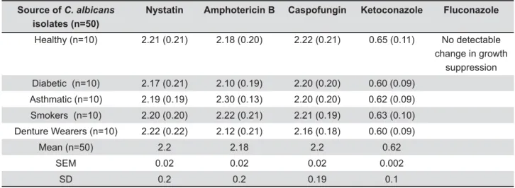
Based on the equation PAFE=T-C, the mean
in vitro PAFE (hours) on 50 oral isolates of C.
albicans following 1 h exposure and subsequent
removal of nystatin, amphotericin B, caspofungin and ketoconazole was 2.20, 2.18, 2.20 and 0.62, respectively (Table 1). Hence, in summary, brief exposure to nystain, amphotericin B and caspofungin was able to suppress the growth of C. albicans isolates giving a PAFE of approximately 2 h whereas it was approximately ½ h for ketoconazole. Fluconazole failed to elicit a detectable PAFE.
Mean hemolysin production (hemolytic index) of the 50 C. albicans isolates, following limited '" *+ of antifungal agents, drug removal and subsequent production of hemolysin production is shown in Table 2. Mean hemolysin production of the unexposed \@ * exposure to nystatin, amphotericin B, caspofungin, # \V\\ 1.37 and 1.42, respectively. Hence, as shown in +V* ! activity was observed following the exposure to all of these drugs, with a mean percentage reduction of 12.27, 13.47, 13.33, 8.53 and 4.93 for nystatin, amphotericin B, caspofungin, ketoconazole and
"!;"\ ?
The analysis of the difference on PAFE and the suppressive effect on hemolysin production elicited
+! * revealed
that the PAFE and the suppressive effect elicited by the polyene and echinocandin antifungal agents *!! ;"\? = addition, the PAFE and the hemolysin suppressive effect elicited by the two azoles were also ! ;"\? with ketoconazole having a superior effect than
Source of C. albicans isolates (n=50)
Nystatin Amphotericin B Caspofungin Ketoconazole Fluconazole
Healthy (n=10) 2.21 (0.21) 2.18 (0.20) 2.22 (0.21) 0.65 (0.11) No detectable
change in growth suppression
Diabetic (n=10) 2.17 (0.21) 2.10 (0.19) 2.20 (0.20) 0.60 (0.09)
Asthmatic (n=10) 2.19 (0.19) 2.30 (0.13) 2.20 (0.20) 0.62 (0.09)
Smokers (n=10) 2.20 (0.20) 2.22 (0.21) 2.21 (0.19) 0.63 (0.10)
Denture Wearers (n=10) 2.22 (0.22) 2.12 (0.21) 2.16 (0.18) 0.60 (0.09)
Mean (n=50) 2.2 2.18 2.2 0.62
SEM 0.02 0.02 0.02 0.002
SD 0.2 0.2 0.19 0.1
Table 1- The post-antifungal effect (PAFE) (in hours) of 50 oral C. albicans isolates following 1 hour exposure and subsequent
C. albicans obtained either from
< "" effect on hemolysin production elicited by the two polyenes and the echinocandin agent were not *!
Finally, when comparing the clinical isolates from diabetics, asthmatics, smokers, denture wearers and healthy individuals, there were no difference in either PAFE or hemolysin production on exposure to sublethal concentrations of any of the antifungals.
DISCUSSION
C. albicans is by far the major aetiological agent of oral candidosis8. Hemolysin is one of C. albicans recently recognized, putative virulent factors. In particular, the secretion of hemolysin facilitates hyphal invasion, which is critical for disseminated candidal infection1,12-14. As described above, it is known that PAFE, the capacity of Candida to recover and grow after an antifungal-insult, is another important measure of the virulence of the yeast4-7. We, along with others, have shown that more
Source of C. albicans isolates
(n=50)
Unexposed controls
Nystatin Amphotericin B Caspofungin Ketoconazole Fluconazole
Healthy 1.49 1.31 1.29 1.29 1.37 1.42
(n=10) -0.02 -0.02 -0.05 -0.05 -0.02 -0.02
Diabetic 1.5 1.3 1.27 1.3 1.38 1.43
(n=10) -0.03 -0.03 -0.06 -0.05 -0.03 -0.02
Asthmatic 1.5 1.33 1.31 1.32 1.38 1.43
(n=10) -0.02 -0.05 -0.07 -0.06 -0.03 -0.03
Smokers 1.51 1.34 1.33 1.29 1.36 1.43
(n=10) -0.02 -0.06 -0.07 -0.07 -0.02 -0.04
Denture Wearers 1.5 1.3 1.29 1.3 1.37 1.42
(n=10) -0.03 -0.07 -0.08 ().05) -0.03 -0.02
Mean (n=50) 1.5 1.32 1.3 1.3 1.37 1.42
SEM 0.003 0.007 0.009 0.008 0.004 0.004
SD 0.02 0.05 0.06 0.06 0.03 0.03
p value p<0.001 p<0.001 p<0.001 p<0.001 p<0.001
Table 2- Mean (SD) hemolysin activity (hemolytic index) of 50 oral C. albicans isolates following 1 hour exposure and
C.
albicans obtained either from healthy individuals, diabetic patients, asthmatic patients, smokers or denture wearers. SD:
Standard deviation. SEM: Standard error of mean
Source of C. albicans Isolates (n=50)
Nystatin Amphotericin B Caspofungin Ketoconazole Fluconazole
Healthy 12.08 13.42 13.42 8.05 4.7
Diabetic 13.33 15.33 13.33 8 4.66
Asthmatic 11.33 12.66 12 8 4.66
Smokers 11.26 11.92 14.57 9.93 5.3
Denture Wearers 13.33 14 13.33 8.66 5.33
Mean 12.27 13.47 13.33 8.53 4.93
SEM 0.46 0.58 0.41 0.37 0.16
SD 1.02 1.3 0.91 0.83 0.35
Table 3- Summary of the mean percentage suppression of hemolysin activity in 50 oral C. albicans isolates following 1 hour
!" " of hemolysin activity in 10 oral C. albicans obtained from either healthy individuals, diabetic patients, asthmatic patients,
virulent and resistant Candida will have a low PAFE, whereas susceptible and less virulent organisms will have higher PAFEs4-7.
The present results indicate that brief exposure to sublethal concentrations of nystatin, amphotericin B, caspofungin and ketoconazole elicited PAFEs ranging from approximately 30 to 120 min with the C. albicans isolates tested. A notable exception tested isolates. The observed PAFE may be related to the pharmacodynamics of these antimycotics on
Candida. For instance, both polyenes (nystatin and amphotericin B) bind to the sterol components in the yeast cell wall and make it more penetrable5,9.
Furthermore, investigations have shown internally collapsed cells with an intact cell wall leaving “ghosts-like” cells, and deflated Candida cells following exposure to subcidal concentrations of polyenes8. Caspofungin, on the contrary, acts by
inhibiting the enzyme responsible for biosynthesis _\+! of the fungal cell wall2. In addition, caspofungin
has shown excellent growth-inhibitory activity against Candida at low concentrations2. As for
;# ? ! alter the fungal cell membranes by blocking the \ _! " + ! ergosterol. The consequent depletion of ergosterol \_!_ alterations in a number of membrane-associated functions4,9. Considering the foregoing wide range
pharmacodynamic properties of the agents we have studied, it is tempting to speculate that the structural changes of the cell wall, as well as the metabolic effects elicited by the drugs, would all contribute to the suppression of growth eliciting J"! a detectable PAFE response in spite of this drug being remarkably more effective in vivo than ketoconazole in the management of candidal infection9. Interestingly, many workers have shown
+ !! C. albicans isolates in vitro is much less than that of ketoconazole4,9,18. Hence, the later could be one
reason for the absence of detectable PAFE following '"
The exposure of C. albicans isolates to the foregoing drugs was also capable of suppressing ! " ! * varying levels ranging from 5% to 13.5%. A previous single study has also shown a reduction of hemolysin activity of C. albicans, as well as Candida tropicalis isolates, following exposure to ! " & 1. It is
* '+! which these drugs modulate changes in hemolysin production in Candida species. However, taken
collectively, * +
understanding of pharmacodynamics of these antifungal agents against two virulent determinants (i.e., PAFE and hemolysin production) incriminated in the pathogenesis of C. albicans infections. In addition, as hemolysins are known to mediate host invasion, particularly by facilitating hyphal penetration of the epithelium, it can be speculated that, by suppressing hemolysin production, these antifungals could also minimize yeast dissemination from and within the oral and periodontal tissues.
* +! polyenes and echinocandins and azoles, as well as their suppressive effect on hemolysin production
of Candida, as seen in this study, may well be
due to the differences in the pharmacodynamics of the different antimycotic groups. Polyenes and echinocandins are fungicidal against C. albicans whereas azoles are fungistatic9. This could be the
foremost explanation for the exquisite sensitivity of C. albicans to nystatin, amphotericin B and caspofungin compared to the azoles. In addition, we also noted a significant difference in the PAFE and hemolysin production when the yeasts were exposed to the two azoles. As previously mentioned, others have shown that the growth
+ !! C. albicans
in vitro is much less than that of ketoconazole4,9,18.
These observations may be related to the fact that # is a triazole. Arguably, therefore, the differences in the mode of activity between the two azoles may # variations in PAFE and hemolysin production.
# !* one to document the antifungal-induced PAFE using a wide array of antifungal agents and the suppression of hemolysin production during the PAFE period, covering the largest number (n=50) of oral C. albicans isolates obtained from a single geographic locale. Testing with a larger number of sessile isolates encased in a Candida + * scenario instead of using only planktonic isolates as used in this study is warranted to further elucidate *The exact cellular mechanisms by which antifungals interfere with hemolysin activity in Candida also remain to be elucidated.
CONCLUSIONS
Brief and transient exposure to sublethal concentrations of antifungal drugs may induce a PAFE and modulate hemolysin production of oral
ACKNOWLEDGEMENTS
The study was partially supported by Kuwait University Research Grant No. DB 01/14. The authors thankfully acknowledged the technical advice and support in laboratory procedures for carrying out this project given by Ms. Rachel Chandy and Ms. Preethi John of Health Sciences Center, Kuwait University, Kuwait.
REFERENCES
1- Anil S, Hashem M, Vellappally S, Patil S, Bandara HM, Samaranayake LP. Sub-inhibitory concentrations of antifungals suppress hemolysin activity of oral Candida albicans and Candida tropicalis isolates from HIV-infected individuals. Mycopathologia. V\~\|%V|_\@
2- Bizerra FC, Melo AS, Katchburian E, Freymüller E, Straus AH, Takahashi HK, et al. Changes in cell wall synthesis and ultrastructure during paradoxical growth effect of caspofungin on four different Candida species. Antimicrob Agents Chemother. V\\~@@%V_\
3- Clinical and Laboratory Standards Institute. Method for antifungal disk diffusion susceptibility testing of yeasts: approved }_!%$$J~V
4- Ellepola AN, Chandy R, Khan ZU. Post-antifungal effect and adhesion to buccal epithelial cells of oral Candida dubliniensis
isolates subsequent to limited exposure to amphotericin B, # = $ V\~ % 10.1111/jicd.12095. Epub ahead of print.
5- Ellepola AN, Joseph BK, Chandy R, Khan ZU. The postantifungal effect of nystatin and its impact on adhesion attributes of oral
Candida dubliniensis }! V\~@|%@_ 6- Ellepola AN, Joseph BK, Khan ZU. The postantifungal effect and phospholipase production of oral Candida albicans from smokers, diabetics, asthmatics, denture wearers and healthy individuals following brief exposure to subtherapeutic concentrations of ' }! V\~@|%@@_
7- Ellepola AN, Khan ZU, Joseph BJ, Chandy R, Philip L. Prevalence of Candida dubliniensis among oral Candida isolates in patients attending the Kuwait University Dental Clinic. Med Princ Pract. V\\~V%V|\_
8- Ellepola AN, Samaranayake LP. The effect of limited exposure to antifungal agents on the germ tube formation of oral Candida albicans }\~V|%V\_
9- Ellepola AN, Samaranayake LP. Oral candidal infections and ! $& }V~\\%\|V_
10- Ellepola AN, Samaranayake LP. Inhalation and topical steroids, %V\~|%V\\_ 11- Haynes K. Virulence in Candida species. Trends Microbiol. V\~%@\_
12- Luo G, Samaranayake LP, Yau JY. Candida species exhibit differential in vitro hemolytic activities. J Clin Microbiol. V\~%V|\_
13- Manns JM, Mosser DM, Buckley HR. Production of a hemolytic factor by Candida albicans==\~V%@\@_ 14- Pendrak ML, Yan SS, Roberts DD. Sensing the host environment: recognition of hemoglobin by the pathogenic yeast
Candida albicans& & "!V~V%\_@ \@_ J $ $ < & phenotypic evaluation of Candida albicans strains isolated from ++ * +" " }}! V\V~@%|_|@
16- Soysa NS, Ellepola AN. The impact of cigarette/tobacco # % V@~\\%V_ 73.
17- Soysa NS, Samaranayake LP, Ellepola AN. Diabetes mellitus as + ! +}V~V%@@_ 9.