w w w . j c o l . o r g . b r
Journal
of
Coloproctology
Original
Article
Use
of
endoanal
ultrasound
as
complimentary
evaluation
for
detection
of
anal
sphincter
injury
after
vaginal
birth
Ahmad
Izadpanah
a,
Ensieh
Izadpanah
b,
Mehrzad
Lotfi
b,
Alamtaj
Samsami
c,
Alireza
Safarpour
a,
Mohammad
Rezazadehkermani
d,∗aShirazUniversityofMedicalSciences,ColorectalResearchCenter,Shiraz,Iran
bShirazUniversityofMedicalSciences,SchoolofMedicine,DepartmentofRadiology,Shiraz,Iran
cShirazUniversityofMedicalSciences,SchoolofMedicine,DepartmentofObstetrics&Gynecology,Shiraz,Iran
dShirazUniversityofMedicalSciences,SchoolofMedicine,DepartmentofGeneralSurgery,Shiraz,Iran
a
r
t
i
c
l
e
i
n
f
o
Articlehistory:
Received6November2016 Accepted21April2017 Availableonline18May2017
Keywords:
Endosonography Vaginaldelivery Sphincterinjury Manometery
a
b
s
t
r
a
c
t
Purpose:Analsphincterinjuryafterdeliveryisthemainfactorinthepathogenesisoffecal incontinence.Clinicalobviousandspecificinjurytoanalcanalsphincterisseenin3%of vaginaldeliveries.Therearemanywomenwhodonothaveaclearandspecificlacerationbut theyaredamagedbysphinctermusclesofanalcanal.Thepurposeofthepresentstudyisto investigatethefrequencyofoccultanalsphincterinjuryaftervaginaldeliverybyEndo-anal sonography.
Methods:Fiftywomenwithfirstpregnancywereassessedat27–33weeksofpregnancy,and at6weeksand6monthsaftervaginaldeliverybyquestionnaire,examinationandEndo-anal sonography. Womenage,durationofdelivery,theeffectofepiduralanesthesia, episiot-omyandbirthweightwerestudiedandEndo-analsonographyresultswererecorded.Anal manometrywasperformedforallmothersbeforedeliveryand5oneswithsphincterinjury at6monthsand3yearsafterdelivery.
Results:Five (10%) patients, with mean age 29.4±6.5years, mean neonatalweight of 3874±287,andmean durationofdelivery11.6±1.51h,hadsignsofsphincterinjuryin Endo-analsonography.Theinjurywaspersistedatsixmonthsafterdelivery.Also, signifi-cantdifferenceswereseenbetweenanalmanometrybeforedeliveryand6monthsand3 yearsafterdelivery(p=0.006formeansqueezingpressure)inthefivemothers.
Conclusion: Endo-analsonographymightbeagoodscreeningtoolforearlydetectionof post-partumanalsphincterdamages.However,furtherprospectivecostbenefitstudiesshould beperformedtoproposeitasastandardofcare.
©2017SociedadeBrasileiradeColoproctologia.PublishedbyElsevierEditoraLtda.This isanopenaccessarticleundertheCCBY-NC-NDlicense(http://creativecommons.org/
licenses/by-nc-nd/4.0/).
∗ Correspondingauthor.
E-mail:[email protected](M.Rezazadehkermani).
http://dx.doi.org/10.1016/j.jcol.2017.04.006
Uso
da
ultrassonografia
endoanal
como
avaliac¸ão
complementar
para
a
detecc¸ão
de
lesão
do
esfíncter
anal
após
parto
vaginal
Palavras-chave:
Ultrassonografiaendoanal Partovaginal
Lesãoesfinctérica Manometria
r
e
s
u
m
o
Finalidade:Alesãodeesfíncteranalapósopartoéofatorprincipalnapatogêneseda incon-tinênciafecal.Observa-seumalesãoclínicaóbviaeespecíficaaoesfíncternocanalanal em3%dospartosvaginais.Emmuitasmulheresnãosepercebeumalacerac¸ãonítidae específica,mashouvelesãonosmúsculosesfinctéricosdocanalanal.Afinalidadedesse estudoéinvestigarafrequênciadelesãoocultadeesfíncternocanalanalemseguidaao partovaginalpormeiodaultrassonografiaendoanal.
Métodos: Cinquentamulheresprimíparasforamavaliadasnoperíodode27-33semanasde gestac¸ãoetambéma6semanase6mesesapósopartovaginalpormeiodequestionário, exameeultrassonografiaendoanal.Foramanotadosaidadedaspacientes,adurac¸ãodo parto,oefeitodaanestesiaepidural,episiotomiasepesodobebêaonascer;tambémforam registradososresultadosdaultrassonografiaendoanal.Antesdoparto,todasasgestantes foramsubmetidasaumexamedemanometria;e5mãescomlesãoesfinctéricatambém passaramporesseprocedimentoa6mesese3anosapósoparto.
Resultados: Cinco(10%)pacientes,commédiadeidade=29,4±6,5anos,pesomédiodobebê aonascer=3874±287gramasedurac¸ãomédiadoparto=11,6±1,51horas,apresentavam sinaisdelesãoesfinctéricaaoexameporultrassonografiaendoanal.Seismesesapósoparto, aslesõespersistiam.Tambémforamobservadasdiferenc¸assignificativasentrea manome-triaanalantesdopartoea6mesese3anosapósoparto(p=0,006paramédiadepressão decontrac¸ão)nascincomães.
Conclusão: Aultrassonografiaendoanalpodeserumbominstrumentodetriagemparaa detecc¸ãoprecocedelesõesdoesfíncteranalnopós-parto.Contudo,éimportantequesejam realizadosnovosestudosprospectivosedecusto-benefício,paraqueessatécnicapossaser propostacomopadrãoterapêutico.
©2017SociedadeBrasileiradeColoproctologia.PublicadoporElsevierEditoraLtda.Este ´eumartigoOpenAccesssobumalicenc¸aCCBY-NC-ND(http://creativecommons.org/
licenses/by-nc-nd/4.0/).
Introduction
Everyyear,50millionwomenduringpregnancy,childbirth,or afterthatarestrickenwithcomplicationsand15%ofwomen livewithchroniccomplicationsanddisabilities.1Oneofthese complicationsisanalsphincterinjuries.Somerisk-factorsare proposedwhichassociatewithincreaseofanalinjuriesare: inductionoflabor,epiduralanalgesia,birthweightmorethan 4kg,persistentoccipitoposteriorposition,primparity,second stageofdeliverylongerthan1handuseofforcepsindelivery.2 Studies showed that women who experience perineal traumacomplainfromurineandstoolincontinence,painful intercourse,bleeding,lastingpainandpelvicmuscles weak-ness.Theseproblemsarelessinwomenwhohavehealthy perineum.3 Pain, urinary incontinence,sexual dysfunction, and hemorrhoids are some problems that last up to one yearafterdeliveryandappearaschroniccomplications.4 It isreportedthat85%ofwomenhavesomedegreesofperineal damageafterdelivery,andsomeofthemneedfuturesurgical intervention.4,5
Postpartumbleedingduetolargecutepisiotomy,extension oflacerationsandadelayinrepairofepisiotomycan endan-germothers’health.4Itisalsostatedthatanalcanalsphincter injuryaftervaginaldeliveryisconsideredasthemainfactor ofpathogenesisoffecalincontinenceandinsomecasesgas
incontinence,inyoungandhealthywomen.Pelvisfloor dur-ingavaginaldeliveryduetostretchingofperineumbyheadof embryoisatriskoftraumawhichcancauseanteriorportion lacerationsinsphincters.6Obviousandspecificclinicalinjury tothesphincterofanalcanal(lacerationsofgrade3and4)is seenin3%ofvaginaldeliveries.Theamountofthisinjuryin theUnitedStatesisreportedupto18%.Theamountofthis clinical injuryhas beenless incasesofmediolateral episi-otomy(0.4–2.5%)andismoreincasesofmidlineepisiotomy (19%).2
Inaddition,therearewomenwhodonothaveany lacera-tionbuttheirsphincterisinjured.Thiskindofinjuryiscalled occultanal sphincterinjuriesthat arenotobviousinthese womenand canbedetectedbyEndo-analsonography.The amountoftheinjuryhasbeenreportedindifferentstudies from9%to35%.2Analsphinctercomplexcouldbeevaluated withvariousmethodssuchasmanometry,electromyography, MRI, andEndo-analsonography.Endo-analsonographyhas acceptableaccuracyindetectingsphinctercomplexinjuries.6 Thepositionofsphincterinjuryinthismethodisreportedas aclockfacesothat12o’clockpositionislocatedatthe ante-riormidlineandinjuryisobservedasdefectanddisruptionof sphincters.7
todetecttherateofpostpartumanalsphincterinjuriesina randomsampleofwomeninsouthofIran.
Methods
ThiscrosssectionaldescriptivestudywasapprovedbyEthics CommitteeofShirazUniversityofMedicalScienceandall par-ticipantswerecompletelyawarefromtheirpresenceinthe project.
All womenwitha firstpregnancy at27 weeksup to33 weeksofpregnancy(average30weeks)wereenrolled.Patients whoreferredtouniversityaffiliatedhospitalsinclude Zaina-biehandHafezhospitalstoevaluatebeforedeliveryandthey werereferredbyobstetricianstoparticipateinthisproject. Motherswithcurrentanal diseaseorpreviousanal surgery wereexcludedfromthestudy.Finally50womenwithmean age26years(16–37years)wereincludedandassessedby ques-tionnaire,examinationandEndo-analsonographyinaverage 30weeksofgestationand6weeksand6monthsafter vagi-naldelivery.Requiredinformationwasrecordedintheform ofpre-designedquestionnaire.
The questionnaire contains information including patients’ageandsymptoms,Endo-analsonographyfindings, durationofdelivery,typeofanesthesia,episiotomy,andbirth weight.Themethodoffilling outthe questionnairewas as face-to-face questions, examination and performance of Endo-analsonography.
Theefficiencyconditionofanalcanalsphincters,gasand stoolcontrolwasspecifiedinthesepatientsjustbefore deliv-eryandafterthat.
Inordertofunctionalevaluationofanalsphincter, manom-etrywasperformedforallmothers beforedeliveryandfive oneswithsphincterinjury,detectedinEndo-analsonography, atsixmonthsandthreeyearsafterdelivery.
Endo-analsonographywasdoneusingBKMedicalClassI typeBUltrasoundScannerwith12MHzprobe. Two dimen-sionalscanwasdonebyradiologistandcolorectalconsultant surgeonrecheckedthosepatientswithreportofanydamages intheirultrasound.NospecificBowelprepwasusedeitherin Endo-analsonographyormanometry.Manometrywasdone bycolorectalsurgeryfellowsusingSphincterometerSystem Machine(Germany).
Thepatients’datawasenteredinSPSSsoftware(version 20,SPSS,Chicago,IL,USA)and statisticalanalysiswas per-formed. Mean,maximumandminimumindiceswere used fordescribingofdata.Somestatisticaltestssuchaschisquare andMann–Whitneytestwereused.pvaluegreaterthan0.05 acceptednullhypothesis.
Results
Ofthe84individualswhowereinitiallyexamined,50women werefinallyinvolvedinthestudyandcompletelyinvestigated. They all had normalexaminationatfirst visitand didnot haveanyclinicalcomplaints;alsoallhadnormalEndo-anal sonography.
Theresultsshowthatthelowestageinthestudywas16 years and themaximum agewas37 years,withmean age of26.3±5.6years.Duration ofdeliverywas alsocalculated from thetimeofentrancetohospitaluntilbabybirth.The minimumandmaximumtimesfordeliverywere6and15h,
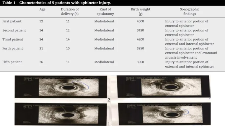
Table1–Characteristicsof5patientswithsphincterinjury.
Age Durationof delivery(h)
Kindof episiotomy
Birthweight (g)
Sonographic findings
Firstpatient 32 11 Mediolateral 4000 Injurytoanteriorportionof
externalsphincter
Secondpatient 34 12 Mediolateral 3420 Injurytoanteriorportionof
externalsphincter
Thirdpatient 24 14 Mediolateral 4200 Injurytoanteriorportionof
externalandinternalsphincter
Forthpatient 21 10 Mediolateral 3850 Injurytoanteriorportionof
externalsphincterandlevatorani muscleinvolvement
Fifthpatient 36 11 Mediolateral 3900 Injurytoanteriorportionof
externalandinternalsphincter
respectively.Thelowestandheaviestweightswere2550gand 4200g,respectively,whilethemeanweightwasreportedas 3342±484.9g(Table1).Also,mediolateralepisiotomywas per-formedfor45(90%)of50patients.
Theresultsshowedthatepiduralanalgesiawasusedonly for3patientswhoweredeliveredinHafezhospitaland94% ofpatientshaddonetheirvaginaldeliverywithoutany anes-thesiaand/oranalgesia.
Intheinvestigationscarriedoutbysonography,anumber of45individualshadnormalsonographyintheirfirst postpar-tumvisitwhichwasperformed6weeksafterdelivery.Also, anyfinding basedonsphincterinjurywasnotfoundinthe individualsatthesecondtimeofsonography.
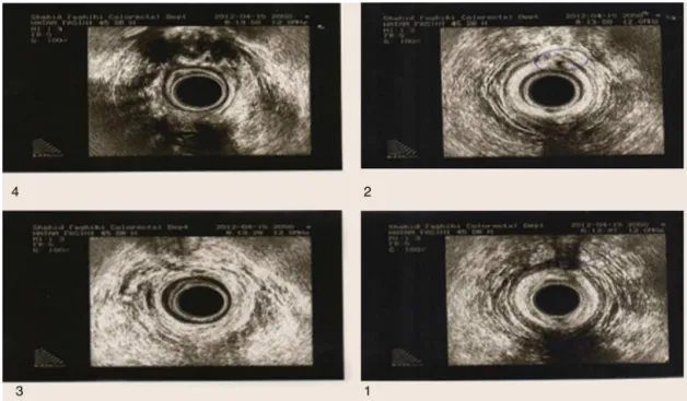
Justfivepatients(meanage29years)withmeanduration ofdelivery11.5handaveragebirthweight3340g,without hav-ing any symptoms and clinical complaintshad findings of
Fig.2–Injurytoanteriorportionofexternalsphincterofthesecondpatient(bluecircleshowssiteofinjury).
Fig.4–Injurytoanteriorportionofexternalsphincterand levatoranimuscleinvolvementoftheforthpatient.
sphincterinjuryintheirfirst postpartumEndo-anal sonog-raphy. The patients’ characteristics are listed in Table 1. Sonographywasperformedforthese5patientsforthethird time after 6 months and persistent damages were seen
(Figs.1–5).
Also, significant differences were seen between anal sphinctermanometry before deliveryand 6months and 3 yearsafterdelivery(p=0.006formeansqueezingpressure)in thefivemothers(Table2).
Discussion
Inthepresentstudyoccultanalsphincterinjuriesaftervaginal deliverywereinvestigated.Analsphincterinjuryaftervaginal
Fig.5–Injurytoanteriorportionofexternalandinternal sphincteroffifthpatient(bluecircleshowssiteofinjury).
deliveryis consideredasthe mainand mostcommon fac-tor inpathogenesisoffecalincontinence inhealthy young women. Theseinjuriesareseeneitherasobviousand spe-cificinjuryintermsofclinical(lacerationofgrade3and4), or as occultanal sphincterinjuries which are not obvious afterdeliverybutcanbedetectedbyEndo-analsonography.2 Sultan et al. in their studies concluded that sonographic findings for external sphincter injury has 100% accu-racy comparedto manometry (70%) and electromyography (75%).8
Table2–Manometryfindingsinfivepatientswithsphincterinjurybeforedeliveryandsixmonthsandthreeyearsafter delivery.
Patients Beforedelivery Sixmonthafterdelivery Threeyearsafterdelivery
MRPa MSPb MRPa MSPb MRPa MSPb
1 72 118 65 93 61 80
2 51 89 39 71 34 62
3 57 96 32 61 28 45
4 63 105 56 75 52 70
5 55 82 31 67 28 74
MRP,meanrestingpressure;MSP,meansqueezingpressure.
a pvalueofMRPdifferences:0.09.
b pvalueofMSPdifferences:0.006.
12%byCortonetal.inwomenwithprimparity.Thisfrequency ofEndo-analsonographyinresultsofthesewomenafter72h9 isconsistent with resultsofthe present study. Carlos Bel-monteet al.in2001ina study on98 womenwithvaginal deliverystatedthat20patientshadclinicalinjury(laceration ofgrade3) after delivery.They alsoreported that 28 (29%) patientshadsonographicevidenceofsphincterinjury with-outanyclinicalsymptoms.10Martinezetal.in2003,inastudy ontraumacausedbyvaginaldeliveryinwomen,statedthat 22patientswithmeanage43yearswerefacedwith compli-cationinsecondaryfecalincontinence,andscarinperineum wasfoundintheirtissueexamination.Also,injuryto ante-riorportionofexternalsphincterwasobservedin16 (73%) patientsandinjuriestoanteriorportionofbothexternaland internalsphincterswereobservedin6(23%)patients.7William etal.intheirstudiesexpressedthattheyhadobserved13(29%) individualswithpostpartumtrauma.Theseinjuriesincluded external sphincter injury in 5 (11%), puboanalis injury in 9 (20%), transverse perineii injury in 3 (7%), and structure injuryin4patients.11 Inthepresent studyanteriorportion ofexternalsphincterinjurycompare toanteriorportion of bothsphincterswasobservedinmostofpatientsandisin accordancewithpastresearches.
Theaverageage ofparticipantsinourstudywashigher thanaverageageofallthestudiedpeoplethatcanbean indi-cationofhighagedmotherinprimparityasariskfactorfor sphincterinjuries,whichiscompatiblewithresultsof Mar-tinezetal.studiesin20037;theaverageageof30yearswas reportedinresultsofWilliamsetal.11;butaccordingto sta-tisticaltests,theobtainedp-valuesinthepresentstudywere notsignificantandfurtherstudieswithlargersamplesizeare needed.
The average duration of delivery in these women was 11.5h,whichishigherthantheaverageofdurationintotal studiedwomenthatwas10.5h.Itisinaccordancewith previ-ousstudiesthatconsideredadeliverywithprolongedsecond stageasariskfactor.2However,theobtainedp-valuesinthe presentstudywerenotstatisticallysignificant,whichitcould beduetotheinability ofthestudyinevaluationofvarious phasesofdelivery.So, the argumentinthis casewould be avoided.
Theaveragebirthweightwas3874gwhichishigherthan theaverageweightoftotalnewbornsthat was3340g.This proveshighweight ofnewbornasarisk factorfor sphinc-ter injuries duringdelivery. p-Value was significant in this
case.Pastresearchesonperinealtraumabyvaginal deliver-iesalsostatedthataveragebirthweightis3245.7gexpressed thathighbirthweightisassociatedwithanalinjuriesin vagi-naldeliveries,12whichagreeswiththepresentstudy.Inthe presentstudy,episiotomywasalsoperformedforallwomen duringdelivery.Performanceofepisiotomyduringdeliveryisa definiteriskfactorforsphincterinjuries,particularlyfor obvi-ousinjurycases.Asinstudiescarriedout,it wasstatedin the past thatreducing the useofepisiotomy inperformed researcheswassupportedandshowedthatharmofroutine episiotomyusageisgreaterthanitsbenefits.13Alsostatedthat episiotomyisonetheriskfactorsofreductionofnormalaction ofmuscles,andithasroleinurinarystressincontinenceand increaselacerationofanalsphincters.14Inthepresentstudy episiotomymethodwasusedforthese5patientsthatwere notrelevanttoanalsphincterdamage.
Theresultsofthisstudyislimitedbecauseofsmallnumber ofpatientsandauthorssuggestsamestudywithlargenumber ofpatientsinfuture.
Conclusions
Likepreviousreportsitseemsthatvaginaldeliveryisarisk factor for anal sphincter injuries. Whether these injuries were symptomaticor asymptomatic Endo-anal sonography shouldbedonefortreatmentplanning.Hereweshowedthat screening Endo-anal sonography could find asymptomatic analsphincterdamagesthatwouldbecomesymptomaticin later ages. In order to propose Endo-analsonography as a screeningtoolweneedfurthercostbenefitstudies.
Conflicts
of
interest
Theauthorsdeclarenoconflictsofinterest.
Acknowledgments
r
e
f
e
r
e
n
c
e
s
1. MinistryofHealthandMedicalEducation:OfficeofFamily
HealthandPopulation.MaternityHealthOffice.Guidelinefor
obstetric&deliveryservicesinbabyfriendlyhospital.1sted.
TehranChaharsooyeHonarPublishing;2006[Articlein
Persian].
2. FowlerGE.Obstetricanalsphincterinjury.JAssocChart
PhysiotherWomen’sHealth.2009;104:12–9.
3. ThompsonJF,RobertsCL,CurrieM,EllwoodDA.Prevalence
andpersistenceofhealthproblemsafterchildbirth:
associationswithparityandmethodofbirth.Birth.
2002;29:83–94.
4. MacArthurC,GlazenerCM,WilsonPD,HerbisonGP,GeeH,
LangGD,etal.Obstetricpracticeandfaecalincontinence
threemonthsafterdelivery.BJOG.2001;108:678–83.
5. OmidvarA.Behaviorchangetechniques,cognitive-behavioral
therapy.Mashhad:PublicationLearnsMotivation.First
Printing;2006.
6. ZeelhaA,AbdulH,RaneeT.Ultrasoundimagingoftheanal
sphinctercomplex.BrJRadiol.2012;85:865–75.
7. MartínezHernándezMagroP,VillanuevaSáenzE,Jaime
ZavalaM,SandovalMunroRD,RochaRamírezJL.Endoanal
sonographyinassessmentoffecalincontinencefollowing
obstetrictrauma.UltrasoundObstetGynecol.2003;22:616–21.
8.SultanAH,KammMA,HudsonCN,ThomasJM,BartramCI.
Anal-sphincterdisruptionduringvaginaldelivery.NEnglJ
Med.1993;329:1905–11.
9.CortonMM,McIntireDD,TwicklerDM,AtnipS,SchafferJI,
LevenoKJ.Endoanalultrasoundfordetectionofsphincter
defectsfollowingchildbirth.IntUrogynecolJ.2013;24:
627–35.
10.Belmonte-MontesC,HagermanG,Vega-YepezPA,
Hernández-de-AndaE,Fonseca-MoralesV.Analsphincter
injuryaftervaginaldeliveryinprimiparousfemales.Dis
ColonRectum.2001;44:1244–8.
11.WilliamsAB,BartramCI,HalliganS,SpencerJA,NichollsRJ,
KmiotWA.Analsphincterdamageaftervaginaldelivery
usingthree-dimensionalendosonography.ObstetGynecol.
2001;97Pt1:770–5.
12.ZareeOB,PashaH,JamaliB.Rytgnexerciseinfluenceandnot
touchingtheperineumareperinealtraumaduringchildbirth.
JFamilyHealth.2013;1:29–34[ArticleinPersian].
13.HartmannK,ViswanathanM,PalmieriR,GartlehnerG,Thorp
JJr,LohrKN.Outcomesofroutineepisiotomy:asystematic
review.JAMA.2005;293:2141–8.
14.SingerrelloLB,HarlowBL,ChekosAK,RepkeJ.Postpartum
sexualfunctioninganditsrelationshiptoperinealtrauma:a
retrospectivecohortstudyofprimiparouswomen.AmJ