Diagnostic validity of different cephalometric analyses
for assessment of the sagittal skeletal pattern
Maheen Ahmed1, Attiya Shaikh2, Mubassar Fida3
Introduction: Numerous cephalometric analyses have been proposed to diagnose the sagittal discrepancy of the craniofacial structures. Objective: This study aimed at evaluating the reliability and validity of different skeletal analyses for the identifica-tion of sagittal skeletal pattern. Methods: A total of 146 subjects (males = 77; females = 69; mean age = 23.6 ± 4.6 years) were included. The ANB angle, Wits appraisal, Beta angle, AB plane angle, Downs angle of convexity and W angle were used to assess the anteroposterior skeletal pattern on lateral cephalograms. The sample was classified into Class I, II and III groups as determined by the diagnostic results of majority of the parameters. The validity and reliability of the aforementioned analy-ses were determined using Kappa statistics, sensitivity and positive predictive value (PPV). Results: A substantial agreement was present between ANB angle and the diagnosis made by the final group (k = 0.802). In the Class I group, Downs angle of convexity showed the highest sensitivity (0.968), whereas ANB showed the highest PPV (0.910). In the Class II group, ANB angle showed the highest sensitivity (0.928) and PPV (0.951). In the Class III group, the ANB angle, the Wits appraisal and the Beta angle showed the highest sensitivity (0.902), whereas the Downs angle of convexity and the ANB angle showed the highest PPV (1.00). Conclusion: The ANB angle was found to be the most valid and reliable indicator in all sagittal groups. Downs angle of convexity, Wits appraisal and Beta angle may be used as valid indicators to assess the Class III sagittal pattern.
Keywords:Diagnosis. Cephalometry. Reliability. Validity.
1 Bakhtawer Amin Medical and Dental College, Dental Section, Department of
Orthodontics (Multan, Pakistan).
2 Liaquat College of Medicine and Dentistry (Karachi, Pakistan). 3 The Aga Khan University, Department of Surgery, Section of Dentistry
(Karachi, Pakistan).
Submitted: March 03, 2017 - Revised and accepted: August 01, 2017
» The authors report no commercial, proprietary or financial interest in the products or companies described in this article.
DOI: https://doi.org/10.1590/2177-6709.23.5.075-081.oar
How to cite: Ahmed M, Shaikh A, Fida M. Diagnostic validity of different cephalometric analyses for assessment of the sagittal skeletal pattern. Dental Press J Orthod. 2018 Sept-Oct;23(5):75-81.
DOI: https://doi.org/10.1590/2177-6709.23.5.075-081.oar
Contact address: Maheen Ahmed
The Aga Khan University Hospital, Section of Dentistry, Department of Surgery P.O. Box 3500, Stadium Road, Karachi 74800, Pakistan
E-mail: [email protected] / [email protected]
Introdução: numerosas análises cefalométricas foram propostas para diagnosticar a discrepância sagital das estruturas cranio-faciais. Objetivo: este estudo teve como objetivo avaliar a confiabilidade e validade de diferentes análises esqueléticas para a identificação do padrão esquelético sagital. Métodos: foram incluídos 146 indivíduos (homens = 77; mulheres = 69; idade média = 23,6 ± 4,6 anos). O ângulo ANB, a avaliação de Wits, o ângulo Beta, o ângulo do plano AB, o ângulo de convexidade de Downs e o ângulo W foram utilizados para avaliar o padrão esquelético anteroposterior em cefalogramas laterais. A amostra foi classificada nos grupos Classe I, II e III, conforme os resultados diagnósticos da maioria dos parâmetros. A validade e a confiabilidade das análises acima mencionadas foram determinadas usando estatísticas Kappa, sensibilidade e valor preditivo positivo (VPP). Resultados: foi encontrada uma concordância significativa entre o ângulo ANB e o diagnóstico feito pelo grupo final (k = 0,802). No grupo Classe I, o ângulo de convexidade de Downs mostrou a maior sensibilidade (0,968), enquanto o ANB apresentou o maior VPP (0,910). No grupo Classe II, o ângulo ANB mostrou a maior sensibilidade (0,928) e o maior VPP (0,951). No grupo Classe III, o ângulo ANB, a avaliação de Wits e o ângulo Beta apresentaram a maior sensibilidade (0,902), enquanto o ângulo de conve-xidade de Downs e o ângulo ANB apresentaram o maior VPP (1,00). Conclusão: o ângulo ANB foi considerado o indicador mais válido e confiável em todos os grupos sagitais. O ângulo de convexidade de Downs, a avaliação de Wits e o ângulo Beta podem ser usados como indicadores válidos para avaliar o padrão sagital de Classe III.
INTRODUCTION
Variations in the normal craniofacial development in sagittal, vertical or transverse planes may result in
different malocclusions.1 However, malocclusions in
the sagittal plane have major esthetic, psychological and functional implications and are usually on top
of the orthodontic problem list.2,3 A sagittal skeletal
malocclusion may result from discrepancies in max-illary or mandibular growth. A more anteriorly po-sitioned mandible with respect to the maxilla may result in a prognathic or concave profile; whereas, a relatively anteriorly positioned maxilla as compared to the mandible results in a retrognathic or con-vex profile. The skeletal discrepancies in the sagit-tal plane are best evaluated on radiographs in which both the morphology of different skeletal structures and their relationship to the surrounding tissues can be accurately assessed. Standardized lateral cepha-logram has established itself as the classical tool to diagnose the sagittal discrepancies in the skeletal,
dental and soft tissues.4
After the standardization of the cephalogram by
Broadbent,5 the diagnosis of the anteroposterior
skel-etal problems has become a straightforward process. Various cephalometric analyses have been proposed for the evaluation of the sagittal skeletal discrepancies.
Downs6 described the AB plane angle and Downs
angle of convexity to assess the anteroposterior jaw
dysplasia. In 1953, Riedel7 introduced the ANB
an-gle, which was later popularized by Steiner.8 Studies
have indicated that these angular measurements are sensitive to small changes in the position of nasion and sella turcica, length of the anterior cranial base
and the vertical growth pattern.9,10 To overcome this
limitation, Jacobson10 proposed the Wits appraisal,
which employed the occlusal plane as the reference. However, the reproducibility and reliability of Wits appraisal has been questioned due to the variations in inclination and difficulties in identification of the
functional occlusal plane.11 Hence, several other
pa-rameters have been and are still being introduced to overcome the shortcomings of the existing cephalo-metric analyses for an accurate diagnosis of sagittal discrepancies. Recently, the Beta angle and W angle have been proposed to evaluate the anteroposterior jaw dysplasia, but their diagnostic performance and
validity have not yet been investigated.12,13
In the past, multiple researchers have correlated various cephalometric analyses for assessing
antero-posterior jaw disrepancy.14-18 Ahmed et al19 reported
the diagnostic accuracy of various cephalometric skel-etal parameters for assessing the skelskel-etal facial vertical pattern. However, to our knowledge, no such study has evaluated the reliability of anteroposterior skeletal dysplasia parameters. This has resulted in numerous parameters that need to be analyzed during cephalo-metric analysis, which is not only time-consuming, but sometimes may also provide conflicting results. Thus, this study aimed to identify the skeletal param-eters that more accurately identified the sagittal skel-etal pattern of an individual — since preference may be given to those analyses which are precise, consis-tent and reliable. This may not only improve the ef-ficiency of the treatment planning process, but may also establish a reliable criteria for the classification of subjects into different sagittal malocclusion groups for research purposes.
MATERIAL AND METHODS
Data was collected retrospectively from the den-tal records of patients attended at the denden-tal clinics of the authors. The sample size was calculated using the OpenEpi software (version 3.0) based on the
find-ings of Gul-e-Erum and Fida.16 The alpha was taken
as 0.05 and power of the study as 80% to calculate the sample size. Results have proposed a sample size with a minimum of 38 subjects in each group. As the subjects were divided into three groups based on ver-tical facial pattern, a minimum of 114 subjects were required. However, to increase the power of the study, a maximum number of subjects were included. A to-tal of 198 subjects aged between 18 and 35 years (99 males and 99 females; mean age = 23.6 ± 4.6 years), having good quality lateral cephalograms were includ-ed. Patients with previous history of any orthodontic treatment, growth disturbance or facial trauma were excluded. Since variations in vertical growth pattern may be a confounding factor, only subjects with nor-mal vertical growth pattern were included. This was determined when all the three vertical dysplasia pa-rameters — FMA, SN-GoGn and PFH-TAFH —
in-dicated a normodivergent growth pattern.9
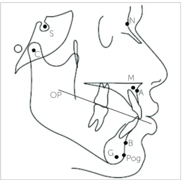
deter-Figure 1 - Cephalometric landmarks and the occlusal plane.
Figure 2 - Cephalometric parameters. Figure 3 - Cephalometric parameters.
1 = Beta angle: The angle formed by A-CB and AB line (normal range = 27° to 35°). 2 = W angle: The angle between the perpendicular line from point M to S-G line and the M-G line (normal range = 51° to 55°).
3 = Downs angle of convexity: The angle between N-point A and point A-Pog (normal range = -8.5° to 10°).
mine the anteroposterior skeletal jaw discrepancy. The cephalograms were manually traced by the main investigator, the skeletal landmarks were identified and the following parameters were measured, as fol-lows (Fig 1, 2, 3):
» ANB angle: the angle formed by point A,
Na-sion and point B (normal range = 0o to 4o).8
» Wits appraisal: the linear distance between AO
and BO (perpendicular drawn from point A and B on to functional occlusal plane) (normal
range = -1mm to +1mm).10
» AB plane angle: the angle formed by AB plane
and N-pog line (normal range = - 9o to 0o).6
» Beta angle: the angle formed by A-CB and AB
lines (normal range = 27o to 35o).12
» W angle: the angle between the perpendicular
line from point M to S-G line and the M-G line
(normal range = 51o to 55o).13
» Downs angle of convexity: the angle between
N-point A and N-point A-Pog (normal range = -8.5o
to 10o).6
The norms of each skeletal analysis as established in literature were used to classify subjects as Class I,
Class II and Class III.6,8,10,12,13 Fifty subjects were
ex-cluded from the study as they were found to have a similar sagittal skeletal pattern as determined by all the parameters. Each of the remaining 146 subjects (males = 77; females = 69) had at least one parameter giving conflicting diagnosis of the sagittal skeletal pat-tern. The final diagnosis of the anteroposterior growth pattern of the remaining subjects was based on the re-sults of the majority of the analyses. This enabled to S
OP
M
G
A
Pog N
B C
1 = ANB angle
2 = Wits appraisal
3 = AB plane angle
1
divide the remaining subjects into Class I, Class II and Class III anteroposterior groups. The final classifica-tion of the subjects resulted in the following groups:
» Class I: n = 63. » Class II: n = 42. » Class III: n = 41.
‘Correctly diagnosed cases’ were labeled when a specific skeletal analysis in a subject matched the final diagnosis. These were then used to assess the diag-nostic accuracy of each parameter.
Thirty cephalograms were retraced and randomly reanalyzed by the main investigator. The errors were
calculated according to Dahlberg’s formula20 and the
coefficient of reliability (ICC). The Dahlberg’s error ranged from 0.103 to 0.890, while the results for the ICC showed a high correlation between the two sets of readings (Table 1).
SPSS for Windows (version 20.0, SPSS Inc. Chi-cago) was used for data analysis. The anteroposterior skeletal analyses were evaluated using the Pearson’s correlation. Kappa statistics were applied to assess the level of agreement between the skeletal analyses and the final diagnosis made from the ‘correctly di-agnosed cases’. The validity in terms of sensitivity and Positive predictive value (PPV) were determined from the two by two tables. A p-value < 0.05 was taken as statistically significant.
RESULTS
The sample comprised 146 subjects (69 females, mean age = 20.67 ± 4.8 years; 77 males, mean age = 21.98 ± 4.8 years). The means and standard de-viations of each parameter in all three sagittal maloc-clusions are shown in Table 2.
Correlation between the different skeletal analyses was determined using Pearson’s correlation. A strong correlation was present between the ANB angle and
Wits appraisal (r = 0.831, p < 0.01), and ANB angle and
Downs angle of convexity(r = 0.823, p < 0.01) (Table 3).
Kappa statistics assessed the agreement among diagnostic criteria of different cephalometric anal-yses. A substantial agreement was present between the ANB angle and the final group (k = 0.802,
p < 0.01) (Table 4).
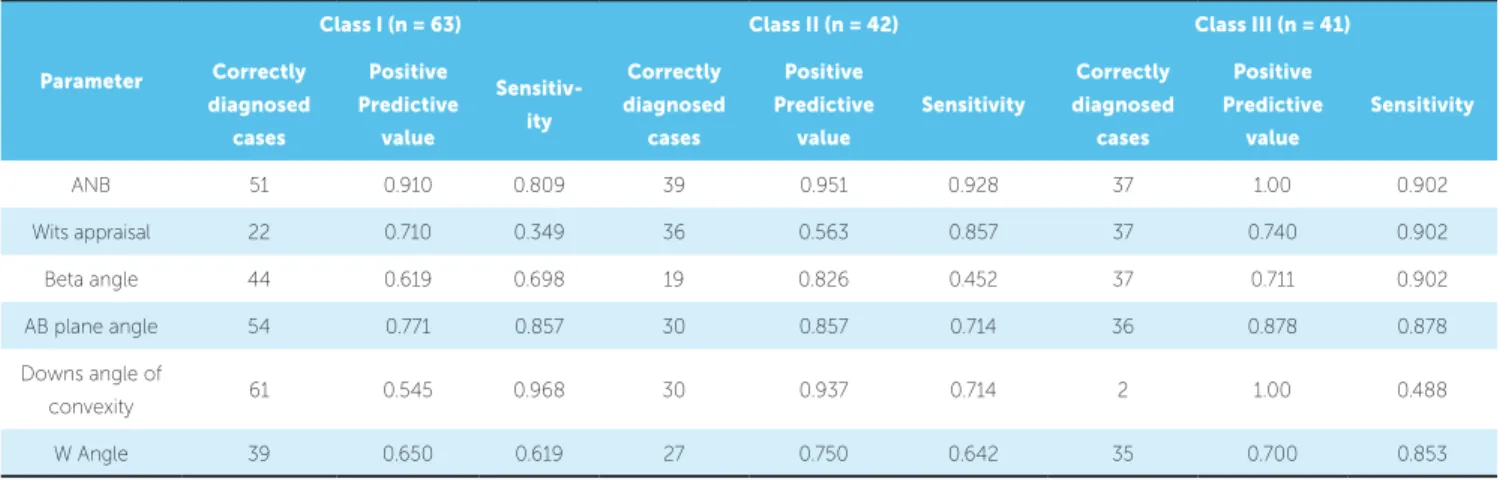
PPV and sensitivity of each diagnostic parameter were also calculated for each group separately. In the Class I group, Downs angle of convexity showed the highest sensitivity (0.968), whereas the ANB angle showed the highest PPV (0.910). In the Class II group, the ANB angle showed the highest sensitiv-ity (0.928) as well as the highest PPV (0. 951). In the Class III group, the ANB angle, Wits appraisal and the Beta angle showed the highest sensitivity (0.902), whereas the Downs angle of convexity and the ANB angle showed the highest PPV (1.00) (Table 5).
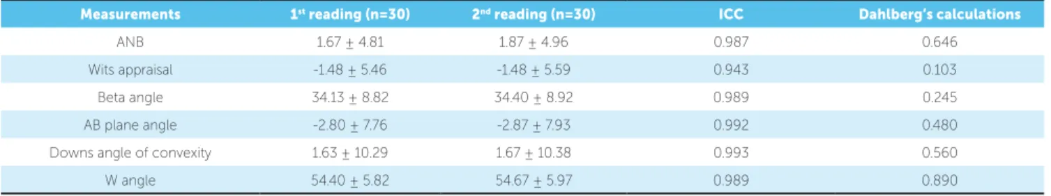
Table 1 - Intraclass Correlation Coefficient.
ICC: Intraclass correlation coefficient. n=30.
Measurements 1st reading (n=30) 2nd reading (n=30) ICC Dahlberg’s calculations
ANB 1.67 ± 4.81 1.87 ± 4.96 0.987 0.646
Wits appraisal -1.48 ± 5.46 -1.48 ± 5.59 0.943 0.103
Beta angle 34.13 ± 8.82 34.40 ± 8.92 0.989 0.245
AB plane angle -2.80 ± 7.76 -2.87 ± 7.93 0.992 0.480
Downs angle of convexity 1.63 ± 10.29 1.67 ± 10.38 0.993 0.560
Table 2 - Mean value of cephalometric parameters.
Parameter
Class I
n = 63 mean ± SD
Class II
n = 42 mean ± SD
Class III
n = 41 mean ± SD
ANB 1.30 ± 1.76 6.45 ± 1.31 -2.17 ± 2.52
Wits appraisal 0.389 ± 3.01 4.36 ± 3.78 -6.30 ± 5.24
Beta angle 32.49 ± 5.43 26.31 ± 4.03 43.54 ± 4.75
AB plane angle -5.14 ± 3.5 -10.48 ± 4.12 3.20 ± 3.51
Downs angle of convexity 4.00 ± 3.94 11.29 ± 3.65 -3.66 ± 3.12
W angle 53.83 ± 3.94 49.45 ± 2.52 58.46 ± 2.54
Table 3 - Correlation among different skeletal analyses to assess sagittal growth pattern.
n = 146. Pearson correlation: weak correlation (± 0.01 < r < ± 0.5); moderate correlation (± 0.5 < r < ± 0.8); strong correlation (± 0.8 < r < ± 1) *p < 0.05; ** p < 0.01.
ANB Wits Appraisal Beta Angle AB Plane Angle Down’s Angle of
Convexity W Angle
ANB 1 0.831** -0.775** -0.783** 0.823** -0.704**
Wits appraisal 1 -0.730** -0.625** 0.634** -0.654**
Beta angle 1 -0.694** -0.680** 0.636**
AB plane angle 1 -0.792** 0.568**
Downs angle of convexity 1 -0.678**
W angle 1
Table 4 - Assessment of agreement among diagnostic criteria of skeletal analyses.
n = 146; Kappa Statistics. *p < 0.05; ** p < 0.01
Parameter
Class I Class II Class III Kappa
P-value
n = 63 n = 42 n = 41 n =146
ANB 56 53 37 0.802** 0.000
Wits appraisal 31 64 51 0.489** 0.000
Beta angle 71 23 52 0.511** 0.001
AB plane angle 70 35 41 0.724** 0.000
Downs angle of convexity 112 32 2 0.397** 0.000
W Angle 60 36 50 0.530** 0.0401
Table 5 - Assessment of positive predictive value and sensitivity of various parameters to assess sagittal discrepancy.
n = 146.
Parameter
Class I (n = 63) Class II (n = 42) Class III (n = 41)
Correctly
diagnosed cases
Positive
Predictive value
Sensitiv-ity
Correctly
diagnosed cases
Positive
Predictive value
Sensitivity
Correctly
diagnosed cases
Positive
Predictive value
Sensitivity
ANB 51 0.910 0.809 39 0.951 0.928 37 1.00 0.902
Wits appraisal 22 0.710 0.349 36 0.563 0.857 37 0.740 0.902
Beta angle 44 0.619 0.698 19 0.826 0.452 37 0.711 0.902
AB plane angle 54 0.771 0.857 30 0.857 0.714 36 0.878 0.878
Downs angle of
convexity 61 0.545 0.968 30 0.937 0.714 2 1.00 0.488
DISCUSSION
In Orthodontics, great importance has been ad-vocated to the cephalometric assessment of the jaw relationship in the sagittal plane. Since the advent of
lateral cephalometry by Broadbent5,various analyses
have been proposed to assess the anteroposterior jaw
relationship.6,8,10-12 In borderline cases, several
skele-tal analyses may show conflicting results, and a clear cut diagnosis regarding the sagittal skeletal pattern is not possible. This study aimed to concise the pro-cess of diagnosis to minimal skeletal parameters by evaluating the diagnostic accuracy of the most com-monly used analyses.
A ‘final diagnosis’ of the anteroposterior skeletal pattern was based on the results of majority of the parameters. This ‘final diagnosis’ was then treated as gold standard. The diagnostic accuracy of the in-cluded skeletal parameters was then compared using kappa statistics, PPV and sensitivity.
In the present study, all the analyses showed significant correlation with each other. A strong positive correlation was present between the Wits appraisal and ANB angle (r = 0.831), and the ANB angle and Downs angle of convexity (r = 0.823).
Ishikawa et al14 reported a strong correlation
be-tween AB plane angle and Downs angle of convex-ity (r = -0.86), AB Plane angle and the ANB angle (r = -0.95), and the ANB angle and Downs angle of convexity (r = 0.97). The variations in results may be due to differences in sample size and inclusion of only Class I subjects. In another study by
Gul-e-Erum and Fida,16 a strong correlation was
report-ed between AB plane angle and ANB (r = 0.749). The present study reported similar findings.
The strength of the correlation does not indi-cate whether the specific parameter can precisely diagnose the skeletal anteroposterior parameter. Hence, in the present study, to compare the diag-nostic agreement between various skeletal analy-ses and the final diagnosis, Kappa statistics were applied. A substantial agreement was present be-tween the final group and ANB angle (k = 0.802). The Kappa statistic explains the variation in
diag-nosis that may occur simply as a result of chance.21
Hence, the ANB angle was found to be the most reliable indicator in precisely assessing the sagittal skeletal pattern of a patient.
It is of prime importance for an analysis to diagnose a certain parameter with consistency and accuracy. Hence the sensitivity of each parameter was determined to validate their diagnostic accuracy. Downs angle of convexity showed the highest sensitivity in the Class I group (0.968), whereas ANB angle was found to be the most sensitive parameter in Class II group (0.928). In the Class III group, ANB angle, Wits appraisal and Beta angle (0.902) were found to have the highest sen-sitivity in evaluating the sagittal growth pattern. Thus in evaluating the sagittal growth pattern with precision in an individual , Downs angle of convexity and the ANB angle may be used as valid indicators in Class I and Class II subjects. In the Class III group, ANB angle, Wits appraisal and Beta angle may be used to accurately assess the sagittal growth pattern of an individual.
In the present study, to confirm whether a cer-tain parameter can accurately depict the skeletal pat-tern, the positive predictive values (PPV) were also calculated for each group separately. The ANB angle yielded the highest PPV in Class I (0.910) and Class II (0.951) sagittal groups. In the Class III sagittal group, ANB angle and Downs angle of convexity showed the highest PPV (1.00). Thus, the ANB angle in all three sagittal groups has a high probability for cor-rectly diagnosing the anteroposterior jaw dysplasia. In addition, if Downs angle of convexity is indicating a Class III jaw relationship in a particular individual, then it is highly likely to be true and may not need to be verified by other analyses.
A number of studies have indicated that the hyper-divergent or hypohyper-divergent vertical growth pattern
may affect the sagittal jaw relationship.9,10 This may
reduce the accuracy and precision in evaluating the diagnostic accuracy of the existing sagittal jaw dys-plasia parameters. In our study, the ANB angle was seen to accurately determine the anteroposterior jaw dysplasia in normodivergent subjects.
CONCLUSION
All the skeletal parameters showed a significant correlation with each other. The ANB angle was found to be the most reliable and valid indicator in as-sessing the anteroposterior jaw relationship in all
sag-ittal groups. Hence, it may be used to precisely and accurately assess the sagittal jaw discrepancy. In ad-dition, Downs angle of convexity, Wits appraisal and Beta angle may be used as valid indicators to assess the Class III sagittal growth pattern.
1. Proffit WR, Fields HW, Sarver DM. Contemporary Orthodontics. 5th ed.
St. Louis: Mosby Elsevier, 2007.
2. Azuma S, Kohzuki M, Saeki S, Tajima M, Igarashi K, Sugaware J. Beneficial effects of orthodontic treatment on quality of life in patients with malocclusion. Tohoku J Exp Med. 2008 Jan;214(1):39-50. 3. Bernabé E, Tsakos G, Oliveira CM, Sheiham A. Impacts on daily
performances attributed to malocclusions using the condition-specific feature of the oral impacts on Daily Performances Index. Angle Orthod. 2008 Mar;78(2):241-7.
4. Devereux L, Moles D, Cunningham SJ, McKnight M. How important are
lateral cephalometric radiographs in orthodontic treatment planning? Am J Orthod Dentofacial Orthop. 2011 Feb;139(2):175-81.
5. Broadbent H. A new x-ray technique and its application to orthodontia: the introduction of cephalometric radiography. Angle Orthod. 1981 Apr;51(2):93-114.
6. Downs WB. Variations in facial relationships: their significance in treatment and prognosis. Am J Orthod. 1948 Oct;34(10):812-40.
7. Riedel RA. The relation of maxillary structures to cranium in malocclusion and in normal occlusion. Angle Orthod. 1952 July;22(3):142-5.
8. Steiner CC. Cephalometrics for you and me. Am J Orthod. 1953
Oct;39(10):720-55.
9. Freeman RS. Adjusting ANB angles to reflect the effect of maxillary position. Angle Orthod. 1981 Apr;51(2):162-71.
10. Jacobson A. The Wits appraisal of jaw disharmony. Am J Orthod. 1975 Feb;67(2):125-38.
11. Haynes S, Chau MNY. The reproducibility and repeatability of the Wits analysis. Am J Orthod Dentofacial Orthop. 1995 June;107(6):640-7. 12. Baik CY, Ververidou M. A new approach of assessing sagittal discrepancies:
the Beta angle. Am J Orthod Dentofacial Orthop. 2004 July;126(1):100-5.
REFERENCES
13. Bhad WA, Subash N, Umal HD. A new approach of assessing sagittal dysplasia: the W Angle. Eur J Orthod. 2013 Feb;35(1):66-70. 14. Ishikawa H, Nakamura S, Iwasaki H, Kitazawa S. Seven parameters
describing anteroposterior jaw relationships: postpubertal prediction accuracy and interchangeability. Am J Orthod Dentofacial Orthop. 2000 June;117(6):714-20.
15. Bošković-Brkanović T, Nikolić Z. Correlation between five parameters for the assessment of sagittal skeletal intermaxillary relationship. Serbian Dent J. 2007;54(4):231-9.
16. Gul-e-Erum, Fida M. A comparison of cephalometric analyses for assessing sagittal jaw relationship. J Coll Physicians Surg Pak. 2008;18(11):679-83. 17. Doshi Jigar R, Trivedi K, Shyagali T. Predictability of yen angle & appraisal of
various cephalometric parameters in the assessment of sagittal relationship between maxilla and mandible in Angle’s Class II malocclusion. People J Sci Res. 2012 Jan;5(1):1-8.
18. Singh AK, Ganeshkar SV, Mehrotra P, Baghchandani A. Comparison of different parameters for recording sagittal maxillo mandibular relation using natural head posture: a cephalometric study. J Orthod Sci. 2013 Jan;2(1):16-22.
19. Ahmed M, Shaikh A, Fida M. Diagnostic performance of various cephalometric parameters for the assessment of vertical growth pattern. Dental Press J Orthod. 2016 Aug;21(4):41-9.
20. Dahlberg G. Statistical methods for medical and biological students. Br Med J. 1940 Sept;14(2):358-9.