rev bras hematol hemoter. 2016;38(3):274–275
w w w . r b h h . o r g
Revista
Brasileira
de
Hematologia
e
Hemoterapia
Brazilian
Journal
of
Hematology
and
Hemotherapy
Images
in
Clinical
Hematology
Gingival
swelling
associated
with
hypoplasminogenemia
Eric
T.
Stoopler
∗,
Faizan
Alawi
UniversityofPennsylvania,SchoolofDentalMedicine,Philadelphia,UnitedStates
a
r
t
i
c
l
e
i
n
f
o
Articlehistory:
Received24March2016 Accepted14April2016 Availableonline3May2016
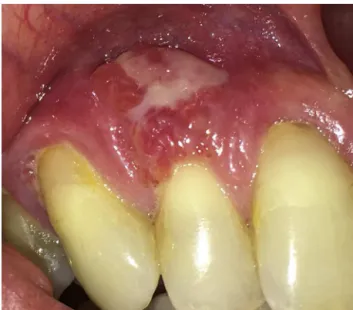
Amiddle-agedwomanpresentedforevaluationofgingival swelling.Shewaspreviouslydiagnosedwith hypoplasmino-genemiaandcurrent plasminogenlevelwas29%(reference value:78–130%). Intraoralexaminationrevealed swelling of the right maxillary gingiva (Figure 1). Biopsy with routine histopathologic analysis revealed fibrinoid deposits and a mixedinflammatoryinfiltratewithinthelaminapropria, con-sistentwithhypoplasminogenemia(Figure2).
Hypoplasminogenemia (type 1 plasminogen deficiency) is commonly associated with ligneous conjunctivitis and ligneous gingivitis.1–3 An oral mucosal biopsy can aid in the diagnosis of this rare condition as histopatho-logic evidence of fibrin deposition is highly suggestive of hypoplasminogenemia.2,3
∗ Correspondingauthorat:DepartmentofOralMedicine,UniversityofPennsylvaniaSchoolofDentalMedicine,240South40thStreet,
Philadelphia,PA19104,USA.
E-mailaddress:[email protected](E.T.Stoopler).
Figure1–Swellingoftherightanteriormaxillarygingiva.
http://dx.doi.org/10.1016/j.bjhh.2016.04.006
revbrashematolhemoter.2016;38(3):274–275
275
A
B
*
*
*
*
*
Figure2–Amorphousfibrinoiddeposits(*)inthegingivallaminapropria.(A)Hematoxylin–eosin(magnification×100),(B)
trichrome(magnification×40).
Conflicts
of
interest
Theauthorsdeclarenoconflictsofinterest.
Acknowledgements
TheauthorsthankDr.SophiaElmuradi forassistancewith clinicalphotography.
r
e
f
e
r
e
n
c
e
s
1.SchusterV,HugleB,TefsK.Plasminogendeficiency.JThromb
Haemost.2007;5(12):2315–22.
2.ScullyC,GokbugetAY,AllenC,BaganJV,EfeogluA,ErsevenG,
etal.Orallesionsindicativeofplasminogendeficiency
(hypoplasminogenemia).OralSurgOralMedOralPatholOral
RadiolEndod.2001;91(3):334–7.
3.ScullyC,GokbugetA,KurtulusI.Hypoplasminogenaemia,