Article
Printed in Brazil - ©2015 Sociedade Brasileira de Química 0103 - 5053 $6.00+0.00
A
*e-mail: [email protected]
Bioactive Cyanopeptides Produced by
Sphaerocavum
brasiliense
Strains
(Cyanobacteria)
Maria Estela Silva-Stenico,a Janaina Rigonato,a Adriana Sturion Lorenzi,a
Maria Teresa Paiva Azevedo,b Célia Leite Sant’Annac and Marli Fátima Fiore*,a
aLaboratório de Ecologia de Cianobactérias, Centro de Energia Nuclear na Agricultura (CENA),
Universidade de São Paulo (USP), 13400-970 Piracicaba-SP, Brazil
bBioalgas, 04304-000 São Paulo-SP, Brazil
cInstituto de Botânica do Estado de São Paulo, 04301-012 São Paulo-SP, Brazil
The halogenated compounds aeruginosin and cyanopeptolin (protease inhibitors) are well documented in several cyanobacteria. In this work, the presence of genetic coding for aeruginosin and cyanopeptolin in Sphaerocavum genus and their characterization by mass spectrometry and potential toxic effects were investigated. Three strains of Sphaerocavumbrasiliense (CCIBt 3094, CCIBt 3096 and CCIBt 3316) isolated from different reservoirs in São Paulo State, and one (CCIBt 3179) from Rio Grande do Norte State were selected for this study. Fragments of aerA-aerB
and mcnC-mcnE gene regions (aeruginosin and cyanopeptolin, respectively) were amplified by polymerase chain reaction (PCR) and sequenced. Compounds were characterized by mass spectrometry. All the studied strains presented aeruginosin and cyanopeptolin synthetase genes. Mass spectrometry showed the presence of several aeruginosin and cyanopeptolin variants. The
Allium test showed that the ethanolic extract promoted toxic and cytotoxic actions in acute and chronic treatments. These naturally occurring protease inhibitors can play an important role in the physiopathology of human diseases.
Keywords: aeruginosin, cyanopeptolin, protease inhibitor, mass spectrometry, Allium test,
cyanobacteria, Sphaerocavum genus
Introduction
Cyanobacteria are known to produce important bioactive compounds, including those with protease inhibition activity.1 Numerous protease inhibitor variants produced
by cyanobacteria have been described, with such names as aeruginosin, aeruginopeptin, aeruginoside, agardhipeptin, anabaenopeptin, anabaenopeptilide, brunsvicamide, cyanopeptolin, cyanostatin, dehydroradiosumin, hofmannolin, insulapeptolide, microcin, microcistilide, microginin, micropeptin, microviridin, oscillapeptin, nodulapeptin, nostocyclin, nostoginin, nostopeptin, oscillaginin, oscillamide, oscillapeptilide, planktocyclin, planktopeptin, spumigin, scyptolin, schizopeptin, somamide, spiroidesin, symplostatin and tasipeptin.2,3
These molecules present significant biological activities, since they act against several different proteases that are
involved in a wide variety of physiological processes in the human body, such as cell cycle progression, food digestion, angiogenesis, blood clotting, blood pressure regulation,
antigen presentation, inflammation and apoptosis.4
Consequently, the characterization of novel protease inhibitors derived from natural product biosynthetic pathways is of great relevance.
Results showing toxicological studies for molecules considered non-toxic are scarce, thus it is imperative that such compounds should be characterized for cytotoxicity, genotoxicity and mutagenicity. The United Nations Environment Programme (UNEP) recommended assays with vascular plants.5 The Allium cepa test is one of the
analyses used to determine cytotoxicity and is an excellent model for detecting environmental mutagens.6
The aim of this study was to evaluate the presence of genetic coding for aeruginosin and cyanopeptolin in strains of Brazilian genus Sphaerocavum and peptides characterized by mass spectrometry. The Allium cepa test system was also used to evaluate cytotoxicity, genotoxicity and mutagenicity.
Experimental
Cyanobacterial strains and growth conditions
Sphaerocavum brasiliense CCIBt 3094 and CCIBt 3316 were isolated from Billings reservoir, CCIBt 3096 from Garças Lake, all of them from São Paulo State and CCIBt 3179 from a reservoir in Rio Grande do Norte State, Brazil. The strains were cultivated in 300 mL of ASM-1 liquid medium8 at 25 oC under constant illumination by
white fluorescent light (40 µmol photons m-2 s-1) for 20 days.
Molecular analyses
Cells of S. brasiliense CCIBt 3094, CCIBt 3096, CCIBt 3179 and CCIBt 3316 in the late logarithmic phase were harvested and total genomic DNA extracted according to Fiore et al.9 In order to investigate the presence of microcystin
synthetase, one region of each of mcyA (MSF/MSR),10
mcyB (FAA/RAA),11 mcyD (mcyDF/mcyDR),12
mcyE (mcyEF2/mcyER4)12 and mcyG (mcyGF/mcyGR)13
genes was chosen. A microcystin producing strain Microcystis aeruginosa SPC77714 was used as a positive
control. Aeruginosin and cyanopeptolin synthetase gene regions were amplified using the primer sets aerA_F/aerB_R and mcnC_F/mcnE_R,15 respectively. All
resulting polymerase chain reaction (PCR) products were cloned into the pGEM®-T Easy Vector System (Promega)
according to the manufacturer’s protocol, followed by DH5α
E. coli competent-cell transformation,16 and recombinant
plasmids were purified by the alkaline lysis method.17 DNA
sequencing was performed using T7 and SP6 anchored in the pGEM®-T Easy Vector primers, and the Big Dye Terminator
v.3.1 Cycle Sequencing Kit (Applied Biosystems). The cycle sequencing reaction was performed in a Techne TC-412 thermal cycler (Barloworld Scientific), and the PCR condition was 25 cycles of 95 °C for 20 s, 50 °C for 15 s and 60 °C for 60 s. The sodium acetate/ethanol purified reaction was resuspended in HiDi formamide (Life Technologies), and the samples read in an ABI PRISM 3100 Genetic Analyzer (Life Technologies). The sequenced fragments were assembled into contigs using the Phred/Phrap/Consed software package program (Philip Green, University of Washington), and only bases with a quality > 20 were considered. Products
in 5 µL samples of PCR mixture were visualized after electrophoresis on 1% agarose gels by staining with SYBR Green I (Molecular Probes, Life Technologies) (presented in the Supplementary Information).
Phylogenetic analyses
The DNA sequences of aerA-aerB and mcnC-mcnE
regions obtained herein were compared with sequences available in the National Center for Biotechnology Information (NCBI) database using the BLAST tool (http://www.ncbi.nlm.nih.gov/BLAST). Cyanobacterial reference sequences retrieved from GenBank were included in the alignment, which was visually refined and used to generate phylogenetic trees. Phylogenetic trees topology analyses were conducted using neighbor-joining (NJ) and maximum likelihood (ML). NJ was performed using
PAUP 4.0,18 and ML was implemented by the MEGA
version 5.0 program package.19 The best-fitting evolutionary
model GTR+G+I was selected using ModelTest under the Akaike information criterion.20 The robustness
of the trees was estimated by bootstrap percentages using 1,000 replications. The accession numbers for
aerA-aerB and mcnC-mcnE nucleotide sequences from
S. brasiliense CCIBt 3094 were KP769528 and KP769524; for CCIBt 3096, were KP769529 and KP769525; for CCIBt 3179, were KP769530 and KP769526, and for CCIBt 3316 were KP769531 and KP769527, respectively.
Metabolite extraction
After growth, cyanobacterial cultures were harvested by centrifugation at 9,000 × g at room temperature. Lyophilized cells (20 mg) were extracted three times in 50 mL of methanol, using glass beads (φ 3 mm).21 The
suspension was agitated for 1 min and after that, centrifuged at 9,000 × gat room temperature. The supernatant was collected and evaporated. The dried extract was dissolved in sterile distilled water (10 mL) and applied to a pre-conditioned C18 solid phase extraction (SPE) cartridge
(Applied Separation). The cartridge was washed with water (2 × 10 mL) and cyanopeptides were eluted with 70% methanol.
Mass spectrometry (MS)
Agilent 6410 Triple Quadrupole equipped with Agilent 1200 Series Binary Pump SL pumping system and Agilent 1200 Autosampler (Agilent Technologies). Data acquisition and analysis were performed using Agilent MassHunter Workstation software. The electrospray ionization (ESI) source was employed in positive mode, and set as follows: drying gas was nitrogen heated at 300 oC, gas flow was
10 L min-1, capillary voltage was held at 5000 V. The
MS/MS parameters were set as follows: dwell time was 200 ms, fragmentor voltage was set at 200 V, the collision gas was nitrogen and collision energy was set according to Table 1. The MS system was operated in full scan and ion precursor mode. The mobile phase A consisted of ultrapure water (Milli-Q) and mobile phase B consisted of methanol, both containing 0.1% (v/v) of formic acid. The flow rate was 0.3 mL min-1. Molecular ions were scanned in the range
of 100 to 1,800 Da.
Allium test
The total cyanobacterial biomass was obtained in 600 mL culture and separated by centrifugation at 9,000 ×g for 5 min. Cells were collected and successive extractions with methanol, using glass beads (φ 3 mm) to disrupt cells, were performed. The organic extracts were subjected to agitation for 1 min followed by centrifugation at 9,000 × g at 4 ºC. The supernatant was then collected and evaporated. For extracellular metabolites, the supernatant was concentrated to 100 mL and subjected to dichloromethane (DCM) extraction using the same volume. The DCM extract was concentrated on a rotary evaporator. The remaining aqueous phase was dried and extracted with ethanol. All the extracts were dried and kept at −20 oC until analysis. Seeds of onion
(Allium cepa) were pre-soaked for 48 h in petri dishes containing moistened cotton with distilled water and kept at 25 ± 1 ºC. An amount of 10 µg of methanol, ethanol and dichloromethane extracts was diluted in Milli-Q water. After root growth of the seeds, they were transferred to petri dishes containing cotton soaked with resuspended extracts. The negative control (Milli-Q water) was also included in the experiment. Root growth and morphological alterations were evaluated for toxicity. For cytotoxicity analysis, the mitotic index was obtained. Five roots were collected after 24 h and 48 h of treatment, fixed in Carnoy solution (3:1, ethanol:glacial acetic acid) and prepared by staining with 2% orcein and crushing the meristematic region. Meristematic cells of five roots (1,000 cells per root, 5,000 in total) were analyzed by optical microscopy at 400× magnification and observed for each treatment, taking into account the number of cells in each phase of mitosis. The mitotic index (MI) was obtained by dividing the number of cells in mitosis by the
total number of analyzed cells. The mitotic phase’s index was calculated by the sum of cells in each phase divided by the sum of cells in mitosis. The genotoxicity was evaluated by observation of chromosomal damage. The statistical analysis was performed using the BioEstat5.3 program.22
Trypsin test
The colorimetric Trypsin activity assay kit (Abcam AB 102531) was used for this test and performed according to the protocol described by the manufacturer. Trypsin EC 3.4.21.4 was tested against extracts of S. brasiliense CCIBt 3094, CCIBt 3096, CCIBt 3096 and CCIBt 3179 on a 96-well microplate. The extract of Cylindrospermopsis raciborskii 339 was used as the negative control. Each assay was performed in duplicate.
Results and Discussion
Molecular evaluation of microcystin, aeruginosin and cyanopeptolin sequences
The microcystin synthetase genes (mcyA, mcyB, mcyD, mycE and mcyG) were chosen as the target sequences to detect the presence of the mcy gene cluster in S. brasiliense strains. Considering the mcy genes amplification for M. aeruginosa SPC777 (positive control), the specific primers for the microcystin synthetase genes failed to produce amplicons for all S. brasiliense strains.
Previously, we reported the presence of aeruginosin and cyanopeptolin synthetase genes for S. brasiliense CCIBt3094, CCIBt3096 and CCIBt3316.4 In this study
all four Sphaerocavum strains evaluated presented positive PCR results for aeruginosin and cyanopeptolin synthetase genes. Fragments of 988 bp were obtained from the intergenic region between aerA and aerB genes, and 586 bp between mcnC and mcnE genes. The aerJ and mcnD genes were not found in the obtained sequences.
The phylogenetic tree constructed using the aeruginosin sequences showed that all the Sphaerocavum strains grouped in a clade together with a Brazilian M.aeruginosa (NPCD-1) strain, a non microcystin producer, while the phylogeny constructed for cyanopeptolin sequences showed that the Sphaerocavum strains grouped in a well-defined clade with a M.aeruginosa PCC7806, isolated from Braakman reservoir, Netherlands, as the closest lineage (Figure 1).
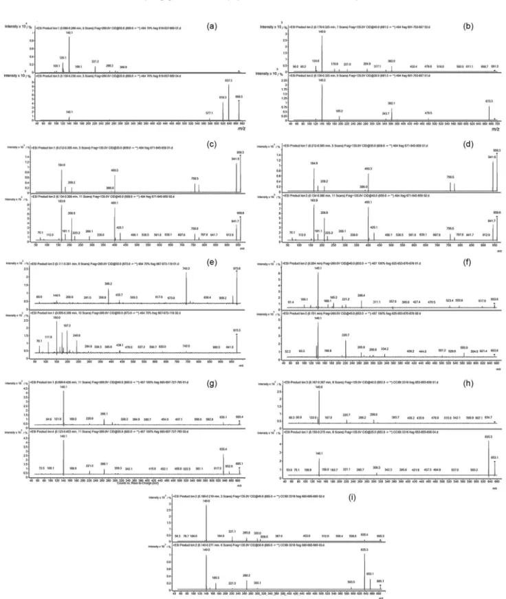
Mass spectrometry
several protease inhibitors in the purified extract. These compounds were identified as aeruginosin (m/z 669 and 691), micropeptin (m/z 945 and 959) and cyanopeptolin (m/z 973) for S. brasiliense CCIBt 3094. Strains of
S. brasiliense CCIBt 3096 and CCIBt 3316 showed
ions at m/z 653 and 685 corresponding to aeruginosin. Although the molecular results showed that S. brasiliense CCIBt 3179 has the genes that encode for aeruginosin and cyanopeptolin, the presence of these compounds, analyzed by mass spectrometry, was not confirmed. Figure 2 shows the fragmentation ions observed for these protease inhibitors, according to different energy collision used. Microcystin molecules were not observed by mass spectrometry analysis.
According to the fragmentation data generated by mass spectrometry (Figures 2a to 2i), aeruginosin (m/z 669, 653 and 685) presented the ions at m/z 140, corresponding to the Choi immonium-(acid 2-carboxy-6-hydroxyoctahydroindole), m/z 221 (Leu-Choi fragment), m/z 266 (Choi-Argininal-CH3N2-H2O + H) and m/z 309
(Choi-Argininal-NH2 + H), all indicative of aeruginosin.
Aeruginosin m/z 669 presented one chlorine (m/z 284) in the portion and aeruginosin (m/z 691) is characterized by
2-carboxy-6-hydroxyoctahydroindole (Choi) moiety and also presented one chlorine. Micropeptin (m/z 945) is well identified by the ion m/z 150 (MTyr-immonium), m/z 209 (Ahp-Leu-H2O + H) and m/z 386 (Ahp-Leu-MTyr-H2O + H).
The other micropeptin (m/z 959) is represented by the
ions m/z 181 (Ahp-Leu-H2O-CO + H) and m/z 209
(Ahp-Leu-H2O + H). Cyanopeptolin (m/z 973) was
supported by the ions m/z 70 (Arg fragment) and m/z 150 (MTyr-immonium). Table 1 shows the assignment ions for aeruginosin, cyanopeptolin and micropeptin according to the collision energy used to fragment all the molecules.
Allium cepa test
Different extracts obtained from Sphaerocavum
strains were evaluated for their potential toxic effects in Allium cepa. Morphological alterations in roots such as tumor and hook formations and meristem tip necrosis were not observed after treatments (Figure 3).
On the other hand, the ethanolic extract caused an inhibition of the roots growth in acute and chronic treatments (Figure 4a). This effect was reflected in the mitotic index, where a decrease in the number of cells in mitosis was observed (Figure 4b). In this case, metaphase was the most affected mitotic phase. Thus, both acute and chronic treatments with ethanolic extract promoted a reduction of cells in this phase (Figure 4c).
An interesting finding was that the roots treated with dichloromethane extract after 48 h did not present cells in mitosis demonstrating a cytotoxic effect (Figure 5), even though the roots continued growing, probably by cell elongation. Chromosomal damage was not observed for all treatments.
Trypsin test
Trypsin (EC 3.4.21.4) was tested against the extracts listed in Table 2. All the extracts were positive for the presence of protease inhibitor, except for S. brasiliense CCIBt 3179 and C. raciborskii 339 extracts. Extracts from S. brasiliense CCIBt 3094, CCIBt 3096, and CCIBt 3316 tested for trypsin inhibition showed inhibitory activity from 56 to 77%.
Sphaerocavum is a tropical and subtropical genus
proposed by Azevedo and Sant’Anna7 from samples
collected from eutrophic reservoirs in São Paulo State, and also from Uruguai. Since the genus was proposed,
S. brasiliense was reported for eutrophic lakes in
different parts of the world.24,25Sphaerocavum brasiliense
CCIBt 3094, isolated from Billings reservoir, in São Paulo State, is the type species and belongs to the family Figure 1. Maximum likelihood topology of aeruginosin in (a) and
Merismopediaceae, subfamily Merismopedioideae due to the two plains of cell division and slightly flattened, but not flat colonies.
Table 1. Assignment of cyanopeptide fragmentations
Cyanobacteria Collision energy / V
Fragmentation
ions / m/z Fragment assignment Chemical structure Reference
S. brasiliense
CCIBt 3094
20
669 −
Aeruginosin structure and ions assignments
adapted from Welker and von
Döhren2
637 M-CH3OH
619 M-CH3OH-H2O + H
577 M-C2H3O
50
309 Choi-Argininal-NH2 + H
291 Choi-Argininal-NH2-H2O + H
266 Choi-Argininal-CH3N2-H2O + Ha
284 Cl-Hpla-Leu-CO + H 221 (Leu-Choi) fragment 140 Choi-immonium ion 100 Argal-fragment
30
691 −
673 M-H2O + H
344 CH3OH-H2O + H
284 Cl-Hpla-Leu-CO + H 221 (Leu-Choi) fragment 140 Choi-immonium ion
40 691 −
30 673 M-H2O + H
40
284 Cl-Hpla-Leu-CO + H 221 (Leu-Choi) fragment 140 Choi-immonium ion
S. brasiliense
CCIBt 3096
40
653 −
266 Choi-Argininal-CH3N2-H2O + Ha
221 (Leu-Choi) fragment
169 Choi + H
140 Choi-immonium ion 100 Argal-fragment
35
685 −
309 Choi-Argininal-NH2 + H 291 Choi-Argininal-NH2-H2O + H
266 Choi-Argininal-CH3N2-H2O + Ha
221 (Leu-Choi) fragment 140 Choi-immonium ion 100 Argal-fragment
S. brasiliense
CCIBt 3316
25 653 −
635 M-H2O + H
40
266 Choi-Argininal-CH3N2-H2O + Ha
221 (Leu-Choi) fragment 140 Choi-immonium ion
685 −
653 M-CH3OH
635 653-H2O
593 653-CH3OH-H2O + H
309 Choi-Argininal-NH2 + H
266 Choi-Argininal-CH3N2-H2O + Ha
221 (Leu-Choi) fragment
169 Choi + H
Cyanobacteria Collision energy / V
Fragmentation
ions / m/z Fragment assignment Chemical structure Reference
S. brasiliense
CCIBt 3094
30
945 −
Micropeptin structure and ions assignments
adapted from Czarneck et al.23
927 M-H2O + H
386 (Ahp-Leu-MTyr)-H2O + H
209 (Ahp-Leu)-H2O + H
150 MTyr-immonium
35
959 −
941 M-H2O + H
386 (Ahp-Leu-MTyr)-H2O + H
40 209 (Ahp-Leu)-H2O + H
181 (Ahp-Leu)-H2O-CO + H
50
973 −
Cyanopeptolin structure and ions assignments
adapted from Welker and von
Döhren2
386 (Ahp-Leu-MTyr)-H2O + H
60
209 (Ahp-Leu)-H2O + H
181 (Ahp-Leu)-H2O-CO + H
150 MTyr-immonium
70 Arg fragment
aCH
3N2-ureido-group of argininal.
Table 1. Assignment of cyanopeptide fragmentations (cont.)
Figure 3. Allium cepa roots treated with extract of Sphaerocavum brasiliense CCIBt 3094 for 24 h and 48 h. Legend: C: negative control; Sl: solvent; Ex: extract.
peptides inhibit serine proteases26 making them an important
molecule for the development of drugs against various human diseases. Aeruginosins have been tested against several proteases, such as thrombin, trypsin and plasmin. Processes of trypsin inhibition are considered of interest for the treatment of pancreatic disorders such as pancreatitis.27
Cyanopeptolins are known as trypsin, chymotrypsin, elastase, plasmin, kallikrein and factor XI inhibitors28 and
can be used to treat asthma and viral infections.29 In addition,
some cyanopeptolin variants are known as specific elastase inhibitors being critical for the treatment of emphysema,
which is mediated by the action of excessive elastase.30 The
high level of trypsin inhibition observed for the extracts of S. brasiliense CCIBt 3094, CCIBt 3096, and CCIBt 3316 demonstrates a great potential for bioprospection of these strains. Sphaerocavum brasiliense CCIBt 3179 presented no inhibitory activity at the tested concentration. MS analysis of the extract of this strain presented no protease inhibitor production, which suggests that despite the positive results in molecular analysis for aeruginosin and cyanopeptolin synthetase genes, they were somehow not expressed.
Genotoxicity studies are important for testing new drugs,31-33 and they must be performed in the early stages
in order to predict potential genotoxic and/or carcinogenic activity and to assist in obtaining new chemical structures less toxic.31,34 Plant bioassays were validated for testing
complex mixtures for in situ monitoring and laboratory testing of the genotoxicity of environmental pollutants.35
Herein, the Sphaerocavum ethanolic and dichloromethane extracts showed a potential cytotoxic effect in the Allium test. This fact could be linked to the several biocompounds that are produced by cyanobacteria. However, they did not present genotoxic effects.
Table 2. Cyanobacterial extracts with inhibitory trypsin activity
Sample Extract concentration / µg Trypsin activity / (mU mL-1) Inhibition / %
Negative control (C. raciborskii 339) 44.6 174.59 0
S. brasiliense CCIBt3094 42.8 25.66 76
S. brasiliense CCIBt3316 53.5 47.09 56
S. brasiliense CCIBt3096 27.0 23.90 77
S. brasiliense CCIBt3179 27.2 129.80 0
Figure 4. Toxicity and cytotoxicity of S.brasiliense extracts in A. cepa
system. Root growth in (a), mitotic index in (b) and mitotic index phase of meristematic cells treated for 24 and 48 h with ethanolic, dichloromethane and methanolic extracts in (c).
Figure 5. Allium cepa test for potential cytotoxic, genotoxic and mutagenic effects caused by S. brasiliense extracts. Negative control in (a), and dichloromethane extract after 48 h treatment in (b). P = prophase, A = anaphase, T = telophase.
or di-chlorinated molecules.15 The presence of chlorine in
aeruginosin and cyanopeptolin reveals the potential for inhibition activities.36 In this study cyanopeptides produced
by the genus Sphaerocavum were found as chlorinated molecules which make them attractive drug targets.
Conclusions
Protease inhibitors are important biomolecules in many biological processes and are related to several human
diseases such as thrombosis or cancer. Therefore, efforts have been made to characterize these protease inhibitors by identifying their structure, biosynthesis, physiology, bioactivity, toxicology and factors that regulate their production. New peptides, as well as new congeners of such biomolecules are of great interest. This study presented information on the chemical characterization, molecular genetics and biological activity of the peptide protease inhibitors aeruginosin and cyanopeptolin produced by
Sphaerocavum genus. The report of metabolites with
pharmacological potential produced by Sphaerocavum
shows the great potential of these cyanobacteria in bioprospecting studies. Also, no microcystin was observed for the evaluated strains, making these strains very promising for drug discovery.
Supplementary Information
Supplementary information (SYBR Green-stained agarose gel of polymerase chain reaction (PCR) products obtained from CCIBt 3094, 3096, 3179 and 3316 aeruginosin specific primers) is available free of charge at http://jbcs.sbq.org.br as PDF file.
Acknowledgements
and The Brazilian Council for Technological and Scientific Development (CNPq) (Project No. 559720/2009-2). M. F. Fiore would like to thank CNPq for a research fellowship (308299/2009-4). M. E. Silva-Stenico and A. S. Lorenzi were the recipients of post-doctoral fellowship from FAPESP (Project No. 2010/09867-9 and Project No. 2008/53627-2, respectively). We also thank Prof Valdemar Tornisielo for LC-MS/MS analysis support in the Ecotoxicology Laboratory, Center for Nuclear Energy in Agriculture, University of São Paulo, Brazil and Watson A. Gama for the Sphaerocavum microphotograph.
References
1. Silva-Stenico, M. E.; Silva, C. S. P.; Lorenzi, A. S.; Shishido, T. K.; Etchegaray, A.; de Lira, S. P.; Moraes, L. A. B.; Fiore, M. F.; Microb. Res.2011, 166, 161.
2. Welker, M.; von Döhren, H.; FEMS Microbiol. Rev. 2006, 30, 530.
3. Chlipala, G. E.; Mo, S.; Orjala, J.; Curr. Drug Targets2011,
12,1654.
4. Silva-Stenico, M. E.; Lorenzi, A. S.; Silva, C. S. P.; Rigonato, J.; Fiore, M. F.; Oecol. Austr. 2012, 16, 183 (ISSN 2177-6199). 5. Laughinghouse, H. D.; Prá, D.; Silva-Stenico, M. E.; Rieger, A.;
Frescura, V. D. S.; Fiore, M. F.; Tedesco, S. B.; Sci. Total Environ.2012, 432, 180.
6. Tedesco, S. B.; Laughinghouse, H. D. In Environmental Contamination; Srivastava, J. K., ed.; InTech Publisher: Rijeka, 2012, p.137-156.
7. Azevedo, M. T. P.; Sant’Anna, C. L.; Algol. Stud. 2003,109, 79.
8. Gorham, P. R.; Mclachlan, J. R.; Hammer, V. T.; Kim, W. K.;
Verh.- Int. Ver. Theor. Angew. Limnol. 1964, 15, 796.
9. Fiore, M. F.; Moon, D. H.; Tsai, S. M.; Lee, H.; Trevors, J. T.;
J. Microb. Methods2000, 39, 159.
10. Tillett, D.; Parker, D.; Neilan, B. A.; Appl. Environ. Microbiol.
2001, 67, 2810.
11. Neilan, B. A.; Dittmann, E.; Rouhiainen, L.; Bass, R. A.; Schaub, V.; Sivonen, K.; Borner, T.; J. Bacteriol.1999, 181, 4089.
12. Rantala, A.; Fewer, D. P.; Hisbergues, M.; Rouhiainen, L.; Vaitomaa, J.; Börner, T.; Sivonen, K.; PNAS2004, 101, 568. 13. Fewer, D. P.; Rouhiainen, L.; Jokela, J.; Wahlsten, M.;
Laakso, K.; Wang, H.; Sivonen, K.; BMC Evol. Biol.2007, 7, 183.
14. Sant’Anna, C. L.; Carvalho, L. R.; Fiore, M. F.; Silva-Stenico, M. E.; Lorenzi, A. S.; Rios, F. R.; Konno, K.; Garcia, C.; Lagos, N.; Neurotoxic. Res.2011, 19, 389.
15. Cadel-Six, S.; Dauga, C.; Castets, A. M.; Rippka, R.; Bouchier, C.; de Marsac, N. T.; Welker, M.; Mol. Biol. Evol.
2008, 25, 2031.
16. Sambrook, J.; Fritsch, E. F.; Maniatis, T. In Molecular Cloning: a Laboratory Manual, 2nd ed.; Cold Spring Harbor Laboratory
Press: Cold Spring Harbor, 1989.
17. Birnboim, H. C.; Doly, J.; Nucleic Acids Res. 1979, 7, 1513. 18. Swofford, D. L.; PAUP*; Phylogenetic Analysis Using
Parsimony (*and Other Methods); Duke University, United States of America, 2003.
19. Tamura, K.; Peterson, D.; Peterson, N.; Stecher, G.; Nei, M.; Kumar, S.; Mol. Biol. Evol.2011, 28, 2731.
20. Posada, D.; Crandall, K. A.; Bioinformatics1998, 14, 817. 21. Silva-Stenico, M. E.; Cantúsio Neto, R.; Alves, I. R.; Moraes,
L. A. B.; Shishido, T. K.; Fiore, M. F.; J. Braz. Chem. Soc.2009,
20, 535.
22. Ayres, M.; Ayres Junior, M.; Ayres, D. L.; Santos, A. A. S.;
BioEstat, Aplicações Estatísticas nas Áreas das Ciências Biomédicas, 5th ed.; Sociedade Civil Mamirauá: Belém, 2007.
23. Czarnecki, O.; Henning, M.; Lippert, L.; Welker, M.; Environ. Microbiol.2006, 8, 77.
24. Wood, S. A.; Crowe, A. L. M.; Ruck, J. G.; Weak, R. G.;
N. Z. J. Bot. 2005, 43, 479.
25. Vardaka, E.; Moustaka-Gouni, M.; Cook, C. M.; Lanaras, T.;
J. Appl. Phycol.2005, 17, 391.
26. Hanessian, S.; Ersmark, K.; Wang, X.; del Valle, J. R.; Blomberg, N.; Xue, Y.; Fjellström, O.; Bioorg. Med. Chem. Lett.2007, 17, 3480.
27. Ersmark, K.; del Valle, J. R.; Hanessian, S.; Angew. Chem. Int.
2008, 47, 1202.
28. Gademann, K.; Portmann, C.; Blom, J. F.; Zeder, M.; Jüttner, F.;
J. Nat. Prod.2010, 73, 980.
29. Singh, R. K.; Tiwari, S. P.; Rai, A. K.; Mohapatra, T. M.;
J. Antibiot.2011, 64, 4011.
30. Mal, H.; Crestani, B.; Aubier, M.; Fournier, M.; Med. Sci.1999,
15, 833.
31. Gollapudi, B. B.; Krishna, G.; Mutat. Res.2000, 455, 21. 32. Hartmann, A.; Elhajouji, A.; Kiskinis, E.; Poetter, R.; Martus,
H. J.; Fjallman, A.; Frieauff, W.; Suter, W.; Food Chem. Toxicol.
2001, 39, 843.
33. Kiskinis, E.; Suter, W.; Hartmann, A.; Mutagenesis2002, 17, 37.
34. Snyder, R. D.; Green, J. W.; Mut. Res.2001, 488, 151. 35. Gopalan, H. N. B.; Mutat. Res., Fundam. Mol. Mech. Mutagen.
1999, 426, 99.
36. Eustáquio, A. S.; Gust, B.; Luft, T.; Li, S. M.; Chater, K. F.; Heide, L.; Chem. Biol.2003, 10, 279.
Submitted: March 6, 2015 Published online: August 7, 2015