Vol. 7, No. 3, 2004 Nucleation and Growth Microstructural Study of Ti Films on 304 SS Substrates 479
Materials Research, Vol. 7, No. 3, 479-482, 2004. © 2004
*e-mail: [email protected]
Nucleation and Growth Microstructural Study of
Ti Films on 304 SS Substrates
Rogério de Almeida Vieira*, Maria do Carmo de Andrade Nono
Associated Laboratory for Sensor and Materials (LAS), National Institute for Space Research (INPE), São José dos Campos - SP, Brazil
Received: December 27, 2003; Revised: May 03, 2004
Coating of steel surfaces with titanium films has been studied with the objective to protect them against corrosion, and to create an intermediate film for CVD diamond and TiN film deposition. In this work, the nucleation, growth mechanisms and microstructural formation of the titanium films deposited on 304 stainless steel (304 SS) substrate are presented and discussed. The titanium films of variable thickness were obtained by vapour phase deposition produced by electron beam. The surfaces of these samples were observed by scanning electron microscopy. The cross sections of these samples were observed by using an atomic force microscope. The Ti film-304 SS interfaces were analyzed by X-ray diffraction. The results showed that titanium films have a columnar growth. The Ti film-304 SS interface had a residual compression stress at room temperature due to the inter-diffusion process.
Keywords:Ti film nucleation and growth, microstructural study, electron beam
1. Introduction
Steel materials with modified surfaces are widely used in a large variety of important technological applications including cutting tools, machine components and molding dies, where their surfaces can be submitted to physical and/or chemical erosion1-3. Therefore, adequate coatings that are well bonded on steel surfaces are desirable in such applica-tions to enhance the mechanical performance and to increase the lifetime against abrasion and corrosion. As a conse-quence of surface smoothness, hardness and chemical in-ertness, the use of diamond polycrystalline films as protec-tive coatings for steel materials is highly desirable and tech-nologically relevant. However, nucleation of the diamond phase is extremely difficult on these material surfaces mainly because of the high diffusivity of carbon ions into steel substrates at temperature used to obtain CVD diamonds, which consumes the carbon species needed for nucleation of diamond phase4-7. In order to enhance nucleation den-sity, the growth of polycrystalline diamond films and the adhesion of these coatings on steel, the creation of a carbon diffusion barrier near or on the surface of the substrate is required. This barrier can be obtained by using an interme-diary layer of a material that has a low diffusion coefficient for carbon, such as a thin deposited film or a surface
modi-fied by ion implantation1,4,8-10. On the other hand, polycrystalline CVD diamond films have a good adhesion on titanium surfaces7,9,11-14.
Titanium nitride (TiN) has been widely employed in cutting tools to extend their working life and in decorations for its golden color. As a protective coating, TiN layers have been studied extensively15,16. TiN films have been particu-larly attractive due to their potential applications ranging from hard and corrosion-resistant films to diffusion barrier layers in advanced integrated-circuit devices17. Studies on the TiN properties have solved significant problems, such as the reduction of the usefulness of the TiN films for cor-rosion-resistant films and for diffusion barriers in the films15-18. However, the adherence of TiN films on steel substrate is not well known yet. Nowadays, many studies are focused on the deposition of adherent diamond coat-ings and TiN films on steel materials to improve their wear and corrosion resistance4-7,9,10,16,19.
480 Vieira & Nono Materials Research
structural phase have been investigated using a scanning electron microscopy (SEM), an atomic force microscope (AFM) and an X-ray diffraction (XRD). The titanium films were deposited by electron beam device.
2. Experimental Details
In preparation for titanium nucleation and growth, 304 SS substrates of 15 mm diameter and 3 mm thick were mechanically ground with abrasive papers, polished with 1µm alumina, and ultrasonically cleaned in an acetone bath before being put in the evaporation chamber.
Deposition of titanium film on 304 SS substrate was carried out by electron beam evaporation device. The films were deposited at an incident angle of approximately 87°. The vapour source subtended an angular spread of 3° due to the substrate-source separation of 42 cm and the melt diameters of approximately 2 cm. The accelerating voltage and the electron beam current were 4keV and 150 mA, re-spectively. After the samples chamber was evacuated to 6.75 × 10⁻⁷ Torr, the deposition was done at a substrate tem-perature of 200 °C by using a halogen lamp. The polished substrates were heated at 200 °C for 5 min in vacuum to remove adsorption gases on the surfaces before deposition. During titanium nucleation and growth, the base pressure was hold at less than (1.5-2.25) × 10-6Torr. Under these con-ditions, the substrate temperature was about 200 °C, which was reached without additional heating. The deposition tem-perature was measured by a thermocouple, embedded 1.0 mm below the deposition surface. A quartz crystal thick-ness monitor (CTM) was used to maintain a nominal depo-sition rate of 2 Å/s. Due to the substrate location and orien-tation, the film growth rate normal to the substrates surface was approximately 1Å/s that was recorded by the CTM. This electron beam provided a controlled stepper motor that rotated the substrates. To evaluate the nucleation and the growth of titanium films on 304 SS substrate, titanium was deposited on three growth thickness: 0.5 µm, 1.5 µm and 3.0 µm. In order to obtain samples of the films at incremen-tal stages of growth, the deposition was periodically paused, the substrates were allowed to cool slowly to room tem-perature and then the system was vented with dry N2, and portions of the substrates removed for later analysis.
The surface morphology of the deposited films were investigated by scanning electron microscopy (SEM), JEOL model JSM-5310a, attached to an energy dispersive spectrometry (EDS). The study of the crystalline structure of the samples before and after titanium films deposition was obtained by X-ray diffraction (XRD) with Cu Kα ra-diation 40 kV / 20 mA. Using the wavelength of Cu Kα the 2θ interval was chosen from 30° up to 90° with a step size of 0.02°. The cross-section of the Ti film-304 SS substrate system was observed by atomic force microscopy.
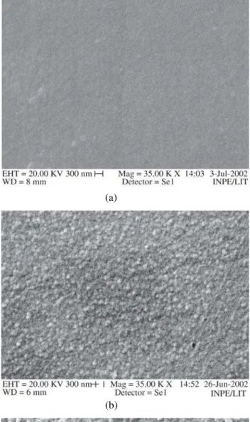
Figure 1. Surface morphology of the nucleation and growth
tita-nium films on 304 SS substrates with different deposition thick-ness: a) 0.5 µm; b) 1.5 µm; c) 3.0 µm.
(a)
(b)
Vol. 7, No. 3, 2004 Nucleation and Growth Microstructural Study of Ti Films on 304 SS Substrates 481
3. Results and Discussion
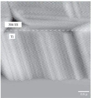
Figure 1a, b and c shows the surface morphology of the titanium nucleation and the growth with different deposi-tion thickness from 0.5 µm to 3.0 µm. Results showed that after approximately 0.5 µm thick, it can not be observed titanium nucleus on 304 SS substrate as shown in Fig. 1a. Titanium nucleation started on 304 SS substrate after depo-Figure 2. An AFM image of the cross-section observation of the
microstructure of titanium film deposited on 304 SS substrate by electron beam evaporation indicating a columnar growth.
Figure 3. XRD patterns for the 304 SS surface without film and
titanium films deposited with 0.5 µm and 3.0 µm thick by electron beam on 304 SS substrate.
sition for about 1.5 µm thick as shown in Fig. 1b. After approximately 3 µm thick, it can be observed that many titanium crystal clusters were observed on the surface. This shows a heterogeneous microstructure formed by several crystal sizes. As can be seen in Fig. 2, in the cross-section of the Ti film-304 SS system, the Ti film has a columnar growth. These microstructures differences between the film surface and its cross-section result in different mechani-cal behavior.
XRD peaks from the Fe-γ phase (111), (200) and (220) of the 304 SS substrate without film and Ti film-304 SS substrate systems are observed. A detailed analysis of X-ray diffractograms of 304 SS surface without film and tita-nium films grown with 0.5 µm and 3.0 µm thick on 304 SS surface shows some differences in the peak positions and the peak intensity. This can be seen in Fig. 3, where the Fe-γ (111) peaks for the 304 SS substrate and Ti film-304 SS systems are presented. It can be observed that with the increase of the deposition thickness, the peak of Fe-γ (111) decrease in 2θ indicating the diffusion of titanium atoms into the 304 SS substrate. The shifting in the peak positions reveals the presence of stress in the modified 304 SS surface by titanium film. As shown in 20, this stress state is a result of the atomic solid solution presents at the Ti film-304 SS substrate interface.
4. Conclusions
The titanium films deposited on stainless steel substrates have been studied as intermediate layer for the TiN and diamond films grown in steel materials. Only af-ter deposition for about 1.5 µm thickness titanium nuclea-tion started on 304 SS substrate surface. The X-ray dif-fraction results showed that titanium films grown on the 304 SS substrate by using the electron beam device re-vealed the presence of stress. This was related to the com-pression stress due to atomic solid solution presents at the interface20. This residual compression stress is formed as a result of the inter-diffusion process, that is, Ti atoms are present in the steel microstructure and Fe, Cr and Ni at-oms are present in the Ti film.
These results suggest that a titanium film growth on a stainless steel substrate by using the electron beam evapo-ration device has potential applications as interlayer to pro-mote better adherence of TiN and diamond films on steel materials.
Acknowledgements
The authors would like to thank FAPESP for financial support.
References
compo-482 Vieira & Nono Materials Research
nents by ion implantation, Mat. Scienc. Eng., v. 90, p. 373-383, 1987.
2. Perepezko, J.; Brewer, L.; Schaefer, R. Principles under-lying coatings and surface modification science, Mat. Scienc. Eng., v. 70, p. 9-22, 1985.
3. Mattox, D.M.; Greene, J.E.; Buckley, D.H.; Somorjai, G.A. Properties of coated and modified surfaces. Mat. Scienc. Eng., v. 70, p. 79-89, 1985.
4. Nono, M.C.A.; Corat,E.J.; Ueda, M.; Stellati, C.; Barroso, J.J.; Conrad, J.R.; Shamin, M.M.; Fetherston, P. Sridharan, K. Surface modification on 304 SS by plasma immersed ion implantation to improve the adherence of a CVD diamond film, Surf. Coat. Technol., v. 112, p. 295-298, 1999.
5. Davanloo, F.; Park, H.; Collins, C.B. Protective coatings of nanophase diamond deposited directly on stainless steel substrates, J. Mater. Res., v. 11, p. 2042-2050, 1996. 6. Schäfer, L.; Fryda, M.; Stolley, T.; Xiang, L.; Klages, C. P. Chemical vapor deposition of polycrystalline dia-mond films on high-speed steel, Surf. Coat. Technol., v. 116-119, p. 447-451, 1999.
7. Sato, T.; Narumi, S.; Ito, S.; Akashi, K. Diamond deposi-tion on titanium and iron substrates pretreated in N2 -C2H2 plasma, Thin Solid Films, v. 316, p. 29-34, 1998. 8. Mahalingam, P.; Dandy, D.S. A quasi-equilibrium model
for the prediction of interlayer chemistry during dia-mond chemical vapor deposition, Thin Solid Films, v. 322, p. 108-116, 1998.
9. Vieira, R.A. Study 304 SS surface modification with polymeric and titanium films to improve the adherence of CVD diamond films, M.Sc, Thesis, Faculdade de Engenharia Química de Lorena - SP, Brazil (Aug. 2000). 10. Vieira, R.A.; Nono, M.C.A. Nucleation and growth of CVD diamond films on titanium intermediate layers deposited on 304 SS, Acta Microsc., v. 9, p. 279-280, 2001.
11. Peng, X.L.; Clyne, T.W. Formation and adhesion of hot filament CVD diamond films on titanium
substrates.,Thin Solid Films, v. 293, p. 261-269, 1997. 12. Yan, B.; Loh, N.L.; Fu, Y.; Sun, C.Q.; Hing, P. Surface and Interface characterization of diamond coatings de-posited on pure titanium, Surf. Coat. Technol., v. 115, p. 256-265, 1999.
13. Grögler, T.; Zeiler, E.; Hörner, A.; Rosiwal, S.M.; Singer, R.F. Microwave-plasma-CVD of diamond coatings onto titanium and titanium alloys, Surf. Coat. Technol., v. 98, p. 1079-1091, 1998.
14. Buccioni, E.; Braca, E.; Kenny, J.M.; Terranova, M.L. Processing-structure-adhesion relationship in CVD dia-mond films on titanium substrates, Diadia-mond Relat. Mater., v. 8, p.17-24, 1999.
15. Kola, P.V.; Daniels, S.; Cameron, D.C.; Hashmi, M.S.J. Magnetron sputtering of TiN protective coatings for medical applications, Journal of Materials Processing Technology, v. 56, p. 422-430, 1996.
16. Vershinin, N.; Filonov, K.; Straumal, B.; Gust, W.; Wiener, I.; Rabkin, E.; Kazakevich, A. Corrosion be-haviour of the protective and decorative TiN coatings on large area steel strips, Surf. Coat. Technol., v. 125, p. 229-232, 2000.
17. Heuvelman, W.M.; Helderman, P.; Janssen, G.C.A.M.; Radelaar, S. Tin reactive sputter deposition studied as a function of the pumping speed, Thin Solid Films, v. 332, p. 335-339, 1998.
18. Kwak, M.Y.; Shin, D.H.; Kang, T.W.; Kim, K.N. Char-acteristics of TiN barrier layer against Cu diffusion, Thin Solid Films, v. 339, p. 290-293, 1999.
19. Eryilmaz, O.L.; Urgen, M.; Çakir, A.F.; Kazmanh, M.K.; Kahraman, U.H. The effect of the sputter cleaning of steel substrates with neutral molecule source on the adhesion of TiN films, Surf. Coat. Technol., v. 97, p. 488-491, 1997.