ISSN 0101-8205 / ISSN 1807-0302 (Online) www.scielo.br/cam
The sodium pump controls the frequency of action-potential-induced calcium oscillations
SHIVENDRA G. TEWARI*
Systems Science and Informatics Unit, Indian Statistical Institute, 8thMile, Mysore Road, Bangalore 560059, India
E-mail: [email protected]
Abstract. Calcium plays a significant role in a number of cellular processes, like muscle con-traction, gene expression, synaptic plasticity, signal transduction, but the significance of calcium oscillations (CaOs) is not yet completely understood in most of the cell types. It is a widely ac-cepted fact that CaOs are a frequency encoded signal that allows a cell to use calcium as a second messenger while avoiding its toxic effects. These intracellular CaOs are primarily driven by some agonist-dependent pathways or fluctuations in membrane potential. The present mathematical model is of the latter type. The model incorporates expression for all major intracellular ionic species and membrane proteins. Especially, it integrates the coupling effect of sodium pump and Na+/ Ca2+exchanger over CaOs. By varying sodium pump current, it is found that, sodium pump is a key player in modulating intracellular CaOs. The model predicts that the sodium pump can play a decisive role in regulating intercellular cell signaling process. The present study forms the basis for sodium pump controlled intercellular signaling process and requires further experimental verification.
Mathematical subject classification: 34M10, 92C20.
Key words:Na+/Ca2+exchanger, sodium pump, calcium oscillations, membrane potential.
1 Introduction
A number of investigators have performed experiments to study the behavior of cytosolic calcium (Ca2+) and the parameters affecting its behavior [1, 2, 3].
#CAM-365/11. Received: 02/V/11. Accepted: 11/IX/11.
But till date the function of cytosolic Calcium oscillations (CaOs) has not been completely understood in most cell types. CaOs are known to play a key role in a number of mechanisms like activation of extracellular signal regulated kinase (ERK) [4, 5], the contraction of smooth muscle [6], increase in the frequency of synaptic currents [7] and maturation of Xenopus laevis Oocyte [8]. These CaOs are supposed to contain frequency-encoded signals that help in using Ca2+ as a second messenger while avoiding its high intracellular concentrations [9]. Also, in the process of signal transduction, intracellular Ca2+ behaves like a switch and decides whether a particular signal needs to be further propagated or not. The increase in intracellular concentration is facilitated by the opening of transmembrane Ca2+channels which lead to the opening of channels at the intracellular stores. There are mainly Rynodine Receptors (RyRs) or Inositol Triphosphate Receptors (IP3Rs) that are located at the membrane of endoplasmic
reticulum (in neurons) or sarcoplasmic reticulum (in myocytes) which causes an efflux of Ca2+ from the intracellular stores. The release of Ca2+ through
IP3Rs is as a result of some agonist or neurotransmitter binding to its receptor
which can cause via G-protein link to phospholipase C (PLC), the cleavage of phosphotidylinositol (4,5)-bisphosphate (PIP2) to inositol triphosphate (IP3) and
diacylglycerol (DAG). This released IP3is free to diffuse through the cytosol and
binds with IP3R and leading to the subsequent opening of these receptors and
release of Ca2+from the intracellular stores. CaOs can be classified into mainly two types:
1) that is induced by changing membrane potential as in the case of an action potential; and
2) that occur in the presence of voltage clamp. The latter part can be further categorized based on the fact that the oscillatory Ca2+flux is from RyRs or IP3Rs but our focus is on the first type.
Jafri et al. [6] showed CaOs for changing membrane potential of endoplas-mic reticulum (ER), Atri et al. [10] showed CaOs for Ca2+ flux through the IP3Rs in Xenopus laevis Oocyte and determined the intermediate range of IP3
kidney (NRK) fibroblast. Recently, Silva et al. [13] proposed a mathematical model for endothelial cells which incorporated nearly all the important biophys-ical parameters but is unable to exhibit CaOs. Thus, we can say that in all the investigations on CaOs carried out by researchers so far, none of the investigators have tried to incorporate the effect of changing cytosolic Na+and K+ions over CaOs. Thus, in this article, we have proposed a mathematical model governing CaOs for changing membrane potential in the absence of fluxes from the intra-cellular stores. Holmgren et al. [14] determined the three distinct steps of Na+ ion release from the sodium pump with the help of high speed voltage jumps. Here, we have tried to incorporate the impact of these distinct steps of sodium pump over cytosolic CaOs, in case of an action potential. Thus, we have in-corporated L-type Ca2+channel, Na+channel, K+channel, Plasma-Membrane (PM) Ca2+ ATPase, Na+/ Ca2+exchanger (NCX), Na+/ K+ATPase (Sodium
pump), inward rectifier potassium channel (Kir), Ca2+ dependent intermediate
potassium channel (IKCa, Ca2+dependent small potassium channel (SKCa) and
dynamic membrane potential. The gating mechanism of the trasmembrane chan-nels emulate the gating mechanism of the famous Hodgkin and Huxley model [15]. Further, the pumps and proteins are modeled to have realistic gating mech-anism such that they are in agreement with the biological facts. The proposed mathematical model leads to a system of non-linear ordinary differential equa-tions. We have used Euler’s method for the simulation of the proposed model for which a MATLAB script has been written.
2 Mathematical formulation
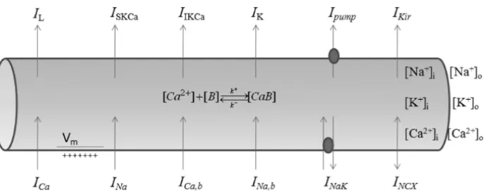
Figure 1 – Schematic diagram of the cell model. The abbreviations representing ionic
concentrations, currents, pumps, proteins and exchangers are defined in this section.
2.1 The Ca2+channel
The L-type Ca2+channel is supposed to have a permeability ratio of 36000:1:18 for Ca2+, Na+and K+ions, respectively [19]. The voltage gated Ca2+channel was modeled using non-linear Goldman-Hodgkin-Katz (GHK) current equation [9, 20], which can be stated as,
IS,L = PSz2S
F2Vm
RT
γi[Si] −γo[So]exp −zsRTF Vm
1−exp −zsF Vm
RT
(1)
where,S is any of the ions,[S]i and[S]oare the intracellular and extracellular concentration of S ion respectively (in mM), PS is permeability (in cm/s) of S ion,zS is its valence,γi,γoare the activity coefficient (a.c.) of the Sion, F is Faradays constant (in Coulombs/moles),Vm is membrane potential (in volts),R is real gas constant (in J/K moles) andTis absolute temperature (in K). The total current through the L-type Ca2+channel is
ICa,t =ICa,L+IN a,L +IK,L.
Further, equation (1) is converted into fluxes (in mM/second), before being used as an expression for individual ionic concentrations, by using Faraday’s constant, Volume of the cytosol (Vcyt) and using the fact that 1 L=10−3m3,
σS,L = −
ISAcap
zSF Vcyt
where,Sis any of the ion andLsignifiesL-type Ca2+channel. In equation (2) there is a negative sign against it because by convention inward current is taken to be negative.
2.2 PM Ca2+ATPase
PM Ca2+ATPase (PMCA) is a P-type ATPase. The energy required to extrude Ca2+ out of the cytosol is met by ATP. The kinetics of this pump follows the enzyme-substrate formulism and hence using Michaelis Menten type kinetics [21], one can formulate the net efflux of Ca2+ions out of the cytosol by,
σpump = −
ˆ
IpumpAcap
zCaF Vcyt
(3)
where,Iˆpumpis the pump current given by the following equation,
ˆ
Ipump = Ipump
[Ca2+]H,pump
[Ca2+]H,pump+KH,pump m
where,[Ca2+]is the intracellular Ca2+concentraton (in mM), H,pump is the Hill’s coefficient for PMCA, KmH,pump is the Ca2+ concentration at which the maximum pump current is halved (in mM).
2.3 Na+/ Ca2+exchanger
This protein is known to play an important role in excitation-contraction cou-pling in cardiac myocytes [22]. In neurons this protein helps in the extrusion of cytosolic Ca2+concentration and hence helps in the modulation of neurotrans-mitter release [23]. It is known thatthe cardiac type3Na+/ 1Ca2+exchanger is dominant in brain [23]. Thus, we have used the same exchange for our model. We know that the amount of energy required to extrude an ion against its con-centration gradient is given by [21, 20, 9],
1S =zSF Vm +RT log
Si
So
(4)
where, S is the extruded ion. Introducing energy barrier,η, and using the fact that
we can write NCX current equation with an allosteric dependence over [Ca2+] as [13],
IN C X =
[Ca2+]H,N C X
[Ca2+]H,N C X +KH,N C X m
gN C X
×[N a
+]3[Ca2+]
oexp ηF VRTm
− [N a+]3
o[Ca
2+]exp
(η−1)F Vm
RT
1+dN C X([N a+]3o[Ca2+] + [N a+]3[Ca2+]o)
(5)
where,[Ca2+]o is the extracellular Ca2+ concentration (in mM),[N a+]is the intracellular Na+ concentration (in mM),[N a+]ois the extracellular Na+ con-centration (in mM),KH,N C X
m is the Ca2+concentration at whichIN C X is halved,
H,N C X is the Hill’s coefficient of NCX, dN C X is constant for saturability of
IN C X,gN C X is the conductance of N C X (in nS).
2.4 Na+/ K+ATPase
Na+ / K+ ATPase (NaK) is also a P-type ATPase which is also known as the
sodium pumpand is a 147 kDa membrane protein [24]. It is known for the extrusion of Na+ ions at an expense of some ATP and inflow of K+ ions. Its formulation is based on the steps given by Holmgren et al. [14], where he used high speed voltage jumps to determine three distinct steps of Na+ ions deocclusion from the pump. The current through the sodium pump has the following form,
ˆ
IN a K =
IN a K
τN a K
kf +
kb
1+
K0.5(0)exp
λF VmRT [N a+]
o
H,N a K (6)
here, IN a K is the scaling factor of NaK current (in μA/cm2), kf (in ms) is the forward (deocclusion) rate constant, kb (in ms) is the backward (occlu-sion) rate constant, K0.5(0) is half activating [N a+]o concentration at 0 mV,
2.5 Cytosolic Ca2+buffers
It is assumed that a single buffer specie is present inside the cytosol and follows the following bi-molecular reaction
[Ca2+] + [B] k
+
⇋
k− [Ca B] (7)
and which can be formulated in terms of the following differential equations,
d[Ca2+]
dt = −k +[
Ca2+][B] +k−[Ca B]
d[B]
dt = −k +[
Ca2+][B] +k−[Ca B]
d[Ca B]
dt =k +[
Ca2+][B] −k−[Ca B]
(8)
If we also assume that there are no sources and no sinks present for buffer. Then lettingBT represent the total buffer concentration, the above equation can be written in the reduced form as,
d[Ca2+]
dt = −k +[
Ca2+][BT − [Ca B]] +k−[Ca B]
d[Ca B]
dt =k +[
Ca2+][BT −Ca B] −k−[Ca B]
(9)
where,k+is the buffer association rate,k−is the buffer dissociation rate,[Ca B]
represents bound buffer concentration.
2.6 Na+and K+channels
To generate action potentials Na+current and K+channels are taken as modeled by Hodgkin and Huxley [15]. The transmembrane current due to Na+ and K+ channels has been modeled using the linear current-voltage relationship derived with the help ofOhm’s Law,
equation (or simply Nernst equation),
VS =1000
RT zSF
log So Si (11)
where, 1000 is used to convert volts into milli-volts. All other symbols have their usual meanings. Here, and in all other instances, individual ionic reversal potential has been determined using Nernst equation at each integration step during runtime.
2.7 Ca2+activated small and intermediate K+channel
The current through SKCa is modeled using a linear current voltage relation as follows,
IS KCa =gS KCaPo,S KCa(Vm−VK)
Po,S KCa =
[Ca2+]niS KCa [Ca2+]nS KCa
i +K nS KCa S KCa
Here,gS KCa is SKCa channel conductance per unit area (in mS/cm 2),P
o,S KCa
is its Ca2+dependent open probability,VK is the reversal potential of K+ions. Similarly, Ca2+ activated intermediate K+ current is modeled using a linear
current voltage relation as follows,
II KCa =gI KCaPo,I KCa(Vm−VK)
Po,I KCa =
[Ca2+]niI KCa [Ca2+]nI KCa
i +K nI KCa I KCa
Here,gI KCa isI KCa channel conductance per unit area (in mS/cm 2), P
o,I KCa
is its Ca2+dependent open probability,VK is the reversal potential of K+ions.
2.8 Inward rectifier K+channel
Inward rectifier K+current is known to contribute to resting membrane potential. It is also modeled using the linear current voltage relationship,
IKir =gKir
s
[K+]
o
Kost
α
where,
α = 0.1
1+exp(0.06(Vm−VK −50))
β = 3 exp(0.0002(Vm−VK +100))+exp(0.0002(Vm−VK −10))
1+exp(−0.06(Vm −VK−50))
and gKir is the Kir conductance (in nS), VK is the reversal potential for K +
given by Nernst equation,[K+]ois the extracellular K+ion concentration. Here,
gKir is converted into mS/cm
2 using Acap before being used in the equation
governing membrane potential.
2.9 Ca2+and Na+leak currents
To balance the net effect ofIN C X andIpump there is supposed to be a Ca2+leak current given by,
ICa,b=gCa,b(Vm−VCa)
where,gCa,bis the Ca2+leak conductance per unit area (in mS/cm2),VCais the reversal potential (in mV) for Ca2+given by Nernst equation. Similarly, we can formulate Na+leak current to balance the net effect of IN C X andIˆN a K,
IN a,b =gN a,b(Vm−VN a)
where,gN a,bis the Na+leak conductance per unit area (in mS/cm2),VN ais the reversal potential (in mV) for Na+given by Nernst equation. The current due to all other ions is considered as leak and is incorporated as,
IL =gL(Vm−VL) (12) where, gL is leak conductance (in mS/cm2) and VL is leak reversal potential assumed to be constant.
2.10 Membrane potential
Like the formulation of Hodgkin and Huxley [15] we have divided the total membrane current into capacitive current and ionic currents. Thus, for capacitive current we have,
Cm
d Vm
where, Iapp is the applied membrane current density (in μA/cm2), Vm is the membrane potential (in mV),Cmis the specific membrane capacitance (μF/cm2),
Ii accounts for all the transmembrane currents discussed earlier andtis time (in ms). The gating mechanism of the transmembrane currents follows Hodgkin and Huxley [15]. Combining equation (1)-(13) we can write the mathematical model governing CaOs with relevance to Na+and K+ions, as in the case of an action potential as,
d[Ca2+]
dt = −σCa+2σN C X −σpump−σCa,b
−k+[Ca2+](BT − [Ca B])+k−[Ca B]
d[Ca B]
dt =k +[
Ca2+](BT − [Ca B])−k−[Ca B]
d[N a+]
dt = −σN a−3σN C X −3σN a K −σN a,b−σN a,L d[K+]
dt = −σK−σS KCa −σI KCa −σKir−σK,L +2σN a K Cm
d Vm
dt =Iapp−mchcvCaICa,t−m 3h I
N a−n4IK
−IN C X − ˆIN a K −IN a,b−ICa,b
−IKir −IS KCa −II KCa − ˆIpump−IL
(14)
In equation (14),
αi, βi (i=m,n,h,mc,hc)
are rate constants which vary with membrane potential but not with time (ms−1) andm,n,h,mc,hcare dimensionless gating variables with values lying between 0 and 1. In this model we assume that fluxes from IP3Rs are absent. This can
be achieved by blocking IP3R channel by using an IP3R antagonist like heparin
[25]. This assumption has been taken to exclude the effect of intracellular stores over CaOs. The initial condition of the system is,
All ionic concentrations are in the units of mM. The ordinary differential equa-tions governing the gating variables (m,n,h,mc,hc) are
dm
dt =αm(1−m)−βmm dn
dt =αn(1−n)−βnn dh
dt =αh(1−h)−βhh dmc
dt =αmc(1−mc)−βmcmc dhc
dt =αhc(1−hc)−βhchc
(16)
Here, m and (1−m) are representing on and off state of the variable m, respectively. Variablesn,h,mc andhc also follow likewise. The mathemati-cal expressions of the voltage-dependent rate constants in equation (14) are as follows,
αn=
0.01(−Vm−60) exp −Vm−60
10
−1;βn=0.125 exp
−Vm −70 80
αm =
0.1(−Vm−45) exp −Vm−45
10
−1;βm =4 exp
−
Vm−70 18
αh =0.07 exp(
−Vm−70 20 );βn=
1
exp −Vm−40 10
+1
αmc =
0.1(−Vm− ˜V −45) exp
−Vm− ˜V−45 10
−1
;βmc =4 exp
−Vm− ˜V −70 18
!
αhc =0.07 exp(
−Vm− ˜V −70
20 );βnc =
1
exp
−Vm− ˜V−40 10
+1
vCa =
KvCa
For the solution of equations (14)-(16), we have used Euler’s method and written a script in MATLAB that has been simulated on an AMD Turion 64×2 machine with 1.6 GHz processing speed and 2.5 GB memory. The time taken per simulation is∼9 sec when simulating for 30 ms using 4000 time steps i.e.
1t=0.0075 ms. The numerical results obtained are used to study the effect of varying transmembrane currents over CaOs which are discussed in the following section.
3 Results and discussion
In all the figures, it is assumed that cytosolic Ca2+ is buffered with 50 μM Ethylene Glycol Tetraacetic Acid (EGTA). The standard biophysical parameters used for simulation of the model are listed in Table 1-4 unless stated along with the figures. Since our main objective is to study CaOs, we have shown results that are pertinent to CaOs only.
Symbol Parameter Value Reference
gN a Na+conductance 120 mS/cm2 [15]
gK K+conductance 36 mS/cm2 [15]
gL Leak conductance 0.3 mS/cm2 [15]
gCa,b Ca2+leak conductance 0.001 mS/cm2 –
gN a,b Na+leak conductance 0.0014 mS/cm2 [17, 18]
gKir Kir conductance 0.95 mS/cm
2 –
gN C X NCX conductance 1.99 nS [13]
gS KCa SKCaconductance 0.62 nS [13]
gI KCa IKCaconductance 1.72 nS [13]
Table 1 – Maximal conductance of channels & leaks.
Symbol Parameter Value F Faraday’s Constant 96487 Coulombs/mole R Real gas constant 8.314 J/K mole T Absolute temperature 293.15 K
zCa Ca2+valence 2
zN a Na+valence 1
zK K+valence 1
Cm Cell capacitance 1μF/cm2
Iapp Applied current density 10μA/cm2
Vcyt Cell Volume 4.2×10⁻⁶μL
Acap Cell Surface area 1.26×10⁻⁵cm2 Table 2 – Some electrophysiological constants.
Symbol Parameter Value Reference
PCa Ca2+Permeability 5.4×10⁻⁴cm/sec [19]
PN a Na+Permeability 1.5×10⁻⁸cm/sec [19]
PK K+Permeability 2.7×10⁻⁷cm/sec [19]
[Ca2+]o Extracellular [Ca2+] 1.8 mM [19]
[N a+]o Extracellular [Na+] 145 mM [19]
[K+]o Extracellular [K+] 5.4 mM [19]
γi,Ca a.c. of[Ca2+] 0.341 [16, 17, 19]
γo,Ca a.c. of[Ca2+]o 1 [16, 17, 19]
γi,N a a.c. of[N a+] 0.75 [16, 17, 19]
γo,N a a.c. of[N a+]o 0.75 [16, 17, 19]
γi,K a.c. of[K+] 0.75 [16, 17, 19]
γo,K a.c. of[K+]o 0.75 [16, 17, 19]
k+ EGTA association rate 1.5μM−1s−1 [11]
k− EGTA dissociation rate 0.3s−1 [11]
BT
Total intracellular buffer
concentration 50μM –
Symbol Parameter Value Reference
VL Leak reversal potential 10.6 mV [15]
Kost Regulatory parameter for Kir 5.4μM [12]
KvCa Ca
2+affinity of Ca2+channel 10μM [12]
˜
V Ca2+channel voltage shift 15 mV –
η NCX energy barrier 0.483 [13]
dN C X NCX constant 3.04×10⁻⁴ [13]
KmH,N C X Ca2+affinity of NCX 0.256×10⁻³ [13]
H,NCX Hill-coefficient of NCX 1.5 [13]
IN a K Scaling factor of sodium pump 1.5μA/cm2 –
kf NaK de-occlusion rate 0.097 ms [14]
kb NaK occlusion rate 1.444 ms [14]
K0.5(0) [N a+]owhen IN a K is halved 6.95×10⁻³mM [14]
λ Electric field dropped
along NaK 0.71 [14]
H,NaK Hill-coeffcient of NaK 1.17 [14]
τN a K Constant forIN a K 0.2834 ms –
Ipump Maximal PMCA current 2.67 pA [13]
KmH,pump Ca2+affinity of PMCA 0.26×10⁻³mM [13]
H,pump Hill-coeffcient of PMCA 1.4 [13]
nS KCa Hill-coeffcient of SKCa 1.6 [13]
KS KCa Ca
2+affinity of SK
Ca 237×10−6mM [13]
nI KCa Hill-coeffcient of IKCa 1.4 [13]
KI KCa Ca 2+
affinity of IKCa 740×10−6mM [13] Table 4 – All other biophysical constants.
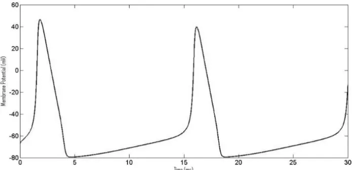
Figure 2 – Changing membrane potential during an action potential in response to an
applied current of 10μAcm−2.
Figure 3 – Behavior of various current densities during action potential (A) Inward
rectifier potassium current,IKir (B) Ca 2+
induced small potassium current, IS KCa (C) Ca2+induced intermediate potassium current,II KCa(D) Na
+/ Ca2+exchanger current,
IN C X(E) PM Ca2+ATPase current,Ipump(F) L-type Ca2+current,ICa(G) Na+/ K+
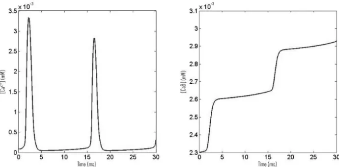
Figure 4 – (A) CaOs in response of an applied current of 10μAcm−2and (B) corre-sponding bound Ca2+concentration.
Figure 5 – CaOs with increasing total buffer concentration BT.
In Figure 5 the effect of increasing total buffer concentration of EGTA is shown. The results are shown for BT =50 μM (dark line) andBT =200μM (broken line). As expected increasing buffer concentration results in lower am-plitude of Ca2+oscillation, which is also evident from Figure 5.
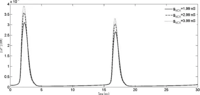
increas-Figure 6 – CaOs with increasing NCX conductance,gN C X.
ing NCX conductance results in higher amplitude of Ca2+oscillation while there is no change resting Ca2+ concentration at more positive membrane potentials as we have used a leak to neutralize the effect of NCX and pump currents.
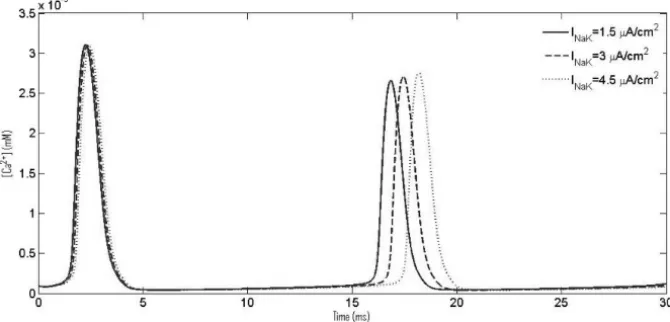
In Figure 7 we observe the results for which the mathematical model was proposed. It is widely believed that cells encode information in the frequency of Ca2+oscillations rather than its amplitude. There are a number of authors who have shown different roles of this ubiquitous sodium pump [26, 27]. Matchkov et al. [27] also experimentally demonstrated that sodium pump plays a significant role in regulating CaOs via regulation of cytosolic Na+ions. Similar philosophy is suggested by our present simulations. We increased pumping rate, IN a K = 1.5μA/cm2 (dark line), 3μA/cm2 (broken line), 4.5μA/cm2 (dotted line), of sodium pump and observed changes in CaOs. It is seen that an increase inIN a K results in an increase in the period of Ca2+oscillations. The changes were quite apparent and are reflected from Figure 7.
In Figures 8-10, the effect of different extracellular concentrations of Ca2+, K+ and Na+are shown over CaOs. The findings of Figure 8 are quite obvious but should be mentioned to show accordance with the biological facts. The curves are shown for [Ca2+]
o=1.8 mM (dark line) and [Ca2+]o=1 mM (broken line). It is apparent from Figure 8 that lowering [Ca2+]
oresults in lower amplitude of
con-Figure 7 – CaOs with increasing NaK current, IN a K.
Figure 8 – CaOs with decreasing extracellular Ca2+concentration,[Ca2+]o.
centration resulted in an increase in frequency of Ca2+ oscillation. Although these findings are also pretty obvious as increasing[K+]oleads to an increase in reversal potential of K+ ions and hence increases the frequency of action potential which in turn increases the frequency of CaOs. In Figure 10, the ef-fect of decreasing[N a+]o concentration is shown. The curves are shown for
Figure 9 – CaOs with increasing extracellular K+concentration,[K+]o.
Figure 10 – CaOs with decreasing extracellular Na+concentration,[N a+]o. obviously does not affect amplitude of action potential. The reason behind the increase in amplitude and latency in Ca2+ oscillation is the change in Na+ ion gradient. This gradient regulates the pumping rate of NCX exchanger decreas-ing the gradient means decreasdecreas-ing the pumpdecreas-ing rate of NCX exchanger. Hence, affecting the net extrusion of Ca2+ ions via NCX exchanger; resulting in an increase in amplitude and latency of CaOs.
biological facts. The obtained results also confirmed the hypothesis of Matchkov et al. [27] that interaction between NCX and Na+/ K+ATPase modulates inter-cellular communication. It was observed that increasing NaK current decreases the frequency of CaOs. The results obtained by previous investigators regarding CaOs have been mainly concerned with membrane potential, inositol triphos-phate (IP3) or ryanodine receptor [6, 28, 29, 10, 11, 12, 13]. None of the earlier
investigators gave much emphasis over this interaction of NCX and sodium pump which in turn effects CaOs. Thus, in this article, we have looked into and demonstrated a novel mechanism which modulates frequency of Ca2+ os-cillation. Here, we have proposed a mathematical model which can be used for problems related to similar cell processes. The results obtained in this paper give new and useful insight for neurologists to look into the paradigm of CaOs at a different perspective. Also, the results obtained are relevant to biomedical scientists for developing protocols for diagnosis and treatment of neurological disorders.
Acknowledgments. The author acknowledges fruitful discussions with Dr. Ronald J. Clarke, School of Chemistry, The University of Sydney, Australia for giving useful insights over the kinetics of Na+/ K+ATPase.
REFERENCES
[1] A.C. Charles, C.C.G. Naus, D. Zhu, G.M. Kidder, E.R. Dirksen and M.J. Sander-son, Intercellular Calcium Signaling via Gap Junctions in Glioma Cells. The Journal of Cell Biology,118(1992), 195–201.
[2] A. Peskoff and G.A. Langer, Calcium Concentration and Movement in the Ventricular Cardiac Cell during an Excitation-Contraction Cycle. Biophys. J.,
74(1998), 153–174.
[3] J. Shuai, J.E. Pearson and I. Parker, Modeling Ca2+Feedback on a Single Inositol 1,4,5-Trisphosphate Receptor and Its Modulation by Ca2+Buffers. Biophys. J.,
95(2008), 3738–3752.
[4] O. Melien, L.S. Nilssen, O.F. Dajani, K.L. Sand, J-G Iversen, D.L Sandnes and T. Christoffersen, Ca2+-mediated activation of ERK in hepatocytes by nore-pinephrine and prostaglandin F2 role of calmodulin and src kinases. BMC Cell Biol.,3(2002).
Cyclic AMP by 2-Methylthioadenosine 5-Diphosphate. The Journal of Pharma-cology and Experimental Therapeutics,311(2004), 334–341.
[6] M.S. Jafri, S.P. Vajda, S. Pasik and B. Gillo, A membrane model for cytosolic calcium oscillations: A study using Xenopus oocytes. Biophys. J.,63 (1992), 235–246.
[7] T.A. Fiacco and K.D. McCarthy, Intracellular Astrocyte Calcium Waves In Situ Increase the Frequency of Spontaneous AMPA Receptor Currents in CA1 Pyrami-dal Neurons. The Journal of Neuroscience,24(2004), 722–732.
[8] L. Sun, R. Hodeify, S. Haun, A. Charlesworth, A.M. MacNicol, S. Ponnappan, U. Ponnappan, C. Prigent and K. Machaca,Ca2+ Homeostasis Regulates Xenopus Oocyte Maturation. Biology of Reproduction,78(2008), 726–735.
[9] J. Keener and J. Sneyd, Mathematical Physiology. Springer,8(1998).
[10] A. Atri, J. Amundson, D. Clapham and J. Sneyd, A Single-Pool Model for Intra-cellular Calcium Oscillations and Waves in the Xenopus laevis Oocyte. Biophys. J.,65(1993), 1727–1739.
[11] J. Wagner and J. Keizer, Effects of Rapid Buffers on Ca2+ Diffusion and Ca2+ Oscillations. Biophys. J.,67(1994), 447–456.
[12] J.M.A.M. Kusters, M.M. Dernison, W.P.M. van Meerwijk, D.L. Ypey, A.P.R. Theuvenet and C.C.A.M. Gielen, Stabilizing Role of Calcium Store-Dependent Plasma Membrane Calcium Channels in Action-Potential Firing and Intracellular Calcium Oscillations. Biophys. J.,89(2005), 3741–3756.
[13] H.S. Silva, A. Kapela and N.M. Tsoukias, A mathematical model of plasma membrane electrophysiology and calcium dynamics in vascular endothelial cells. Am. J. Physiol. Cell. Physiol.,293(2007), C277–C293.
[14] M. Holmgren, J. Wagg, F. Bezanilla, R.F. Rakowski, P. De. Weer and D.C. Gadsby,
Three distinct and sequential steps in the release of sodium ions by the Na+ / K+ ATPase. Nature,403(2000), 898–901.
[15] A.L. Hodgkin and A.F. Huxley,A Quantitative Description of Membrane Current and its Application to Conduction and Excitation in Nerve. J. Physiol.,117(1952), 500–544.
[16] C.H. Luo and Y. Rudy, A model of the ventricular cardiac action potential. Depolarization, repolarization, and their interaction. Circ. Res.,68(1991), 1501– 1526.
[17] C.H. Luo and Y. Rudy, A dynamic model of the cardiac ventricular action po-tential. I. Simulations of ionic currents and concentration changes. Circ. Res.,
[18] C.H. Luo and Y. Rudy, A dynamic model of the cardiac ventricular action po-tential. II. After depolarizations, triggered activity, and potentiation. Circ. Res.,
74(1994), 1097–1113.
[19] T.R. Shannon, F. Wang, J. Puglisi, C. Weber and D.M. Bers, A Mathematical Treatment of Integrated Ca2+ Dynamics Within the Ventricular Myocyte. Biophys. J.,87(2004), 3351–3371.
[20] G.L. Fain, Moleculer and cellular physiology of neurons. Harvard University Press (1999).
[21] D.L. Nelson and M.M. Cox,Lehninger Principles of Biochemistry. W.H. Freeman (2005).
[22] Y. Fujioka, K. Hiroe and S. Matsuoka,Regulation kinetics of Na+-Ca2+ exchange current in guinea-pig ventricular myocytes. J. Physiol.,529(2000), 611–623.
[23] M.P. Blaustein and W.J. Lederer, Sodium / Calcium exchange: its physiological implications. Physiol. Rev.,79(1999), 763–854.
[24] R.J. Clarke and D. J. Kane,Two Gears of Pumping by the Sodium Pump. Biophys. J.,93(2007), 4187–4196.
[25] L.Y. Bourguignon, N. Iida, L. Sobrin and G.J. Bourguignon, Identification of an IP3 receptor in endothelial cells. J. Cell. Physiol.,159(1994), 29–34.
[26] A. Miyakawa-Naito, P. Uhlen, M. Lal, O. Aizman, K. Mikoshiba, H. Brismar, S. Zelenin and A. Aperia, Cell Signaling Microdomain with Na,K-ATPase and Inositol 1,4,5-Trisphosphate Receptor Generates Calcium Oscillations. J. Bio. Chem.,278(2003), 50355–50361.
[27] V.V. Matchkov, H. Gustafsson, A. Rahman, D.M. Boedtkjer, S. Gorintin, A.K. Hansen, E.V. Bouzinova, H.A. Praetorius, C. Aalkjaer and H. Nilsson, Interac-tion Between Na+/K+-Pump and Na+/Ca2+-Exchanger Modulates Intercellular Communication. Circ. Res.,100(2007), 1026–1035.
[28] G.W. De Young and J. Keizer,A single-pool inositol 1,4,5-trisphosphate-receptor-based model for agonist-stimulated oscillations in Ca2+ concentration. Proc. Natl. Acad. Sci. USA,89(1992), 9895–9899.
[29] J. Sneyd, S. Girard and D. Clapham, Calcium wave propagation by calcium-induced calcium release: an unusual excitable system. Bull. Math. Biol.,
![Figure 10 – CaOs with decreasing extracellular Na + concentration, [N a + ] o . obviously does not affect amplitude of action potential](https://thumb-eu.123doks.com/thumbv2/123dok_br/18978220.456071/19.918.117.801.136.848/figure-decreasing-extracellular-concentration-obviously-affect-amplitude-potential.webp)
