Article
J. Braz. Chem. Soc., Vol. 22, No. 5, 943-949, 2011. Printed in Brazil - ©2011 Sociedade Brasileira de Química
0103 - 5053 $6.00+0.00
A
*e-mail: [email protected]
Chemical Constituents from Branches of Maytenus gonoclada (Celastraceae) and
Evaluation of Antimicrobial Activity
Fernando C. Silva,a,c Lucienir P. Duarte,*,a Grácia D. F. Silva,a Sidney A. V. Filho,a,b
Ivana S. Lula,a Jacqueline A. Takahashic and William S. T. Sallumc
aNúcleo de Estudos de Plantas Medicinais and cLaboratório de Biotecnologia e Bioensaios, Departamento de Química, Universidade Federal de Minas Gerais,
Av. Antônio Carlos, 6627, 31270-901 Belo Horizonte-MG, Brazil
bEscola de Farmácia, Universidade Federal de Ouro Preto, Rua Costa Sena, 171, 35400-000 Ouro Preto-MG, Brazil
Seis triterpenos pentacíclicos isolados dos galhos de Maytenus gonoclada (Celastraceae), incluindo todos os dados de RMN do novo composto 3-oxo-12α,29-diidroxifriedelano são aqui relatados. A estereoquímica do novo friedelano foi estabelecida por dados de RMN bidimensional (HSQC, HMBC e NOESY), e sua massa molecular conirmada por espectrometria de massas (ESI). Testes de atividade antimicrobiana usando método de difusão em disco e de macrodiluição foram realizados contra as bactérias Escherichia coli, Citrobacter freundii e Bacillus cereus, e contra o fungo Candida albicans. O triterpeno 3-oxo-12α-hidroxifriedelano mostrou resultado positivo contra C. albicans.
Six pentacyclic triterpenes were isolated from branches of Maytenus gonoclada (Celastraceae) and all NMR data of a new compound 3-oxo-12α,29-dihydroxyfriedelane are herein reported. The stereochemistry of the new friedelane was established by bidimensional NMR (HSQC, HMBC and NOESY) data, and its molecular weight conirmed by ESI mass spectrometry. Antimicrobial activity assays using the method of disk diffusion and macrodilution were carried out against the bacteria Escherichia coli, Citrobacter freundii, and Bacillus cereus, and against the fungi Candida albicans. The triterpene 3-oxo-12α-hydroxyfriedelane showed positive result against C. albicans.
Keywords:Maytenus gonoclada, Celastraceae, 3-oxo-12α-hydroxyfriedelane, 3-oxo-12α ,29-dihydroxyfriedelane, antimicrobial activity
Introduction
Celastraceae family contains many species that have been extensively studied in function of their use in traditional medicine. Some species of Maytenus genus are worldwide distributed and have been used by Africans to treat cancer, by Asian people as an insecticide,1 and by the South American people on the treatment of gastrointestinal diseases.2 The biological activities associated to Maytenus species have been assigned to different classes of secondary metabolites such as phenolic glucosides,3 lavonoids4 and triterpenes.5
Pentacyclic triterpenes (PCTT) have been commonly isolated from species of the Celastraceae family, and some
of them, like 3-oxofriedelane and 3β-hydroxyfriedelane, are considered taxonomic markers of Maytenus genus.4 A large number of pharmacological activities has been associated to triterpenes isolated from species of the Maytenus genus, such as, 3,15-dioxo-21α -hydroxyfriedelane, isolated from Maytenus robusta, that showed antiulcerogenic activity,6 and maytenfolic acid, isolated from Maytenus heterophylla, thatshowed growth inhibitory effect on Candida albicans.7
Maytenus gonoclada Martius, popularly known as “tiuzinho”, can be found in regions of “cerrado” and rupestrian fields of Southeastern and Northeastern Brazil. In our previous studies of the hexane extract from
Chemical Constituents from Branches of Maytenus gonoclada (Celastraceae) J. Braz. Chem. Soc.
944
The present paper reports the phytochemical study of hexane extract from branches of M. gonoclada, which was isolated ive known triterpenes: 3-oxofriedelane (1), 3β-hydroxyfriedelane (2), 3-oxo-12α-hydroxyfriedelane (3), 3,11-dioxofriedelane (4) and 3,16-dioxofriedelane (5).8 In addition, a new compound of the friedelane series, 3-oxo-12α,29-dihydroxyfriedelane (6), was also isolated and its structure was developed by detailed 1H and 13C NMR analysis, including bidimensional (HSQC, HMBC and NOESY) spectral data.
The NMR data of compound 6, as well as the complete hydrogen chemical shifts assigned to compound 5, that have not been described in the literature yet, are herein reported for the irst time.
An evaluation of the antimicrobial activity of the hexane extract is also reported here. The compounds 3-oxofriedelane (1), 3-oxo-12α-hydroxyfriedelane (3), 3,16-dioxofriedelane (5) and 3-oxo-12α ,29-dihydroxyfriedelane (6) were screened by the methods of disk diffusion and macrodilution. The assays were based on the growth inhibition of standard strains of Escherichia coli, Citrobacter freundii, Bacillus cereus, and the yeast
Candida albicans. Minimal inhibitory concentration (MIC), produced by hexane extract and 3-oxo-12α -hydroxyfriedelane (3) were determined. The results showed that 3-oxo-12α-hydroxyfriedelane (3) presented growth inhibition activity.
Experimental
General experimental procedures
Column chromatography (CC) was carried out using silica gel 60 (70-230 Mesh, Merck) and for thin layer chromatography (TLC) it was employed precoated silica gel plates. The detection of spots was made by spraying a mixture (1:1) of vanillin (ethanol solution, 1% m/v) and perchloric acid (aqueous solution, 3% v/v).9 A Mettler FP 80 HT apparatus was used to determine melting points (uncorrected). Elemental analyses were performed on a CHN Perkin-Elmer 2400 apparatus. Optical rotations were measured on a Perkin-Elmer model 341 polarimeter using a 100 mm, 1.0 mL cell tube capacity. Infrared spectra were recorded on a Perkin Elmer, Spectrum One spectrophotometer (ATR). Mass spectrometry was conducted in a LCQFleet (Thermo Scientiic, San Jose, CA) bearing an electrospray ionization (ESI) source, operating in the positive mode. ESI source conditions were: heated capillary temperature of 290 °C, sheath gas (N
2) low rate at 20 (arbitrary units), spray voltage of 4.8 kV, and capillary voltage of 2.0 V.
The 1H and 13C NMR spectra were measured on a Bruker DRX 400 Avance spectrometer at 400 and 100 MHz at 300 K, equipped with inverse detection 5 mm multinuclear head 1H/13C. Each compound was dissolved in CDCl3 or in CDCl3 with 2 drops of pyridine-d5, and transferred to a 5 mm o.d. NMR tube. TMS was used as internal standard (dH = dC = 0). Bi-dimensional (2D) NMR spectra were acquired under standard conditions. Data processing was carried out on SGI workstation using the Bruker (DRX 400) software.
Plant material
Samples of M. gonoclada’s branches were collected in Serra da Piedade, Caeté, Minas Gerais, Brazil, in October 2004. Botanical identiication was provided by Dr. Rita Maria Carvalho-Okano. The voucher specimen (HBCB 60280) was deposited in the Herbarium of the Universidade Federal de Minas Gerais, Belo Horizonte, Minas Gerais, Brazil.
Extraction and isolation
Silva et al. 945 Vol. 22, No. 5, 2011
acetate and methanol in mixtures of increasing polarity. Forty-seven fractions of 10 mL each were obtained yielding the 3-oxo-12α-hydroxyfriedelane (3) (10.3 mg) and 3,16-dioxofriedelane (5) (12.0 mg). Using TLC, the fraction 56 (56.2 mg) (eluted with ethyl acetate) was also characterized as a mixture, and then named as material B.
Material A (234.0 mg) was submitted to silica gel CC (10 g) eluted with pure hexane, chloroform and ethyl acetate, or in mixtures of increasing polarities, providing 73 fractions of 25 mL each. After solvent removal, Fr.A25 (eluted from hexane/chloroform 7:3) gave a white solid (14.1 mg) which was identiied as 3,11-dioxofriedelane (4), and Fr.A27 (eluted from hexane/chloroform 7:3) (10.0 mg) was identiied as 3,16-dioxofriedelane (5). The group of fractions A30-45 eluted from hexane/chloroform 1:1 was puriied by recrystallization (ethanol with drops of acetone) producing a crystalline solid (32.6 mg), which was identiied as 3-oxo-12α-hydroxyfriedelane (3).
Material B (56.2 mg) was submitted to silica gel CC (22.6 g) eluted initially with chloroform/ethyl acetate (1:1) and then with pure ethyl acetate, furnishing 75 fractions of 10 mL each. After solvent evaporation, Fr.B49-69 gave an amorphous solid (9.0 mg) that was identiied as 3-oxo-12α,29-dihydroxyfriedelane (6).
3,16-Dioxofriedelane (5)
Amorphous white solid, mp 218-220 °C. NMR spectral data: See Table 1.
3-Oxo-12α,29-dihydroxyfriedelane (6)
Amorphous white solid, [α]D20 = −23 (CHCl3), mp 250-254 °C. IR (ATR) νmax/cm
-1: 3327, 2920-2850, 1712,
1455, 1389, 1294, 1245, 1218, 1204, 1170, 111, 1056, 1039, 999, 977, 918, 823 and 751. NMR spectral data: See Table 1. MS ((+)-ESI): m/z 423.35 (24%) [M + H – 2H2O]; 405.33 (100%).
Antimicrobial bioassays
The bacteria strains, E. coli ATCC 25723, B. cereus
ATCC 11778, C. freundii ATCC 29935 and the yeast C. albicans ATCC 18804 used in this study were obtained from American Type Culture Collection. The media broth heart infusion (BHI) was purchased from Merck (Darmstadt, Germany) and Biobrás (Montes Claros, Brazil). The strains were maintained on BHI medium and refrigerated at 7 °C. Antimicrobial activity was evaluated using the disk diffusion method, according to the literature.10 The microorganisms were cultivated in medium BHI and incubated for 18 h at 37 °C. Cells were suspended, according to the McFarland protocol, in saline solution
to produce a suspension containing approximately 5×10⁵ CFU mL-1. An aliquot (10 µL) of this suspension was added to 10 mL of sterile antibiotic agar at 40 °C and then, inside a laminar low cabinet, this mixture was poured onto an agar plate. Each tested compound (100 µg) was dissolved in chloroform and put on a paper disk (6 mm diameter), that was dried and placed on the agar plate. Each plate was constituted by 5 sample/disk, together with a disk containing chloramphenicol (30 µg) and another containing only chloroform that were used as positive and negative controls, respectively. The susceptibility of the bacteria and the yeast was determined by the formation of a growth inhibitory zone (mm) of each extract and tested compounds, observed after 18 h of incubation at 37 °C. Experiments were run in triplicate, and the results are presented as mean values of the three measurements. Chloramphenicol and miconazole were purchased from Sigma Chemical Co. (St. Louis, MO). The broth dilution test was used to evaluate the minimum inhibitory concentration (MIC) of growth and the initial inoculums contained 5×10⁵ CFU mL-1.11 Tested compounds were dissolved in dimethyl sulfoxide (DMSO). Sequential dilutions provided the inal concentrations of 512, 256, 128, 64, 32, 16, 8, 4, 2, and 1 µg/mL of tested compounds in BHI medium. Then, 100 µL of the inoculum was added to each tube. After an incubation time of 18 h at 37 °C, the lowest concentration of the tested compounds that inhibited the microorganisms growth (MIC) was visually determined. Tests using DMSO as negative control and chloramphenicol (for bacteria) and miconazole (for
Candida) as positive controls were carried out in parallel. MIC tests were performed in duplicate with full agreement between both results.
Results and Discussion
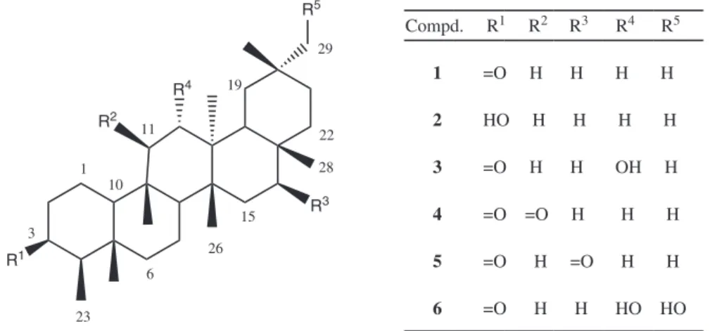
A new triterpene (3-oxo-12α,29-dihydroxyfriedelane,
6) and ive known compounds (Figure 1) were isolated from the phytochemical study of the hexane extract of M. gonoclada branches. The known compounds were isolated in high degree of pureness and they were respectively identiied as 3-oxofriedelane (1),12,13 3β-hydroxyfriedelane (2),14 3-oxo-12α-hydroxyfriedelane (3),8 3,11-dioxofriedelane (4),15 3,16-dioxofriedelane (5).12,16 For the identiication, their physical and spectral data were analyzed and the results compared with previously published data.
Chemical Constituents from Branches of Maytenus gonoclada (Celastraceae) J. Braz. Chem. Soc.
946
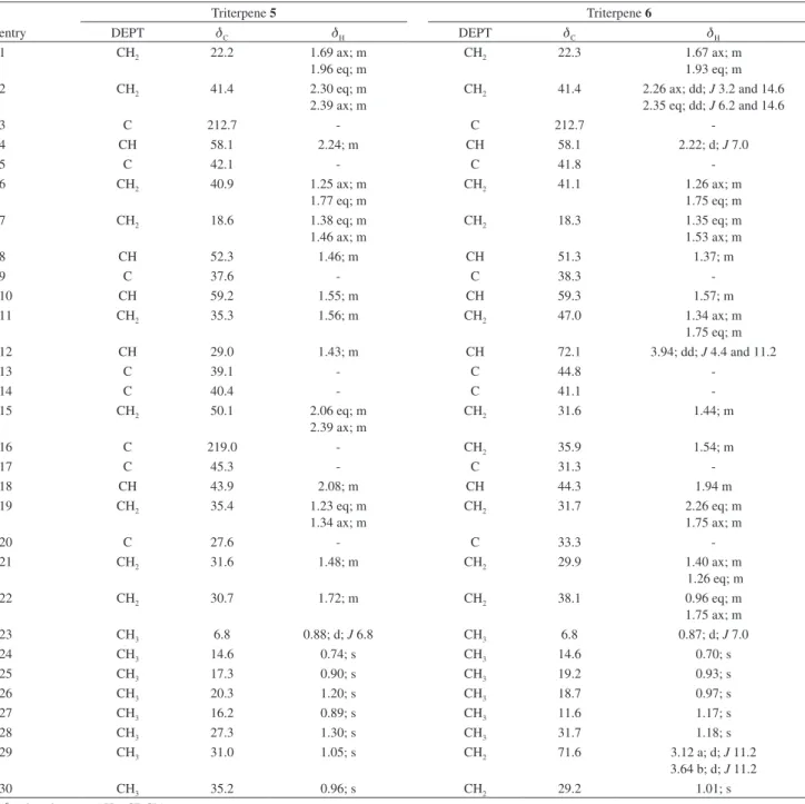
spectral data (HSQC, HMBC and NOESY), the chemical shifts of all hydrogens assigned to compound 5 as well as its correlations were fully established (Figure 2 and Table 1). To the best of our knowledge, it is the irst time that these data are reported.
Compound 6 (Figure 2) was obtained as an amorphous white solid, mp 250-254 °C, [α]D
20 −23, CHCl 3], and showed positive LB test for triterpenes.17 The elemental analysis of compound 6 presented C 78.54% and H 10.97%, compatible with the molecular formula C30H50O3 (MW 458 g mol-1; calculated: C 78.55%; H 10.99%). ESI mass spectrometry conirmed this molecular weight, presenting a fragment m/z 423.35 (24%) [M + H - 2H2O]. This type of fragmentation is described for PCTT. According to Rhourri-Frih,18 in PCTTs like betulin, the loss of two molecules of water occurs due to the presence of two hydroxyl groups, as observed for compound 6. The IR spectra of 6 showed absorption bands: at 3327 cm-1, compatible with the presence of hydroxyl group; at 2920 and 2850 cm-1, characteristic of the alicyclic hydrocarbon; and also at 1712 cm-1, which was attributed to a carbonyl
group. The 13CNMR and DEPT 135 spectra showed 30 signals: seven primary, eleven secondary, ive tertiary and seven quaternary carbons, which were according to the PCTT skeleton. The 1H and 13CNMR data indicated the compound 6 as friedelane derivative,8 containing two hydroxyl groups.
The 1H NMR spectrum of 6 showed hydrogen signals at
dH 0.70 (s), dH 0.93 (s), dH 0.97 (s), dH 1.01 (s), dH 1.17 (s),
dH 1.18 (s) and dH 0.87 (d; J 7.0 Hz) that were associated to seven methyl groups. According to the literature,8 the doublet at dH 0.87 is consistent with methyl group H-23 of members of the friedelane series.
In the 1H NMR spectrum, the hydrogen signals at
dH 3.12 (d; J 11.2 Hz) and dH 3.64 (d; J 11.2 Hz) were observed. Correlations of these signals with the signal of C-29 (dC 71.6) (CH2) were observed in the HSQC contour map. The chemical shifts in this NMR region are typical of carbons attached to hydroxyl group,14,16,19 suggesting the existence of hydroxyl group attached to C-29. Correlations observed in the HMBC contour map, among the signal of H-29 with signals of C-19 and C-30, conirmed the presence
Figure 1. Chemical structures of the triterpenes isolated from branches of M.gonoclada. R1
R2
R3
R4
R5
Compd. R1 R2 R3 R4 R5
1 =O H H H H
2 HO H H H H
3 =O H H OH H
4 =O =O H H H
5 =O H =O H H
6 =O H H HO HO 1 3 6 10 11 15 19 23 22 26 28 29 O O H3C
CH3 CH3
CH3
CH3 H3C
CH3 CH3 H 2 4 23 10 8 12 18 27 29 20 H H H H H 14 24 25 26 H H
Silva et al. 947 Vol. 22, No. 5, 2011
of a hydroxyl attached to C-29. By comparison of spectral data of 6 with similar skeleton reported in the literature,8 it was also possible to locate the signal of C-12 (dC 72.1) and observe, in HMBC map, correlations of H-12 with C-27 (dC 11.6).
The detailed analysis of 2D NMR spectra and comparison with literature8 allowed the assignment of all 1H and 13C chemical data oftriterpene 6. The following sequence of long range correlations (HMBC), H-23 (dH 0.87) C-5 (dC 41.8) H-24 (dH 0.70) C-10 (dC 59.3) H-25 (dH 0.93) C-8 (dC 51.3) H-26 (dH 0.97) C-13
(dC 44.8) H-27 (dH 1.17) C-18 (dC 44.3) H-28 (dH 1.18) was consistent with a friedelane type triterpene skeleton.12,14,16,19
The stereochemistry of triterpene 6 was established by means of data obtained from NOESY spectrum. It was possible to observe nOe correlations between H-23 and H-4ax, H-2eq, H-6eq and H-24. nOe effects were also observed between H-24 and H-1ax, H-6eq and H-25, between H-25 and H-11eq and H-12ax. By these NOESY correlations the establishment of a chair conformation for rings B and C (Figure 3) was enabled.
Table 1.1H and 13C NMR spectral data of 3,16-dioxofriedelane (5) and 3-oxo-12α,29-dihydroxyfriedelane (6)
entry
Triterpene 5 Triterpene 6
DEPT dC dH DEPT dC dH
1 CH2 22.2 1.69 ax; m
1.96 eq; m
CH2 22.3 1.67 ax; m
1.93 eq; m
2 CH2 41.4 2.30 eq; m
2.39 ax; m
CH2 41.4 2.26 ax; dd; J 3.2 and 14.6 2.35 eq; dd; J 6.2 and 14.6
3 C 212.7 - C 212.7
-4 CH 58.1 2.24; m CH 58.1 2.22; d; J 7.0
5 C 42.1 - C 41.8
-6 CH2 40.9 1.25 ax; m
1.77 eq; m
CH2 41.1 1.26 ax; m
1.75 eq; m
7 CH2 18.6 1.38 eq; m
1.46 ax; m
CH2 18.3 1.35 eq; m
1.53 ax; m
8 CH 52.3 1.46; m CH 51.3 1.37; m
9 C 37.6 - C 38.3
-10 CH 59.2 1.55; m CH 59.3 1.57; m
11 CH2 35.3 1.56; m CH2 47.0 1.34 ax; m
1.75 eq; m
12 CH 29.0 1.43; m CH 72.1 3.94; dd; J 4.4 and 11.2
13 C 39.1 - C 44.8
-14 C 40.4 - C 41.1
-15 CH2 50.1 2.06 eq; m
2.39 ax; m
CH2 31.6 1.44; m
16 C 219.0 - CH2 35.9 1.54; m
17 C 45.3 - C 31.3
-18 CH 43.9 2.08; m CH 44.3 1.94 m
19 CH2 35.4 1.23 eq; m
1.34 ax; m
CH2 31.7 2.26 eq; m
1.75 ax; m
20 C 27.6 - C 33.3
-21 CH2 31.6 1.48; m CH2 29.9 1.40 ax; m
1.26 eq; m
22 CH2 30.7 1.72; m CH2 38.1 0.96 eq; m
1.75 ax; m
23 CH3 6.8 0.88; d; J 6.8 CH3 6.8 0.87; d; J 7.0
24 CH3 14.6 0.74; s CH3 14.6 0.70; s
25 CH3 17.3 0.90; s CH3 19.2 0.93; s
26 CH3 20.3 1.20; s CH3 18.7 0.97; s
27 CH3 16.2 0.89; s CH3 11.6 1.17; s
28 CH3 27.3 1.30; s CH3 31.7 1.18; s
29 CH3 31.0 1.05; s CH2 71.6 3.12 a; d; J 11.2
3.64 b; d; J 11.2
30 CH3 35.2 0.96; s CH2 29.2 1.01; s
Chemical Constituents from Branches of Maytenus gonoclada (Celastraceae) J. Braz. Chem. Soc. 948 2 4 23 10 8 12 18 27 29 20 15 24 25 26 O
H3C
CH3 CH3
CH3 CH3 CH3 CH3 HO OH H H H H H H H
Figure 3. NOESY correlations observed for 3-oxo-12α,29-dihydroxyfriedelane (6).
In the NOESY contour map, it was observed that the signal of H-18 (dH 1.94) is correlated with the signals of H-12ax (dH 3.94) and H-26 (dH 0.97). The H-12 (dH 3.94) showed correlations between the signals of H-25 (dH 0.93) and H-26 (dH 0.97), indicating that H-12 is in axial position, and consequently the ring D of this new friedelane structure is in a chair conformation. Other evidence that conirms this hypothesis is the correlation between H-8 (dH 1.37) with H-27 (dH 1.17), which is only possible for ring D in chair conformation.
Correlations between the signal of H-29a (dH 3.12) with the signal of H-30 (dH 1.01), H-21 (dH 1.26) and H-15ax (dH 2.26) were also observed. The signal at dH 3.64 (H-29b) is correlated with the signals of H-27 (dH 1.17) and H-22 (dH 1.75). The NOESY data allowed determining that ring E also has chair conformation (Figure 3).
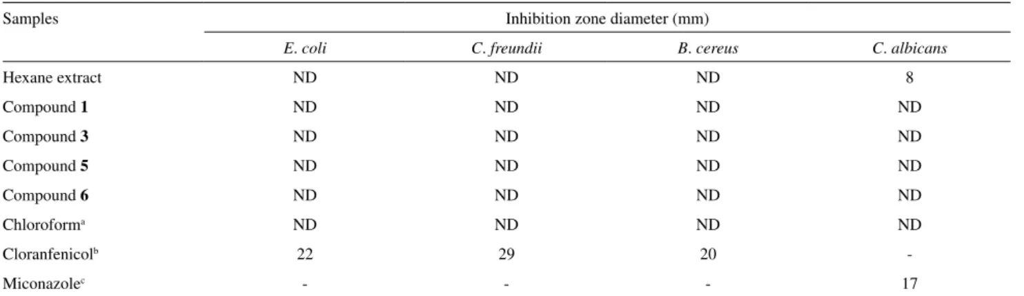
Antimicrobial activities have been described for pentacyclic triterpenes, such as oleananes,20 ursanes,20 friedelanes,21 and lupanes.22 It is speculated that the mechanism of action of triterpenes is due to a disruption on the microorganism’s cellular membrane.20,23 For this reason, the hexane extract and triterpenes 3-oxofriedelane (1), 3-oxo-12α-hydroxyfriedelane (3), 3,16-dioxofriedelane (5) and 3-oxo-12α,29-dihydroxyfriedelane (6) were tested against standard bacteria strains of Escherichia coli, Citrobacter freundii, Bacillus cereus and against the yeast
Candida albicans, using disk diffusion test. The hexane extract was moderately active on disk diffusion test as well as in the broth dilution test, presenting a MIC value of 512 µg/mL. From the tested triterpenes, 3-oxo-12α -hydroxyfriedelane (3) was active in the macrodilution test, presenting a MIC = 512 µg/mL against C. albicans. The activity of 3 was lower in comparison to that was produced by miconazole (16 µg/mL), and was not detected in the
disk diffusion test, probably because of low polarity of this compound.24
Supplementary Information
Spectra (IR, 1D/2D NMR and EM) and Tables of antimicrobial test results are available free of charge at http://jbcs.org.br as PDF ile.
Acknowledgments
The authors thank the Fundação de Amparo a Pesquisa do Estado de Minas Gerais (FAPEMIG, Grants CEX APQ-1863-5.02/07 and PRONEX EDT-479/07), for inancial support. F. C. S. thanks Conselho Nacional de Desenvolvimento Cientíico e Tecnológico (CNPq) for scholarship.
References
1. Schaneberg, B. T.; Green, D. K.; Sneden, A. T.; J. Nat. Prod.
2001, 64, 624.
2. Cordeiro, P. J. M.; Vilegas, J. H. Y.; Lanças, F. M.; J. Braz. Chem. Soc.1999, 10, 523.
3. Sannomiya, M.; Vilegas, W.; Rastrelli, L.; Pizza, C.;
Phytochemistry1998, 49, 237.
4. Da Silva, M. S.; De Sousa, D. P.; Medeiros, V.M.; Folly, M. A. B.; Tavares, J. F.; Barbosa-Filho, J. M.; Biochem. Syst. Ecol. 2008, 36, 500.
5. Lindsey, K. L.; Budesinsky, M.; Kohout, L.; Staden, J. V.;
S. Afr. J. Bot.2006, 72, 473.
6. Andrade, S. F.; Comunello, E.; Noldin, V. F.; Monache, F. D.; Filho, V. C.; Niero, R.; Arch. Pharm. Res. 2008,
Silva et al. 949 Vol. 22, No. 5, 2011
7. Orabi, K. Y.; Al-Qasoumi, S. I.; El-Olemy, M.; Mossa, J. S.; Muhammad, I.; Phytochemistry2001, 58, 475.
8. Oliveira, M. L. G.; Duarte, L. P.; Silva, G. D. F.; Vieira Filho, S. A.; Knupp, V. F.; Alves, F. G. P.; Magn. Reson. Chem. 2007,
45, 895.
9. Wagner, H.; Bladt, S.; Plant Drug Analysis, Springer: Berlin, 1996.
10. Takahashi, J. A.; Castro, M. C. M.; Souza, G. G.; Lucas, E. M. F.; Bracarense, A. A. P.; Abreu, L. M.; Marriel, I. E.; Oliveira, M. S.; Floreano, M. B.; Oliveira, T. S.; J. Med. Mycol. 2008,
18, 198.
11. Lana, E. J. L.; Carazza, F.; Takahashi, J. A.; J. Agric. Food Chem. 2006, 54, 2053.
12. Mahato, S. B.; Kundu, A. P.; Phytochemistry1994, 37, 1517. 13. Agrawal, P. K.; Jain, D. C.; Prog. NMR Spectrosc.1992, 24, 1. 14. Salazar, G. D. C.; Silva, G. D. F.; Duarte, L. P.; Vieira Filho, S.
A.; Lula, I. S.; Magn. Reson. Chem. 2000, 38, 977.
15. Wandji, J.; Wansi, J. D.; Fuendjiep, V.; Dagne, E.; Mulholland, D. A.; Tillequin, F.; Fomum, Z. T.; Sondengam, B. L.; Nkeh, B. C.; Njamen, D.; Phytochemistry2000, 54, 811.
16. Patra, A.; Chaudhuri, S. K.; Magn. Reson. Chem. 1987, 25, 95. 17. Matos, F. J. A.; Introdução a Fitoquímica Experimental, UFC:
Fortaleza, Brasil, 1998.
18. Rhourri-Frih, B. ; Chaimbault, P. ; Claude, B. ; Lamy, C. ; André, P. ; Lafosse, M. ; J. Mass. Spectrom. 2009, 44, 71.
19. Patra, A.; Chaudhuri, S. K.; Rübgger, H.; J. IndianChem. Soc. 1990, 67, 394.
20. Saleem, M.; Nazir, M.; Ali, M. S.; Hussain, H.; Lee, Y. S.; Riaz, N.; Jabbar, A.; Nat. Prod. Rep.2010, 27, 238.
21. Chiozem, D. D.; Trinh-Van-Dufat, H.; Wansi, J. D.; Djama, C. M.; Fannang, V. S.; Seguin, E.; Tillequin, F.; Wandji, J.; Chem. Pharm. Bull. 2009, 57, 1119.
22. Awanchiri, S. S.; Trinh-Van-Dufat, H.; Shirri, J. C.; Dongfack, M. D. J.; Nguenang, G. M.; Boutefnouchet, S.; Fomum, Z. T.; Seguin, E.; Verite, P.; Tillequin, F.; Wandji, J.; Phytochemistry
2009, 70, 419.
23. Zellner, B. D.; Amorim, A. C. L.; Miranda, A. L. P.; Alves, R. J. V.; Barbosa, J. P.; Costa, G. L.; Rezende, C. M.; J. Braz. Chem. Soc.2009, 20, 322.
24. Araújo, F. M.; Passos, M. G. V. M.; Lima, E. O.; Roque, N. F.; Guedes, M. L. S.; Souza-Neta, L. C.; Cruz, F. G.; Martins, D.;
J. Braz. Chem. Soc.2009, 20, 1805.
Submitted: December 22, 2009
Supplementary Information
J. Braz. Chem. Soc., Vol. 22, No. 5, S1-S18, 2011. Printed in Brazil - ©2011 Sociedade Brasileira de Química
0103 - 5053 $6.00+0.00
S
I
*e-mail: [email protected]
Chemical Constituents from Branches of
Maytenus gonoclada
(Celastraceae) and
Evaluation of Antimicrobial Activity
Fernando C. Silva,a,c Lucienir P. Duarte,*,a Grácia D. F. Silva,a Sidney A. V. Filho,a,b
Ivana S. Lula,a Jacqueline A. Takahashic and William S. T. Sallumc
aNúcleo de Estudos de Plantas Medicinais and cLaboratório de Biotecnologia e Bioensaios,
Departamento de Química, Universidade Federal de Minas Gerais, Av. Antônio Carlos, 6627, Pampulha, 31270-901 Belo Horizonte-MG, Brazil
bEscola de Farmácia, Universidade Federal de Ouro Preto, Rua Costa Sena, 171,
35400-000 Ouro Preto-MG, Brazil
Table S1. Antimicrobial activities of hexane extract and compounds 1, 3, 5 and 6 (each concentration at 100 µg/mL)
Samples Inhibition zone diameter (mm)
E. coli C. freundii B. cereus C. albicans
Hexane extract ND ND ND 8
Compound 1 ND ND ND ND
Compound 3 ND ND ND ND
Compound 5 ND ND ND ND
Compound 6 ND ND ND ND
Chloroforma ND ND ND ND
Cloranfenicolb 22 29 20
-Miconazolec - - - 17
aNegative control; bPositive control (bacteria); cPositive control (fungus); ND (Not Detected).
Table S2. Antimicrobial activities of hexane extract and compounds 3
Samples Minimum Inhibitory Concentration (µg/mL)
E. coli C. freundii B. cereus C. albicans
Hexane extract ND ND ND 512
Compound 3 ND ND ND 512
DMSOa ND ND ND ND
Cloranfenicolb 8 4 8
-Miconazolec - - - 16
Chemical Constituents from Branches of Maytenus gonoclada (Celastraceae) J. Braz. Chem. Soc.
S2
Figure S1. IR spectrum of compound 1 (ATR).
Figure S2. 1H NMR spectrum of compound 1 (CDCl
Silva et al. S3 Vol. 22, No. 5, 2011
Figure S4. 13C NMR-DEPT spectrum of compound 1 (CDCl
3 + pyridine-d5, 100 MHz).
Figure S3. 13C NMR spectrum of compound 1 (CDCl
Chemical Constituents from Branches of Maytenus gonoclada (Celastraceae) J. Braz. Chem. Soc.
S4
Figure S6. 1H NMR spectrum of compound 2 (CDCl
3, 400 MHz).
Silva et al. S5 Vol. 22, No. 5, 2011
Figure S8. 13C NMR-DEPT spectrum of compound 2 (CDCl
3, 100 MHz).
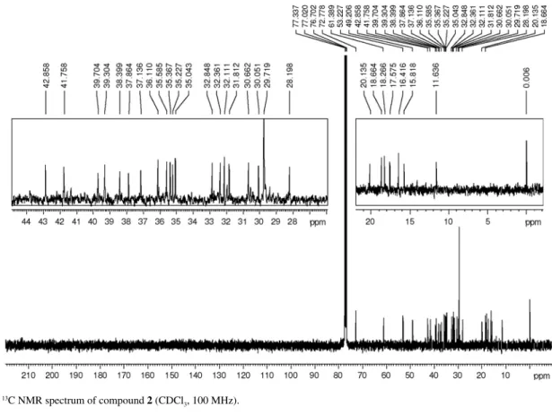
Figure S7. 13C NMR spectrum of compound 2 (CDCl
Chemical Constituents from Branches of Maytenus gonoclada (Celastraceae) J. Braz. Chem. Soc.
S6
Figure S10. 1H NMR spectrum of compound 3 (CDCl
3, 400 MHz).
Silva et al. S7 Vol. 22, No. 5, 2011
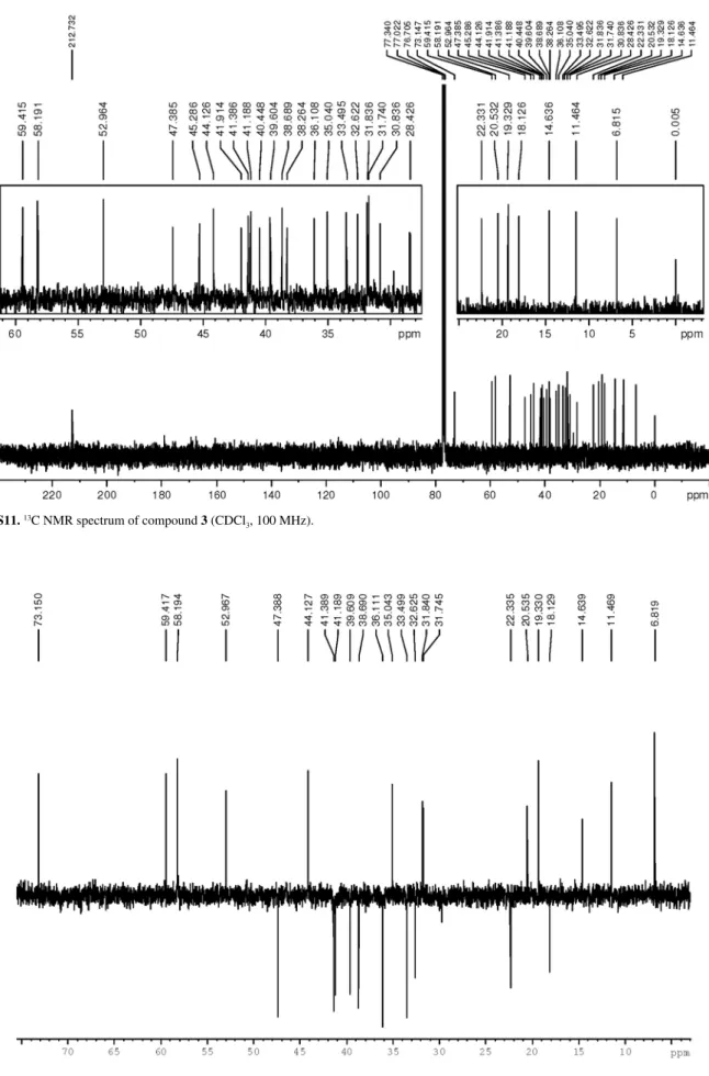
Figure S12. 13C NMR-DEPT spectrum of compound 3 (CDCl
3, 100 MHz).
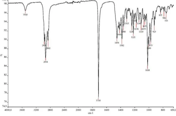
Figure S11. 13C NMR spectrum of compound 3 (CDCl
Chemical Constituents from Branches of Maytenus gonoclada (Celastraceae) J. Braz. Chem. Soc.
S8
Figure S14. 1H NMR spectrum of compound 4 and 5 (CDCl
3 + pyridine-d5, 400 MHz).
Silva et al. S9 Vol. 22, No. 5, 2011
Figure S16. 13C NMR-DEPT spectrum of compound 4 and 5 (CDCl
3 + pyridine-d5, 100 MHz).
Figure S15. 13C NMR spectrum of compound 4 and 5 (CDCl
Chemical Constituents from Branches of Maytenus gonoclada (Celastraceae) J. Braz. Chem. Soc.
S10
Figure S18. 1H NMR spectrum of compound 5 (CDCl
3, 400 MHz).
Silva et al. S11 Vol. 22, No. 5, 2011
Figure S20. 13C NMR-DEPT spectrum of compound 5 (CDCl
3, 100 MHz).
Figure S19. 13C NMR spectrum of compound 5 (CDCl
Chemical Constituents from Branches of Maytenus gonoclada (Celastraceae) J. Braz. Chem. Soc.
S12
Figure S22. HMBC spectrum of compound 5 (CDCl3, 400 MHz).
Silva et al. S13 Vol. 22, No. 5, 2011
Figure S24. IR spectrum of compound 6 (ATR).
Figure S23. 1H, 1H NOESY spectrum of compound 5 (CDCl
Chemical Constituents from Branches of Maytenus gonoclada (Celastraceae) J. Braz. Chem. Soc.
S14
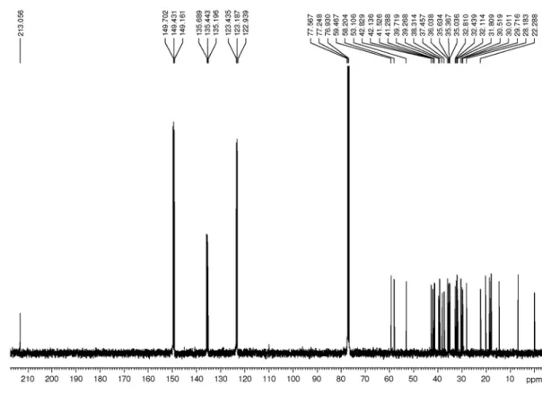
Figure S26. 13C NMR spectrum of compound 6 (CDCl
3 + pyridine-d5, 100 MHz).
Figure S25. 1H NMR spectrum of compound 6 (CDCl
Silva et al. S15 Vol. 22, No. 5, 2011
Figure S28. First expansion of HSQC spectrum of compound 6 (CDCl3 + pyridine-d5, 400 MHz).
Figure S27. 13C NMR-DEPT spectrum of compound 6 (CDCl
Chemical Constituents from Branches of Maytenus gonoclada (Celastraceae) J. Braz. Chem. Soc.
S16
Figure S30. First expansion of HMBC spectrum of compound 6 (CDCl3 + pyridine-d5, 400 MHz).
Silva et al. S17 Vol. 22, No. 5, 2011
Figure S32. First expansion1H, 1H NOESY spectrum of compound 6 (CDCl
3 + pyridine-d5, 400 MHz).
Chemical Constituents from Branches of Maytenus gonoclada (Celastraceae) J. Braz. Chem. Soc.
S18
Figure S34. ESI-mass spectrum of compound 6.
Figure S33. Second expansion1H, 1H NOESY spectrum of compound 6 (CDCl