www.jped.com.br
ORIGINAL
ARTICLE
Prevalence
and
characterization
of
neonatal
skin
disorders
in
the
first
72
h
of
life
夽
,
夽夽
Flávia
Pereira
Reginatto
a,∗,
Damie
DeVilla
b,
Fernanda
M.
Muller
b,
Juliano
Peruzzo
c,
Letícia
P.
Peres
c,
Raquel
B.
Steglich
b,
Tania
F.
Cestari
a,caUniversidadeFederaldoRioGrandedoSul(UFRGS),ProgramadePós-graduac¸ãoemSaúdedaCrianc¸aeAdolescente,Porto
Alegre,RS,Brazil
bComplexoHospitalarSantaCasadePortoAlegre,DepartamentodeDermatologia,PortoAlegre,RS,Brazil cUniversidadeFederaldoRioGrandedoSul(UFRGS),HospitaldeClínicasdePortoAlegre(HCPA),Departamentode
Dermatologia,PortoAlegre,RS,Brazil
Received9May2016;accepted16June2016 Availableonline19November2016
KEYWORDS Newborn; Neonatology; Childhealthservices; Childhealth;
Infantcare; Skinmanifestations
Abstract
Objective: To determine the prevalence of neonatal dermatological findings and analyze
whetherthereisanassociationbetweenthesefindingsandneonatalandpregnancy
charac-teristicsandseasonality.
Methods: NewbornsfromthreematernityhospitalsinaBraziliancapitalcitywererandomly
selectedtoundergodermatologicalassessmentbydermatologists.
Results: 2938neonates ageduptothreedaysoflifewererandomlyselected,ofwhom309
wereexcludedduetoIntensiveCareUnitadmission.Ofthe2530assessedneonates,49.6%were
Caucasians,50.5%weremales,57.6%werebornbyvaginaldelivery,and92.5%ofthe
moth-ersreceivedprenatalcare.Somedermatologicalfindingwasobservedin95.8%ofneonates;of
these,88.6%hadtransientneonatalskinconditions,42.6%hadcongenitalbirthmarks,26.8%had
somebenignneonatalpustulosis,2%hadlesionssecondarytotrauma(includingscratches),0.5%
hadskinmalformations,and0.1%hadaninfectiousdisease.Themostprevalent
dermatologi-calfindingswere:lanugo,whichwasobservedin38.9%ofthenewborns,sebaceoushyperplasia
(35%),dermalmelanocytosis(24.61%),skindesquamation(23.3%),erythema toxicum
neona-torum(23%),salmonpatch(20.4%),skinerythema(19%),genitalhyperpigmentation(18.4%),
eyelidedema(17.4%),milia(17.3%),genitalhypertrophy(12%),andskinxerosis(10.9%).
夽
Pleasecitethisarticleas:ReginattoFP,DeVillaD,MullerFM,PeruzzoJ,PeresLP,SteglichRB,etal.Prevalenceandcharacterizationof neonatalskindisordersinthefirst72hoflife.JPediatr(RioJ).2017;93:238---45.
夽夽
StudyconductedatthePostgraduatePrograminChildandAdolescentHealth,UniversidadeFederaldoRioGrandedoSul(UFRGS);and attheHospitaldeClínicasdePortoAlegre(HCPA),UniversidadeFederaldoRioGrandedoSul(UFRGS),PortoAlegre,RS,Brazil.
∗Correspondingauthor.
E-mail:[email protected](F.P.Reginatto).
http://dx.doi.org/10.1016/j.jped.2016.06.010
Conclusions: Dermatological findings are frequent duringthe first days oflife and some of
them characterize thenewborn’s skin.Mixed-race newborns andthose whose mothers had
somegestationalriskfactorhadmoredermatologicalfindings.Thegestationalage,newborn’s
ethnicity,gender,Apgaratthefirstandfifthminutesoflife,typeofdelivery,andseasonality
influencedthepresenceofspecificneonataldermatologicalfindings.
©2016SociedadeBrasileiradePediatria.PublishedbyElsevierEditoraLtda.Thisisanopen
accessarticleundertheCCBY-NC-NDlicense(http://creativecommons.org/licenses/by-nc-nd/
4.0/).
PALAVRAS-CHAVE Recém-nascido; Neonatologia; Servic¸osdesaúdeda crianc¸a;
Saúdedacrianc¸a; Cuidadodolactente; Manifestac¸ões cutâneas
Prevalênciaecaracterizac¸ãodasafecc¸õescutâneasneonataisnasprimeiras72horas devida
Resumo
Objetivo: Verificar a prevalência dos achados dermatológicosnos primeiros dias de vidae
analisarseháassociac¸ãocomcaracterísticasneonatais,gestacionaisesazonalidade.
Métodos: Recém-nascidosdetrêsmaternidadesdeumacapitalbrasileiraforamselecionados
aleatoriamenteparaseremsubmetidosaoexamedermatológicorealizadopordermatologistas.
Resultados: Foram selecionados aleatoriamente 2839 neonatos com até 72 horas de vida,
309 foram excluídos por terem sido admitidos em Unidade de Tratamento Intensivo. Dos
2530neonatosexaminados49,6%eramdarac¸abrancae50,5%dosexomasculino.Foi
obser-vadoalgumachadodermatológicoem95,8%dosrecém-nascidos;destes,88,6%tinhamlesões
cutâneastransitóriasneonatal,42,6%marcadenascimento,26,8%tinhampustulosebenigna
neonatal, 2%lesõessecundáriasao trauma,0,5%malformac¸ãocutânea e0,1%doenc¸a
infec-ciosa.Oachado dermatológicomaisfrequente foiolanugo,quefoiobservadoem38,9%dos
neonatos,seguidopelahiperplasiadeglândulassebáceas(35%),melanocitosedérmica(24,6%),
descamac¸ãodapele(23,3%),eritematóxiconeonatal(23%),manchasalmão(20,4%),eritema
dapele(19%),hiperpigmentac¸ãodagenitália(18,4%),edemapalpebral(17,4%),cistosdemília
(17,3%),hipertrofiadagenitália(12%)exerosecutânea(10,9%).
Conclusões: Osachadosdermatológicossãofrequentementeidentificadosnosprimeirosdiasde
vidaemuitosdelescaracterizamapeledorecém-nascido.Osneonatospardoseaquelescujas
mães apresentavamalgum fator deriscogestacional tiverammais achadosdermatológicos.
A idade gestacional, aetnia do neonato,ogênero, oíndice de Apgar, otipode partoe a
sazonalidadeinfluenciaramnapresenc¸ademanifestac¸õescutâneasespecíficas.
©2016SociedadeBrasileiradePediatria.PublicadoporElsevierEditoraLtda.Este ´eumartigo
OpenAccesssobumalicenc¸aCCBY-NC-ND(http://creativecommons.org/licenses/by-nc-nd/4.
0/).
Introduction
The neonatalperiodis atimeof adjustmentwhen patho-logicalandphysiologicalreactionsareoftenconfused;skin changesarecommoninthisperiod.1---4Someauthorsreport
that 57---99.4% of newborns (NB) have some type of skin alteration and 84% have more than one dermatological finding.3,5,6 Thefrequencyoftheseeventsdiffersbetween
differentracialgroups.1,3
Most of these lesionsare transient and specific to the neonatal period, such as lanugo, sebaceous gland hyper-plasia,andskindesquamation.5Birthmarks,suchassalmon
patchanddermalmelanocytosis,areusuallyidentifiedsoon afterbirth,largelyresultingfromthenewbornskin matu-rationordeepeningofskinpigmentationovertime.2Cases
of neonatalbenign pustulosis,which areusuallyobserved within the first weeks of life, include erythema toxicum neonatorum (ETN), transient neonatal pustular melanosis (TNPM),andbenigncephalicpustulosis(BCP).7---9Regarding
thedevelopmentalabnormalitiesthatoccurintheskinand canbeobservedintheneonatalperiod,themostfrequent
are:aplasiacutis,fistulasandcysts,supernumerarynipple, andperimamillar adnexalpolyp.10 Moreover,the
introduc-tionofnewtechnologiesandapproachesinnewborncare,in additiontoskinfragilityatthisage,causeskincomplications tobemorefrequentlyobserved.11
Studies to estimate the frequency of skin disorders in newbornshavebeen performed inseveralcountries; how-ever,thissubjecthasbeenscarcelystudiedinBrazil.Thus, thepresentstudyaimedtoincreasemedicalknowledgeon neonateskin characteristics, aswellastoassociate them withneonatal,pregnancy,andseasonalityfactors.
Materials
and
methods
Pepi4-Random --- Procedures using Random Numbers pro-gram, version 4.0 (WINPEPI, PEPI-for-Windows, NY, USA), wasusedto randomizeeight monthly datesfor one year. Thestudyincludedallneonates bornonthedays random-izedfordatacollection,whosetutorsorparentsagreedto participateandsignedtheinformedconsentform.Neonates admittedtotheintensivecareunit(ICU)wereexcludedfrom thestudyanalysis.
Dermatological assessment was performed by derma-tologistswith standardized training and with the help of dermatoscopy.The re-assessmentswerecarriedoutinthe ClinicalResearchCenterofHCPA.Thestudywasapproved bytheResearchEthicsCommitteeofthethreeinstitutions whereitwasperformed:HCPA---Project1101013;Hospital SantaCasadePortoAlegre---Protocol3513/1,andHospital Fêmina---Project11-065.
Dermatological findings were classified as transient neonatal skin lesions, birthmarks, benign neonatal pus-tulosis, hemangioma or hemangioma precursors, lesions secondarytotrauma,skinmalformation,andinfectious dis-ease.
DatawereanalyzedusingtheSPSSv.20.0program(IBM Corp.Released2013.IBMSPSSStatisticsfor Windows, Ver-sion22.0. NY, USA). Categorical variables were described byfrequenciesandpercentages,andtheprevalencebythe 95%confidenceintervals.Inthesecondaryanalysisofdata, categoricalvariableswereassociatedwitheachotherusing thechi-squared test, and the quantitative variableswere comparedbytheStudent’st-testforindependentsamples. AmultivariablePoissonregressionwithrobustvariancewas performed to evaluate possible associations between dif-ferentstudyfactorsandlesionsandtoadjustforpotential confounders.Thesignificancelevelwassetbetween2%and 5%.
Results
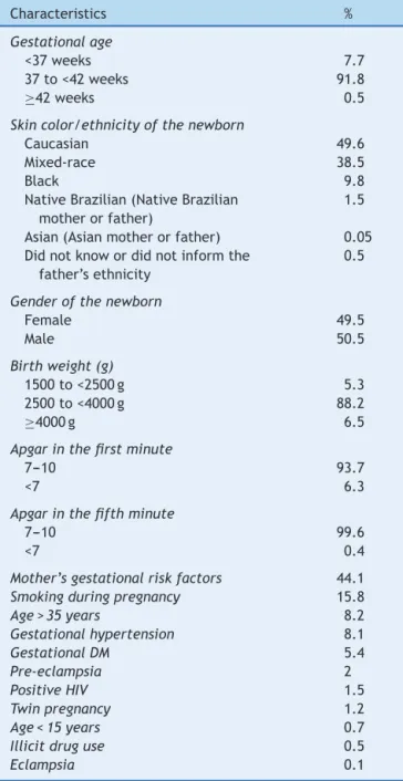
Ofthe2839newbornsselectedtoparticipateinthestudy, 309wereexcludedforhavingbeenadmittedtotheICU.The meangestationalage was38.41 weeks(median39;25---50 and75 percentiles: 38---39 and40, respectively); the pre-dominantethnicitywasCaucasian;and57.6%werebornby vaginaldelivery.Regardingthemothers,92.5%received pre-natalcare, 59.8% were multiparous, and 44.1% had some gestationalrisk factor (GRF). Table 1 shows the neonatal characteristicsandtheGRFs.
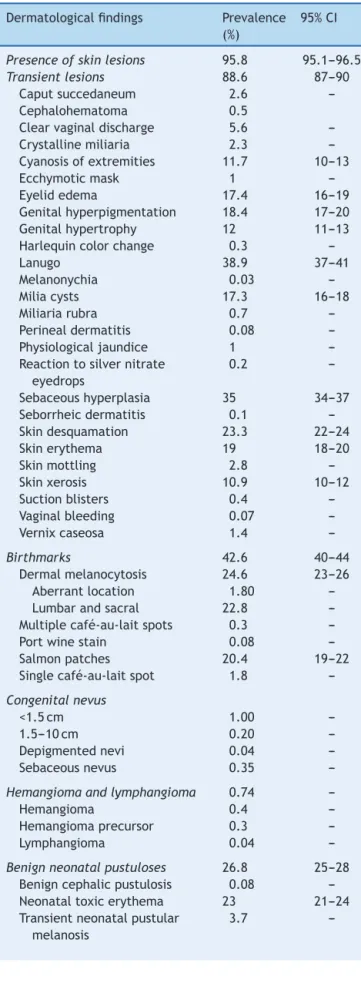
Oftheassessednewborns,95.8%hadsome dermatolog-icalfinding. Of these,88.6% had some transient neonatal skinmanifestations,42.6%hadbirthmarks,26.8%had neona-tal benignpustulosis, 2% hadsome skin lesionssecondary totrauma, 0.5% had skin malformation,and 0.1% had an infectiousdisease(Table2).Ameanof3.23dermatological findingswasobservedperneonatewithsometypeof skin manifestation.
The most common dermatological finding was lanugo, foundin38.9%ofinfants,followedbysebaceous hyperpla-sia(35%),dermalmelanocytosis(24.6%),skindesquamation (23.3%), ETN (23%), salmon patch (20.4%), skin erythema (19%), genital hyperpigmentation (18.4%), eyelid edema (17.4%), milia cysts (17.3%), genital hypertrophy (12%),
Table1 Characteristicsofnewbornswithoutadmissionto
theNICUandgestationalriskfactorsinmaternityhospitals
ofPortoAlegre.
Characteristics %
Gestationalage
<37weeks 7.7
37to<42weeks 91.8
≥42weeks 0.5
Skincolor/ethnicityofthenewborn
Caucasian 49.6
Mixed-race 38.5
Black 9.8
NativeBrazilian(NativeBrazilian
motherorfather)
1.5
Asian(Asianmotherorfather) 0.05
Didnotknowordidnotinformthe
father’sethnicity
0.5
Genderofthenewborn
Female 49.5
Male 50.5
Birthweight(g)
1500to<2500g 5.3
2500to<4000g 88.2
≥4000g 6.5
Apgarinthefirstminute
7---10 93.7
<7 6.3
Apgarinthefifthminute
7---10 99.6
<7 0.4
Mother’sgestationalriskfactors 44.1
Smokingduringpregnancy 15.8
Age>35years 8.2
Gestationalhypertension 8.1
GestationalDM 5.4
Pre-eclampsia 2
PositiveHIV 1.5
Twinpregnancy 1.2
Age<15years 0.7
Illicitdruguse 0.5
Eclampsia 0.1
DM,diabetesmellitus;HIV,humanimmunodeficiencyvirus.
cyanosisoftheextremities(11.7%),andskinxerosis(10.9%) (Figs.1and2).
Table2 Prevalenceofdermatologicalfindingsofnewborns
without admission to the neonatal intensive care unit in
maternityhospitalsofPortoAlegre.
Dermatologicalfindings Prevalence
(%)
95%CI
Presenceofskinlesions 95.8 95.1---96.5
Transientlesions 88.6 87---90
Caputsuccedaneum 2.6
---Cephalohematoma 0.5
Clearvaginaldischarge 5.6
---Crystallinemiliaria 2.3
---Cyanosisofextremities 11.7 10---13
Ecchymoticmask 1
---Eyelidedema 17.4 16---19
Genitalhyperpigmentation 18.4 17---20
Genitalhypertrophy 12 11---13
Harlequincolorchange 0.3
---Lanugo 38.9 37---41
Melanonychia 0.03
---Miliacysts 17.3 16---18
Miliariarubra 0.7
---Perinealdermatitis 0.08
---Physiologicaljaundice 1
---Reactiontosilvernitrate
eyedrops
0.2
---Sebaceoushyperplasia 35 34---37
Seborrheicdermatitis 0.1
---Skindesquamation 23.3 22---24
Skinerythema 19 18---20
Skinmottling 2.8
---Skinxerosis 10.9 10---12
Suctionblisters 0.4
---Vaginalbleeding 0.07
---Vernixcaseosa 1.4
---Birthmarks 42.6 40---44
Dermalmelanocytosis 24.6 23---26
Aberrantlocation 1.80
---Lumbarandsacral 22.8
---Multiplecafé-au-laitspots 0.3
---Portwinestain 0.08
---Salmonpatches 20.4 19---22
Singlecafé-au-laitspot 1.8
---Congenitalnevus
<1.5cm 1.00
---1.5---10cm 0.20
---Depigmentednevi 0.04
---Sebaceousnevus 0.35
---Hemangiomaandlymphangioma 0.74
---Hemangioma 0.4
---Hemangiomaprecursor 0.3
---Lymphangioma 0.04
---Benignneonatalpustuloses 26.8 25---28
Benigncephalicpustulosis 0.08
---Neonataltoxicerythema 23 21---24
Transientneonatalpustular
melanosis
3.7
---Table2 Continued)
Dermatologicalfindings Prevalence
(%)
95%CI
Infectiousdiseases 0.14
---Bullousimpetigo 0.04
---Congenitalsyphilis 0.12
---Skinmalformations 0.54
---Accessorytragusorauricle 0.03
---Cleftlip 0.04
---Congenitaltalipesequinovarus 0.08
---Epispadiasorhypospadias 0.12
---Helicalearmalformation 0.08
---Periaerolaradnexalpolyp 0.40
---Periauricularfistula 0.04
---Supernumeraryfinger 0.04
---Supernumerarynipple 0.08
---Traumalesion 2.0
---Bruisingbypuncturing 0.12
---Claviclefracture 0.03
---Excoriationsbyscratching 1.14
---Traumabyforcepsorscalpel 0.59
---Ahigherprevalence oflanugowasobservedinpreterm newborns(p<0.001).Thefull-terminfantshadmorebenign neonatal pustuloses and milia cysts (p<0.001), whereas post-term newborns had a higher prevalence of transient skinlesions(p=0.001), suchasgenitalhyperpigmentation (p=0.001)andskinxerosis(p=0.02),aswellasbirthmarks (p<0.001), such as Mongolian spots and salmon patches (p<0.001). ETNwaspositivelyassociated withgestational age(p<0.001).
Regarding the newborn’s ethnicity, it was observed thatCaucasian newborns hada higher prevalence of ETN (p<0.001), as well as some transitional lesions, such as miliacysts(p=0.005),skinerythema(p<0.001),sebaceous hyperplasia (p=0.005), and salmon patches (p=0.017). Black newborns showed a higher prevalence of Mongo-lian spots, skin desquamation in the extremities, genital hyperpigmentation, and xerosis (p<0.001). In mixed-race newborns, transientskin findings had ahigher prevalence (p<0.001), such asgenitalhypertrophy (p<0.001). As for gender,itwasobservedthatfemalenewbornshadahigher prevalenceoftransientfindings(p=0.026),whereasmales hadahigherprevalenceofETN(p=0.002)andgenital hyper-pigmentation(p<0.001).
RegardingtheApgar inthefirstminuteoflife,ahigher prevalence of ETN was observed in neonates with Apgar scorebetween8and10(p<0.001);asfortheApgarscore at5minoflife,neonateswithApgarbetween0and3hada higherprevalenceoflanugo.
Figure1 (A)Lanugo;(B)miliacystsonthenasaldorsum;(C)skinerythema;(D)dermalmelanocytosis.
Figure2 (A)Poliosis;(B),melanocyticnevusonthescalp;(C)neonateskindesquamation;(D)hemangioma.
performed more frequently in infants weighing between 2500 and 3999g and in full-term or post-term newborns (p<0.001).
WhenevaluatingtheGFR,itwasobservedthatneonates borntomotherswithsomeriskfactorhadahigher preva-lenceofdermatologicalfindings(p=0.047).Thisdifference wasalsoobservedfortransientneonatallesions(p<0.001). Thenewbornsofmotherswithnoriskfactorshadahigher prevalenceofbirthmarks(0.016)andETN(p<0.001).Infants
Regarding the seasonality, it was observed that there were more transient lesions in summer (p=0.008), such assebaceoushyperplasia(p=0.049)andxerosis(p<0.001). TheprevalenceofETNwashigherinthespring(p=0.004). The prevalence of eyelid edema (p=0.001) and skin ery-thema (p=0.001) was higher in the fall; in the winter months,skindesquamation(p<0.001)andgenital hypertro-phy(p<0.001)weremoreprevalent.
Discussion
Several authors have assessed the prevalence of neona-taldermatologicalfindingsindifferentcountries.However, thereisnohomogeneityregardingtheterminologyandthe design used in the studies, which makes the comparison between them difficult. No other authors evaluated the seasonalityofneonataldermatologicalfindings,whichthis study did, for the first time. Despite the methodological differencesbetweenthestudies,theprevalenceof derma-tologicalfindingsinnewbornswassimilartothatdescribed by other authors (90%). However,it was higher than that reported by authors in Turkey, whofound a frequency of 67.3%ofskinfindingsin1234newbornsupto48hoflife.6
Physiologicaldesquamationisobservedinmostnewborns andit is usuallymoreintense betweenthe sixthand sev-enthdaysoflife.12 Theresultsof thepresentstudy differ
fromotherpublisheddata,astheratewaslowerthanthat reported by authors whoassessed newborns within seven daysoflife,12andhigherthanthatreportedbyauthorswho
evaluatednewbornsupto48hoflife.3Theauthorsbelieve
thatthisdifferencewasduetothefactthatthe dermato-logical examinationwasperformedwithinthe first72hof life,duringwhichthisfindingisusuallylessprevalent.
The prevalence of salmon patch was lower than that reportedby some authors, whoobserved a prevalence of 26---83%,3,13,14 andsimilartothatreportedby others,such
asthestudyperformedinTurkey,whichobserveda preva-lenceof19.2%.15 Theprevalence ofdermal melanocytosis
washigherthanthatdescribedforCaucasianchildren,which wasreportedtooccurinlessthan10%ofnewborns,2,13
prob-ablyduetothemiscegenationcharacteristicsofthestudy population.Boththesalmonpatchanddermal melanocyto-sisshowedapositivecorrelationwithgestationalageand, despitethe smallnumberofpost-term newborns assessed in the present study, it may indicate that the birthmarks areamarkerofskinmaturityintheneonate.Moreover,the differenceobserved withdatapublished by other authors may indicate that the population characteristics have an importantinfluenceonthepresenceofbirthmarks.
Theprevalenceofmiliacystswassimilartothatfoundin Spanishnewborns,of16.6%16;however,itwashigherthan
thatreportedbyAmericanauthors,whichwasof8%.13Milia
cystsweremoreprevalentinfull-termneonates;theycan alsobeanindicatorofskinmaturity.
Alowerprevalenceofvernixcaseosawasobservedwhen comparedwiththatreportedinSpanishnewborns,42.9%.16
Theauthorsbelievethatthisdifferenceisduetothefact thatmostassessednewbornshadalreadyreceivedthefirst bath,whichinthematernityhospitalswherethestudywas carriedoutusuallyoccursbetween24and48hafterbirth.
Ahigher prevalenceof benign neonatalpustuloses was observed in the first 48h of life than expected.17,18 The
prevalenceofETNwassimilartothatreportedina prospec-tive study carried out in Spain with 365 infants, which found a prevalence of 25.3%.19 However,it wasobserved
thatETNoccurredmoreoftenthanexpectedfornewborns withinthefirst48hoflifewhencomparedwithastudy per-formedin California,which showedaprevalence of 7%of ETNinneonateswithupto48hoflife.13Unliketheauthors
who reported that ETN was more prevalent in cesarean sectiondeliveries4,6andotherswhoobservedahigher
preva-lence of ETN in vaginal births,20 no differences between
thetypeof delivery andfrequency of ETNwereobserved in the present study. Furthermore,TNPM prevalence was higherthan expectedfor a predominantly Caucasian pop-ulation,probablyduetothemiscegenationcharacteristics ofthestudypopulation.ETNwaslessprevalentinthewinter months,similartothatreportedbyotherauthorswho indi-catedthatthisconditionismorecommoninhotandhumid climates.21 ItwasalsoobservedthatETNwasmore
preva-lentin infants bornunder optimalconditionsand without GRF,whichmayindicatethatthisisadermatologicalfinding ofhealthynewborns.
Unlikeother studies,20 noassociationsbetween
prema-turity and genital hyperpigmentation or cutis marmorata wereobserved. Incontrast, ahigher frequency of genital hyperpigmentation was observed in post-term newborns; thiscould also be an indicator of skin maturity. No asso-ciation was found between the newborn’s maturity and sebaceoushyperplasiaandskindesquamation,asreported byotherauthors.16
The frequency of neonataldermatological findings was higher in infants of mothers with GRF due to neonatal transitionallesions,whichindicates that certainmaternal comorbiditiesmayinfluencetheskin ofnewborns,suchas diabetesandgestationalhypertension,which were associ-atedwithskinerythemaandgestationalhypertensionwith genitalhypertrophy.Ahigherprevalenceof genital hyper-trophywasalsoobservedinnewbornsofmotherswhohad gestationalhypertension.Other authors found an associa-tionbetween genital hypertrophy and gestationaldisease andmedicationuseduringpregnancy22;however,thedrugs
usedduringpregnancywerenotevaluatedinthisstudy. TheprevalenceofpositiveserologyforHIVinpregnancy was higher than that expected for women of all ages in Brazil,whichis0.42%;itwashigherthanthatreportedby otherauthorsforpregnantwomenbetween15and24years of age treated in Brazilian hospitals (0.7%),23 and it was
alsohigherthanthatreportedinpregnantwomeninother regionsofBrazil.24,25 Althoughtheprevalence ofHIV
posi-tivitywashigh whencomparedtoother regions ofBrazil, thisfacthadnoassociationwithskinlesions.
Theprevalenceofinfectiouslesionsinthepresentstudy waslowerthanthatreportedfornewbornsinneonatalICUs (4%),16 probablybecauseonlyneonatesinrooming-inwere
included.
humidweathercharacteristicofwintermonthsmay precip-itateskindesquamationinneonates.
Dermatologicalfindingsareoftenidentifiedinnewborns, andmanyofthemcharacterize thenewborn’s skin,which justifiesadetaileddermatologicalexamination.24 Theskin
findings most commonly observed in neonates are gen-erally transient and result from the normal physiological responses.Theyareusuallylimitedtothefirstdaysorweeks of life and, therefore, they are rarely evaluated by der-matologists.The correctidentification ofthese findingsis importanttohelptodifferentiatethem frompathological findings,avoidunnecessarydiagnosticandtherapeutic pro-cedures,aswell asdecreasethe concerns of parentsand assistants.4
Thismulticenterstudyofdatacollectedduringoneyear ina city withwell-defined seasons, usingtheappropriate dermatological nomenclature and with skin examinations performed by specialists,provided importantdata onthe prevalenceofdermatologicalfindingsinthefirst72hoflife, aswellastheinfluenceofethnicity,GRF,andseasonality.
A high prevalence of neonatal dermatological findings wasobserved in the first 72h of life in the region where the study was performed, highlighting the importance of adetaileddermatologicalexamination.Theneonate’s eth-nicityandmaternal gestationalriskfactorsinfluencedthe presenceofdermatologicalfindings.Furthermore,an asso-ciationwasobservedbetween specificskin manifestations and certain neonatal characteristics, such as gestational age,ethnicity,gender,andApgarscoreinthefirstandfifth minutesoflife,aswellaswithgestationalcharacteristics, suchasthetypeofdelivery.Themostprevalent dermato-logicalfindingswerealsodistinctinthedifferentseasonsin theyear.
Thedifferencesintheprevalenceofskinmanifestations incomparisonwithother studiesshowthatthepopulation characteristicsandtheperiodduringwhichtheskinofthe newbornwasassessedmayinfluencethepresence of der-matologicalfindings,butthisassociationisnotclear;more specificstudiesarenecessary.
Funding
ThestudywasfundedbyFundodeIncentivoaPesquisado HospitaldeClínicasdePortoAlegre(FIPE).
Conflicts
of
interest
Theauthorsdeclarenoconflictsofinterest.
Acknowledgements
WewouldliketothankVânia NaomiHirakataandDaniela Benzanofor thehigh-quality statisticalanalysisof datain thisarticle.Theauthorswouldalsoliketothankthe physi-cians Ana Carolina Saraiva Camerin, Fabiana de Oliveira Bazanella, Kalyanna Gil Portal, Renata Rosa, Rodrigo Piz-zoni,and Samanta DaianaDe Rossi, for their crucialhelp toenterdataintoExcelspreadsheets.Finally,theauthors wouldlike tothankthe medicaland nursing staffsof the hospitalswherethestudywasperformedandespeciallythe
parentsofnewbornswhoallowedtheirnewborns’skintobe examined,enablingtheperformanceofthisstudy.
References
1.LarraldeM,GiachettiA,BaselgaE,GrecoMF,FriedenL. Enfer-midadesneonatales.In:LarraldeM,AbdadME,LunaPC,editors. Dermatologíapediátrica.2nded.BuenosAires:Journal;2010. p.13---53.
2.MoosaviZ, Hosseini T. One-year survey of cutaneous lesions in 1000 consecutive Iranian newborns. Pediatr Dermatol. 2006;23:61---3.
3.GokdemirG,ErdoganHK,KosluA,BaksuB.Cutaneouslesionsin Turkishneonatesborninateachinghospital.IndianJDermatol VenereolLeprol.2009;75:638.
4.BautistaR,LlopM.Reciénnacido:lesionescutáneasbenignas transitorias.In:BaselgaE,AsínHLE,SabatéJF,editors. Proto-colosdedermatología.2nded.Madrid:AEP;2007.p.309---16. 5.EkizO, GülU,Mollamahmuto˘gluL, GönülM.Skinfindingsin
newbornsandtheirrelationshipwithmaternalfactors: obser-vationalresearch.AnnDermatol.2013;25:1---4.
6.BenjaminLT.Birthmarksofmedicalsignificanceintheneonate. SeminPerinatol.2013;37:16---9.
7.GhoshS.Neonatalpustulardermatosis:anoverview.IndianJ Dermatol.2015;60:211.
8.BrazzelliV,GrassoV,CrociG,FigarT,BorroniG.Anunusualcase oftransientneonatalpustularmelanosis:adiagnosticpuzzle. EurJPediatr.2014;173:1655---8.
9.Reginatto FP, De Villa D, Cestari TF. Afecc¸ões cutâneas neonataisbenignascompresenc¸adepústulas.AnBras Derma-tol.2016;91:124---34.
10.BelletJS.Developmentalanomaliesoftheskin.Semin Perina-tol.2013;37:20---5.
11.FonteneleFC,CardosoMV.Skinlesionsinnewbornsinthe hos-pitalsetting:type,sizeandaffectedarea.RevEscEnfermUSP. 2011;45:130---7.
12.MonteagudoB,LabandeiraJ,Leon-MuinosE,RomarisR, Caban-illasM,Gonzalez-VilasD,etal.Physiologicaldesquamationof thenewborn:epidemiologyandpredisposingfactors.Actas Der-mosifiliogr.2011;102:391---4.
13.KanadaKN,MerinMR,Munden A, FriedlanderSF.A prospec-tive study of cutaneous findings in newborns in the United States: correlationwith race,ethnicity,and gestational sta-tususingupdated classificationandnomenclature. JPediatr. 2012;161:240---5.
14.FerahbasA, UtasS,AkcakusM,GunesT,MistikS.Prevalence ofcutaneousfindingsinhospitalizedneonates:a prospective observationalstudy.PediatrDermatol.2009;26:139---42. 15.MonteagudoB,LabandeiraJ,CabanillasM,AcevedoA,
Leon-MuinosE,ToribioJ.Prevalenceofmiliaandpalatalandgingival cystsinSpanishnewborns.PediatrDermatol.2012;29:301---5. 16.MonteagudoB, Labandeira J, Acevedo A, Ramirez-Santos A,
CabanillasM,CorralesA,etal.Prevalenceandclinicalfeatures ofcongenitalmelanocyticneviin1000Spanishnewborns.Actas Dermo-sifiliogr.2011;102:114---20.
17.GonzalezEcheverria F, Martinez Rodriguez J, Ancin Chandia T,CordobaIturriagaA. Isneonataltoxicerythemaarisk fac-tor in the development of allergy in childhood? An Pediatr. 1997;47:515---20.
18.YamasakiO,ManabeK,MorimotoA,IwatsukiK.Pustular ery-thematoxicumneonatorumin twosiblingsbornto amother with group B streptococcus colonization. Eur J Dermatol. 2011;21:271---2.
20.EkizO, GulU,MollamahmutogluL, GonulM.Skinfindingsin newbornsandtheirrelationshipwithmaternalfactors: obser-vationalresearch.AnnDermatol.2013;25:1---4.
21.LiuC, Feng J, QuR, Zhou H, Ma H, NiuX, et al. Epidemi-ologicstudyofthepredisposingfactors inerythematoxicum neonatorum.Dermatology.2005;210:269---72.
22.Boccardi D, Menni S, Ferraroni M, Stival G, Bernardo L, La Vecchia C, et al. Birthmarks and transient skin lesions in newbornsand their relationship to maternal factors:a pre-liminaryreport from northern Italy. Dermatology. 2007;215: 53---8.
23.MirandaAE,PintoVM,McFarlandW,PageK.HIVInfectionamong youngpregnantwomeninBrazil:prevalenceandassociatedrisk factors.AIDSBehav.2014;18Suppl1:S50---2.
24.Ferezin RI, Bertolini DA, Demarchi IG. Prevalence of posi-tivesorologyforHIV,hepatitisB,toxoplasmosisandrubellain pregnantwomenfromthenorthwesternregionofthestateof Parana.RevBrasGinecolObstet.2013;35:66---70.