In Vivo
Magnetic Resonance Techniques and Drug Discovery
Nicolau Beckmann
Novartis Institutes for BioMedical Research, Discovery Technologies, Lichtstr. 35, WSJ-386.2.09, CH-4002 Basel, Switzerland
Received on 5 October, 2005; accepted on 5 November, 2005
The long and resource intensive process of drug discovery and development is confronted with the basic challenge of providing effective and safe therapies at reasonably low costs. The better the mechanism of a disease is known, the higher the probability to find an appropriate therapy. Also, the better and earlier a disease can be diagnosed and characterized, the higher the chance to be able to interfere in this process with a chemical entity. This reasoning sets the framework for the use of imaging in drug discovery. We discuss the relevance of magnetic resonance imaging and spectroscopy to derive anatomical, functional, metabolic and target-related information in the context of pharmacological researchin vivo.
Keywords: Magnetic resonance; Imaging; Drug discovery
I. INTRODUCTION
The process to discover and to bring a drug to market is complex, costly and long, requiring in average 12 years. It begins with the identification and validation of a potential drug target. High affinity binders are searched for by us-ing high-throughput screenus-ing (HTS). Compounds that have passed some initial selectivity filters are then further evalu-ated. Despite recent significant investments in biology related areas such as genomics and proteomics, and in technology platforms designed to increase the number of compounds as-sayed, the development time and attrition rate in clinical trials remained unchanged.
It is expected that improvements in the chemistry should reduce the attrition rate, thus increasing the success rate and, perhaps, reducing the development costs at the same time [1]. A much better understanding of this structure/performance re-lationship is needed to develop predictive algorithms that will increase the survival rate of compounds in clinical trials. Im-portant features to be considered are absorption, distribution, metabolism, excretion and toxicology (ADMET) – all associ-ated with the molecular structure.
II. IMAGING IN DRUG RESEARCH
Another important attempt in shortening the drug discovery and development process is related to improving the charac-terization of compounds and their effects in early and not yet so costly phases, and to transferring this knowledge into the clinical phase of testing. Intimately linked to this reasoning stand the knowledge about a given disease or disease model, and their early diagnosis and characterization. For sure, the better the etiology of a disease is known, the higher the chance to find an appropriate therapy. Also, the better a disease can be diagnosed and characterized, the higher the chance to be able to interfere in this process with a chemical entity. Advances in the understanding of disease progression at the cellular and molecular levels, which spur the development of drugs that are highly specific for their molecular target, along with progress
in bioanalytical assay technologies constitute an attractive ba-sis for choosing, describing and evaluating new biomarkers.
According to the Food and Drug Administration (FDA), a biomarker is a characteristic that is objectively measured and evaluated as an indicator of normal biological processes, pathogenic processes, or pharmacologic responses to a ther-apeutic intervention [2]. In our context, a biomarker can be considered a bioanalytical readout with diagnostic and prog-nostic quality that can be used for the identification of a pathology, monitoring of its progression, and for the eval-uation of therapeutic interventions. Criteria for validating biomarkers include considerations of mechanistic plausibility, available methods and technologies, and preclinical and clini-cal feasibility. This link between precliniclini-cal and cliniclini-cal stud-ies demands for non-invasive bioanalytical technologstud-ies such as imaging. Validated biomarkers may be used for identifying patient populations, as well as for providing evidence of drug efficacy and potential toxicity (see section 4). The develop-ment of imaging strategies that meet the requiredevelop-ments for use in a clinical setting will facilitate the translation from animal models to human subjects, by minimizing changes in experi-mental paradigms while the model organism is changed [3-6]. What sets imaging biomarkers apart from e.g. analyses from blood serum and urine, used for decades in medicine and in drug development, or recently proposed proteomics biomark-ers, is the fact that imaging readouts tend to be much more closely related to the disease phenotype, thus facilitating di-rect associations between therapy and effect.
III. BENEFITS AND LIMITATIONS OFIN VIVO MR TECHNIQUES
The principal assets of magnetic resonance imaging (MRI) are non-invasiveness, high spatial resolution, of the order of 100µm for rodent studies, and excellent soft tissue contrasting capabilities. The MR signal is governed by a number of pa-rameters, e.g., proton density, relaxation times (T1, T2, T2*),
proton exchange rates, water diffusion, macroscopic motion (blood flow), which depend on the biophysical properties of the tissue. This wealth of information renders MRI a valuable tool for diagnosis, tissue staging and in vivo morphometry, for obtaining physiological and functional readouts, and for deriving metabolic and, to some extent, target-specific tissue characteristics (see section 6 below) in a non-invasive manner. A major limitation of MR is its low sensitivity, which sig-nificantly determines the possible roles of the technique in pharmaceutical research. A simple calculation illustrates the fact that MR is, generally speaking, not suited for directly assessing the distribution of drugs in the organism [7]. A compound of molecular mass 500 administered at a dose of 1 mg/kg and evenly distributed throughout the body will re-sult in an approximately 2µM tissue concentration (neglecting drug elimination). Nuclear medicine techniques such as single photon emission computed tomography (SPECT) or positron emission tomography (PET) and, more recently, near-infrared reflectance fluorescence optical imaging provide the required sensitivity to detect compounds in micromolar concentrations. Yet, these methods are hampered by a relatively low spatial resolution (in best cases, of about 1 mm), which although ac-ceptable in clinical applications turns out to be limiting when studying small animals, and a lack of chemical specificity, being therefore unable to distinguish whether the emitting reporter group is attached to the parent drug molecule or a metabolite. In vivoMR methodologies on the other hand re-quire tissue concentrations in the millimolar range. The sig-nals of a few endogenous metabolites can be observed and until now in exceptional cases only the fate of a drug in the tar-get organ could be monitored using magnetic resonance spec-troscopy (MRS). For instance,19F MRS has been successfully applied to assess the pharmacokinetics of fluorinated drugs [8-11], and13C MRS to detect the distribution of13C labeled compounds in tumors [12,13]. These examples have been es-sentially limited to cancer therapeutics administered at high concentrations [14]. How many of such compounds can be administered at doses sufficiently high to be detectablein vivo by MR is unknown. Moreover, spatial resolution in these stud-ies is poor, significantly inferior to nuclear approaches. There-fore, in general terms, the role of MRI/S in pharmacological research is to study theeffects of a drug on tissue morphol-ogy, physiology and biochemistry rather than to study the fate of the drug itself in the organism; in other words MR methods yieldpharmacodynamic and not a pharmacokinetic readouts [15-19].
Another limitation of MR methods is quantification. While absolute values of structural parameters (e.g. volumes of or-gans) are readily attainable, assessments of absolute physio-logical parameters from MRI data or absolute concentrations
of metabolites are not straightforward. Complex tissue mod-els involving multiple assumptions and approximations are re-quired to translate MRI parameters into relevant biomedical information. For instance, assessment of absolute rates of tis-sue perfusion requires knowledge of the arterial input function [20,21]. Hence, the majority of physiological MRI applica-tions use semiquantitative analysis (i.e., parameter values in the region of interest in relation to a reference tissue). An exemption remains cardiological applications, in which ab-solute values of functional parameters like stroke volume and ejection fraction can be derived by MRI, which is essentially based on morphometric measurements [22].
IV. IN VIVOMR TECHNIQUES IN DRUG RESEARCH
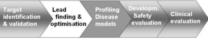
In a simplified view, the drug discovery and development process can be divided in several steps (Fig. 1). In vivo MR techniques allows addressing relevant problems almost throughout the process.
FIG. 1: Simplified view of the drug discovery process. In vivoMR techniques can play important roles in several steps, highlighted in grey. Transgenic mice may be used for target identification and vali-dation, as disease models and for safety evaluations. Non-transgenic animals are used in disease models and for safety assessments. After a compound has been extensively determined to be safe in animal tests, its safety is assessed in healthy volunteers before large clinical efficacy studies can be started in patients.
A. Target Identification and Validation
B. Lead Finding, Validation and Optimization
Once a target has been identified and validated, identifica-tion of a lead compound is the starting point for the develop-ment of new drug candidates. Today large compound libraries of typically 106 compounds are screened using biochemical
and cellular assays that have been made compatible for highly automated HTS. Primary hit compounds displaying activity exceeding a set threshold are further evaluated in secondary screening assays. The most promising hits are then selected for an often lengthy lead optimization program involving the synthesis and testing of compound analogues, during which issues such as target affinity, physico-chemical properties and aspects of compound safety are being addressed. In vivoMR techniques on the other hand do not have any function in this phase.
C. Profiling Compounds in Animal Models of Diseases
Lead optimization results in drug candidate compounds that need to be thoroughly evaluated in animal models of the hu-man disease. The basic aim here is to obtain relevant infor-mation concerning drug efficacy, absorption, distribution, ef-ficacy, metabolism and elimination. Both wildtype and trans-genic animals, mostly small rodents, are used.
In view of a potential translation of methods to clinical drug testing non-invasive readouts of drug efficacy are prefer-able at this stage, yet not mandatory. In fact, the majority of pharmacological studies use invasive procedures. Nev-ertheless, there is an ethical motivation to use noninvasive methods such as imaging techniques, which contribute to the Three Rs (Reduce, Refine, Replace) principles used by Ani-mal Ethics Committees in the governance of aniAni-mal experi-mentation [27]. The use of imaging might be attractive from both an animal welfare and an economical point of view, since the number of animals required for a study can be reduced by up to 80% [15,18], a feature that is especially interesting in chronic studies, and in experiments involving transgenic species. Longitudinal study designs furthermore allow the re-duction of inter-individual variances by using each animal as its own control, thereby enhancing the statistical power of ex-periments.
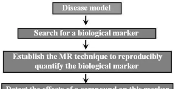
In the past,in vivoMR techniques have most extensively been used during compound profiling [15,17,18]. The gen-eral flow of activities is summarized in Fig. 2. For many dis-eases/disease models, a potential endpoint for the evaluation of the disease status or therapy efficacy is not readily accessi-ble requiring the identification of biomarker, which is indica-tive of the disease status. Such biomarkers should have a clear disease relevance and should have predictive quality both with regard to spontaneous disease progression and potential ther-apy response. In addition, in order to facilitate translational activities, biomarkers used for preclinical studies should also be relevant for the human disease and clinical drug efficacy. Validation/qualification of the MR biomarker involves exten-sive comparison with established, usually invaexten-sive, readouts, in particular histology. In addition, they should correctly
re-produce the well described effects of reference compounds. Only then may they be applied for non-invasive testing of novel drug candidate compounds.
FIG. 2: General flow of activities for testing therapy efficacy in an animal model of disease.
D. Safety Evaluation
The same MR techniques used for evaluation of treatment efficacy in preclinical models of human diseases can also be applied to detect potential safety issues. The advantages of using MRI (and other non-invasive technologies) for toxico-logical studies are that effects can be studiedin vivo(orpost mortem) without the need for tissue dissection, sectioning and staining. Also, the occurrence and progression of poten-tially harmful structural and functional tissue alterations can be monitored in a longitudinal manner. Despite this attractive profile, the use of MRI, and imaging technologies in general, in drug safety studies has received little attention up to now. A main reason is the fact that toxicological studies used for regu-latory authorities must be carried out following the guidelines for Good Laboratory Practice (GLP). Incorporation of MR techniques into routine toxicology programs running under GLP conditions will most likely require separate installations, since GLP compliance is usually not guaranteed in standard biological imaging laboratories. On the other hand, applica-tion of imaging techniques in experimental toxicology does not fall under these restrictions: studies in separate groups of animals aimed at establishing novel toxicological readouts or for internal decision making can be carried out in a straightfor-ward manner. Such studies should be of great value to phar-macological research and will ultimately show whether imag-ing techniques might be used in a broader sense for evaluation of drug safety.
E. Clinical Studies
safety in clinical studies, which can lead to a substantial re-duction in costs and development time [28,29]. More impor-tant is patient management: the time lost for a patient, who is not responsive to treatment, can be significantly reduced. A biomarker that substitutes a clinical endpoint is called a sur-rogate endpoint [30-33]. The most important criteria for valid surrogates are biological plausibility, a documented statistical relationship between the surrogate and an accepted clinical endpoint in epidemiological studies, and demonstration that treatment effects on the surrogate correspond to the clinical outcome.
In early clinical trials biomarkers can be used to demon-strate proof-of-concept of the pharmacological principle and to identify appropriate dose regimens for efficacy studies. both preclinical and clinical investigations are necessary to show a link between disease, pharmacological mechanism, and clinical endpoints.
The acceptance of biomarkers in general, and imaging bio-markers in particular by regulatory agencies is increasing [28], especially for oncology [34,35]. For example, for the 71 on-cology drug approvals by the FDA in the period 1990-2002, end points other than survival were basis for approval in 68% (39 of 57) of applications granted regular approval and for all 14 applications granted accelerated approval [35]. The most common surrogate endpoint used was tumor response as de-termined by changes in tumor volume assessed by MRI or computerized tomography (CT).
The use of non-invasive readouts to assess therapy efficacy facilitates the translation from preclinical to clinical drug de-velopment In this regard, the non-invasive character of MR is a major asset. The potential of using the same readout in the preclinical and clinical phases of drug testing enhances the value of MR in pharmaceutical research. With the recent advances in the field of medical imaging, it is not surprising that the use of imaging biomarkers for the assessment of drug therapies is becoming more common. An imaging readout able to diagnose and characterize a disease state better than conventional methods will sooner or later be incorporated into clinical drug trials.
V. MEASURING AT DIFFERENT SPATIAL SCALES
The criteria imaging biomarkers must meet in order to be accepted by regulatory agencies are multifold. The valida-tion/qualification process may involve not only clinical activi-ties, but also extensive animal experimentation. Hence, trans-ferability of images and protocols from small animal to hu-man scanners and vice-versa becomes an issue. Sharing im-ages from different platforms and manufacturers is necessary (see also section 6). The use of similar acquisition consoles in small animal and whole body systems is an option being evaluated by many manufacturers. Although this is an attrac-tive approach for translational research, since it would consid-erably simplify the comparison of data obtained in different systems, it remains to be shown that protocol optimizations for human imaging are also applicable to e.g. mouse imaging. Some characteristics specifically related to the anatomy and
physiology of every species could render this transition more challenging than a mere change of dimensional parameters in the acquisition protocols.
Spatial resolution constitutes a challenge when performing studies in small rodents. Voxel volumes have to be scaled with anatomical scales in order to accurately represent structures. Typical voxel volumes for a mouse brain study are about 3 nl (100x100x300µm3), while a corresponding voxel in studies of human brain is 2µl (1000x1000x2000µm3). The reduction in voxel volume by almost three orders of magnitude leads to a sensitivity issue: signal-to-noise ration in the mouse image would be reduced by a factor of 25 (square root of the ratio of voxel volumes) provided all other experimental parameters including the detector coil would be identical. Additionally, artifacts may be more severe when studying small structures. For example, effects of magnetic field inhomogeneities at tis-sue interfaces (bone/soft tistis-sue), which are governed by the magnetic field strength and the change in magnetic suscepti-bility, but not by the anatomical dimension of the study object, will be more serious in mouse than in human images. Also, small rodents have higher respiratory and cardiac rates, which pose additional challenges when studying the thorax or ab-domen.
Acquiring high quality images requires in many cases long examination times and interference with the animals’ physiol-ogy. This is often incompatible with the reality ofin vivo phar-macological studies in animals, in which the disease model and/or a compound may profoundly influence the physiology. The primary purpose of MRI in preclinical research is not to generate images of ultimate quality, but to allow the acqui-sition of data from which useful biomedical information can be derived with good reproducibility (and with a reasonable throughput). Obviously, MR techniques must be adapted to the biological situation, rather than to enforce the physiol-ogy for the sake of facilitating image acquisition. Since an MRI session always represents a certain burden for an animal, starting from the anesthesia, a careful balance between image quality and biological constraints should be envisaged [36]. As a general rule, the duration of an imaging session includ-ing animal preparation should be shorter than one hour.
VI. MOLECULAR IMAGING
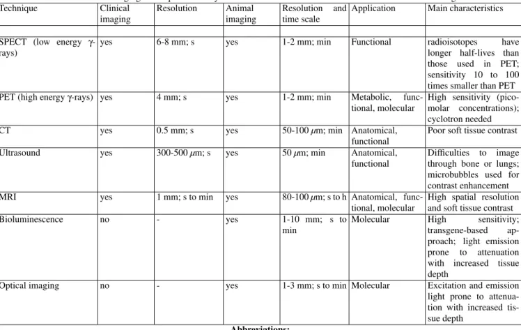
Table 1 –In vivo imaging techniques currently used in the context of biomedical research and/or medical diagnosis. Technique Clinical
imaging
Resolution Animal imaging
Resolution and time scale
Application Main characteristics
SPECT (low energy γ -rays)
yes 6-8 mm; s yes 1-2 mm; min Functional radioisotopes have longer half-lives than those used in PET; sensitivity 10 to 100 times smaller than PET PET (high energyγ-rays) yes 4 mm; s yes 1-2 mm; min Metabolic,
func-tional, molecular
High sensitivity (pico-molar concentrations); cyclotron needed CT yes 0.5 mm; s yes 50-100µm; min Anatomical,
functional
Poor soft tissue contrast
Ultrasound yes 300-500µm; s yes 50µm; min Anatomical, functional
Difficulties to image through bone or lungs; microbubbles used for contrast enhancement MRI yes 1 mm; s to min yes 80-100µm; s to h Anatomical,
func-tional, molecular
High spatial resolution and soft tissue contrast Bioluminescence no - yes 1-10 mm; s to
min
Molecular High sensitivity; transgene-based ap-proach; light emission prone to attenuation with increased tissue depth
Optical imaging no - yes 1-3 mm; s to min Molecular Excitation and emission light prone to attenua-tion with increased tis-sue depth
Abbreviations:
ADMET absorption, distribution, metabolism, excretion and toxicology; CT computerized tomography; FDA Food and Drug Administration; GLP Good Laboratory Practice; HTS high-throughput screening; MR magnetic resonance; MRI magnetic resonance imaging; MRS magnetic
resonance spectroscopy; PET positron emission tomography; SPECT single photon emission computed tomography
The most important imaging techniques potentially suited for providing molecular information in small animals are sum-marized in Table 1. In many respects the techniques are com-plementary; there is no ‘all-in-one’ imaging modality provid-ing optimal sensitivity, specificity and temporo-spatial resolu-tion. Due to its limited sensitivity, MRI is of limited value for detecting molecular processes in vivo; nevertheless, its high spatial resolution provides the exquisite anatomical reference for molecular data obtained with high sensitivity, low reso-lution imaging modalities. This might be achieved by post-processing of data obtained in different imaging sessions or by simultaneous multimodality small animal imaging such as PET-MRI [40,41] and PET-CT [42]. Combining imag-ing data requires compatibility of data formats for the vari-ous modalities as well as sophisticated software tools for im-age coregistration (fusion), data visualization and integration across modalities.
Besides the imaging technique per se, a critical aspect of molecular imaging applications concerns the synthesis of ap-propriate target-specific probes (see [43] for a recent review on target-specific smart MR contrast agents). Most probes synthetized for molecular imaging will be limited to exper-imental research, since the approval process for human use involves similar hurdles as those for registering drugs [44]. Nevertheless, for target validation and assessments of drug
distribution in animals, molecular imaging is going to play a relevant role in drug discovery. In order to take advantage of multimodality small animal imaging, the development of mul-timodal probes (e.g. for optical and MR imaging) is desirable [45].
VII. CONCLUDING REMARKS
The impact that MR techniques may exert on the complex drug discovery process is basically determined by three fac-tors:
(i)the multiparameteric contrast is of high diagnostic value increasing the chances to detect pathological transformation of tissue. Early disease detection enhances the probability of a successful therapeutic intervention;
(ii)accuratein vivomorphometric measurements allow sen-sitive and reproducible assessment of drug effects;
(iii) non-invasiveness allows the design of longitudinal study designs thereby increasing the statistical power.
in-teraction can, in general, not be addressed. Hence, MRI/S pro-vide pharmacodynamic rather than pharmacokinetic informa-tion. However, coupled pharmacokinetic/pharmacodynamic studies could be carried out by combining MR with more sen-sitive molecular imaging approaches, such as PET [40] or op-tical imaging [46]. A crucial step for imaging to be fully inte-grated in drug research concerns the systematic and rigorous qualification of biomarkers via extensive correlation with well characterized and accepted reference data. Finally, standard-ization of imaging acquisition protocols and processing pro-cedures, which will facilitate the comparison of data acquired at different sites, has to be propagated.
This huge task is going to keep researchers and clinicians busy for quite some time. The impact of their work will be the establishment of even more powerful diagnostic and prognos-tic tools that should translate into shortened drug development times. Not only the pharmaceutical industry is going to profit from such an effort, but also the larger medical community and, ultimately, the patients.
Acknowledgement
This contribution is dedicated with profound gratitude to Prof. Horacio Panepucci, with whom I had the great pleasure to work during my MSc studies in S˜ao Carlos. Horacio has in-stilled in me a passion for science, which is the driving force behind my daily activities, and has taught me the value of in-tuition in research. Being in his group, I had the privilege to witness, in March 1984, the formation of the first MR image acquired in the southern hemisphere, a feature that was real-ized primarily due to efforts by Alberto Tann´us. I remember with great affection the long discussions in Horacio’s office and lab, about the physics of imaging. Horacio’s view that the pathway of physics is intimately linked to that of biology profoundly influenced my decisions and gave me sufficient impetus to pursue my own ways elsewhere. I owe Horacio a great deal of thankfulness.
[1] D. B. Kassel. Curr. Opin. Chem. Biol.8, 339 (2004).
[2] NIH-FDA Conference: Biomarkers and Surrogate Endpoints: Advancing Clinical Research and Applications. Abstracts. Dis-ease Markers14, 187 (1998).
[3] G. Duyk. Science,302, 603 (2003).
[4] B. M. Seddon and P. Workman. Br. J. Radiol.76(Spec 2), S128 (2003).
[5] A. Stahl, H. Wieder, M. Piert, H.J. Wester, R. Senekowitsch-Schmidtke, and M. Schwaiger. Mol. Imaging Biol. 6, 214 (2004).
[6] H. Horig and W. Pullman. J. Transl. Med.2, 44 (2004). [7] M. Rudin, N. Beckmann, and M. Rausch. Methods Enzymol.
385, 240 (2004).
[8] D. W. Klomp, H. V. Laarhowen, A. P. Kentgens, and A. Heer-schap. Magn. Reson. Med.50, 303 (2003).
[9] H. W. van Laarhoven, D. W. Klomp, Y. J. Kamm, C. J. Punt, and A. Heerschap. Cancer Res.63, 7609 (2003).
[10] D. A. Hamstra, K. C. Lee, J. M. Tyhewicz, V. D. Schepkin, B. A. Moffat, M. Chen, K. J. Dornfeld, T. S. Lawrence, T. L. Chenevert, B. D. Ross, J. T. Gelovani, and A. Rehemtulla. Mol. Ther.10, 916 (2004).
[11] Y. J. Kamm, A. Heerschap, E. J. van den Bergh, and D. J. Wa-gener. Anticancer Drugs15, 229 (2004).
[12] J. R. Griffiths and J. D. Glickson. Adv. Drug Deliv. Rev.41, 75 (2000).
[13] D. Artemov, M. Solaiyappan, and Z.M. Bhujwalla. Cancer Res.
61, 3039 (2001).
[14] M. Rudin, N. Beckmann, and A. Sauter. NMR Biomed.12, 404 (1999).
[15] M. Rudin, N. Beckmann, R. Porszasz, T. Reese, D. Bochelen, and A. Sauter. NMR Biomed.12, 69 (1999).
[16] M. Rudin, P. Allegrini, N. Beckmann, H.U. Gremlich, R. Kneuer, D. Laurent, M. Rausch, and M. Stoeckli. Ernst Scher-ing Res. Found. Workshop48, 47 (2004).
[17] N. Beckmann, T. Mueggler, P.R. Allegrini, D. Laurent, and M. Rudin. Anat. Rec.265, 85 (2001).
[18] N. Beckmann, B. Tigani, D. Laurent, R. Panizzutti, and M. Rudin. Drug Discov. Today9, 35 (2004).
[19] N. Beckmann, B. Tigani, L. Mazzoni, and J. R. Fozard. Trends
Pharmacol. Sci.24, 550 (2003).
[20] R. M. Weisskoff, D. Chesler, J. L. Boxeman, and B. R. Rosen. Magn. Reson. Med.29, 553 (1993).
[21] M. Rudin, N. Beckmann, and A. Sauter. Magn. Reson. Imaging
15, 551 (1997).
[22] M. Rudin, P. Birgit, K. Umemura, and W. Zierhut. Basic Res. Cardiol.86, 165 (1991).
[23] R. L. Stein. Drug Discov. Today8, 245 (2003).
[24] J. Knowles and G. Gromo. Nat. Rev. Drug Discov.2, 63 (2004). [25] J. Tornell and M. Snaith. Drug Discov. Today7, 461 (2002). [26] B. P. Zambrowicz and A. T. Sands. Nat. Rev. Drug Discov.2,
38 (2003).
[27] C. A. Schuppli, D. Fraser, and M. McDonald. Altern. Lab. Anim.32, 525 (2004).
[28] R. Frank and R. Hargreaves. Nat. Rev. Drug Discov.2, 566 (2003).
[29] J. J. Smith, A. G. Sorensen, and J. H. Thrall. Radiology227, 633 (2003).
[30] W. A. Colburn. J. Clin. Pharmacol.40, 1419 (2000).
[31] L. J. Lesko, M. Rowland, C. C. Peck, and T. F. Blaschke. J. Clin. Pharmacol.40, 803 (2000).
[32] L. J. Lesko and A. Atkinson, Ann. Rev. Pharmacol. Toxicol.41, 347 (2001).
[33] H. H. Pien, A. J. Fischman, J. H. Thrall, and A. G. Sorensen, Drug Discov. Today10, 259 (2005).
[34] G. J. Kelloff and C. C. Sigman, Eur. J. Cancer41, 491 (2005). [35] J. R. Johnson, G. Williams, and R. Pazdur. J. Clin. Oncol.21,
1404 (2003).
[36] L. A. Colby and B. J. Morenko, Comp. Med.54, 623 (2004). [37] M. Rudin and R. Weissleder, Nat. Rev. Drug Discov.2, 123
(2003).
[38] T. F. Massoud and S. S. Gambhir, Genes Dev.17, 545 (2003). [39] I. J. Hildebrandt and S. S. Gambhir, Clin. Immunol.111, 210
(2004).
[40] R. B. Slates, K. Farahani, Y. Shao, P. K. Marsden, J. Taylor, P. E. Summers, S. Williams, J. Beech, and S. R. Cherry. Phys. Med. Biol. 44,2015 (1999).
D. Coplan, A. Biegon, L. Rosenblum, B. Scharf, J. S. Gatley, and N. D. Volkow. J. Nucl. Med.46, 312 (2005).
[42] F. H. Fahey. Neuroimaging Clin. N. Am.13, 659 (2003). [43] E. J. Delikatny and H. Poptani, Radiol. Clin. North Am.43, 205
(2005).
[44] F. A. Jaffer and R. Weissleder, JAMA,293, 855 (2005).
[45] M. Doubrovin, I. Serganova, P. Meyer-Kuckuck, V. Ponomarev, and R.G. Blasberg. Bioconjug. Chem.15, 1376 (2004). [46] V. Ntziachristos, J. Ripoll, L. V. Wang, and R. Weissleder. Nat.