Article
Printed in Brazil - ©2017 Sociedade Brasileira de Química0103 - 5053 $6.00+0.00
*e-mail: [email protected]
Dedicated to Prof Angelo da Cunha Pinto (in memoriam) for his outstand-ing contributions to the field of Organic Chemistry in Brazil. This article was submitted to the special issue dedicated to Professor Angelo da Cunha Pinto and name Angelocunhol is also additional homage.
Angelocunhol: New Erythroxylane Diterpene and Other Compounds from
Simira sampaioana
(Rubiaceae)
Vinicius F. Moreira,*,a Ivo J. C. Vieirab and Raimundo Braz-Filhob
aDepartamento de Química, Instituto Federal de Educação Ciência e Tecnologia Fluminense,
28909-971 Cabo Frio-RJ, Brazil
bCentro de Ciências e Tecnologias, Universidade Estadual do Norte Fluminense Darcy Ribeiro,
28013-600 Campos dos Goytacazes-RJ, Brazil
The first Simira sampaioana (Rubiaceae) phytochemical study allowed the isolation and structural determination of a new erythroxylane diterpene named Angelocunhol, 11β,12α -dihydroxy-2,4(18),15-eritroxilatrien-1-one, together with 14 known compounds: simirane B, harman, maxonine, isomalindine, malindine, sitost-4-en-6-ol-3-one, estigmast-4,22-dien-6-ol-3-one, campest-4-en-6-ol-3-estigmast-4,22-dien-6-ol-3-one, sitost-4-en-3-estigmast-4,22-dien-6-ol-3-one, stigmast-4,22-dien-3-estigmast-4,22-dien-6-ol-3-one, campest-4-en-3-estigmast-4,22-dien-6-ol-3-one, β-sitosterol, stigmasterol, and stigmast-4,22-dien-3-ol from the wood of a specimen of the species. The structures of these compounds were elucidated on the spectroscopic-data analysis basis, mainly
1H and 13C nuclear magnetic resonance (NMR), including 2D experiments (1H-1H correlation
spectroscopy (COSY), nuclear Overhauser spectroscopy (NOESY), heteronuclear multiple-bond correlation-HMBC and heteronuclear single-quantum correlation-HSQC), and high-resolution electrospray mass spectrometry (HRESI-MS).
Keywords: Simira sampaioana, Rubiaceae, diterpene, alkaloid, steroid
Introduction
Species from Simira genus of Rubiaceae family have been investigated mainly due to the biological activities. Many of these species have been used by diverse communities as coloring producers, antifebrile, tonic and purgative substances, and by the phototoxic activities presented by some of their chemical constituents.1-4
Simira sampaioana (synonyms Sickingia sampaioana in the Atlantic Rainforest) is known by its common names “arariba”, “canela-samambaia”, “marfim” and “maiate”, and economic interest is justified by use as timber and for the afforestation of streets.1,5 As the first phytochemical
study involving S. sampaioana, this article describes the isolation and structural characterization of the new diterpene 11β,12α-dihydroxy-2,4(18),15-eritroxilatrien-1-one, named Angelocunhol (1), together with 14 known compounds: simirane B (2), harman (3), maxonine (4), isomalindine (5),
malindine (6), sitost-4-en-6-ol-3-one (7), estigmast-4,22-dien-6-ol-3-one (8), campest-4-en-6-ol-3-one (9), sitost-4-en-3-one (10), stigmast-4,22-dien-3-one (11), campest-4-en-3-one (12), β-sitosterol (13), stigmasterol (14), and stigmast-4,22-dien-3-ol (15) (Figure 1) from the wood of the plant. The structures of all compounds were characterized by 1D and 2D nuclear magnetic resonance (NMR) techniques, and also the high-resolution electrospray ionization mass spectrometry (HRESI-MS), infrared spectroscopy (IR) and comparisons with known compounds available in literature data.
Results and Discussion
Angelocunhol (1) was obtained as yellow oil. Its IR spectrum exhibited bands at νmax 3362 (broad, νOH) and
1649 cm-1 (ν
C=O of an α,β-unsaturated carbonyl group).
The molecular formula was assigned as C20H28O3, based
on the quasi-molecular peak at m/z 339.1934 ([M + Na]+,
calcd. for C20H28O3Na, m/z 339.1951) revealed by the
identified by single signals at dH 1.24 (s, 3H-19), 1.21
(s, 3H-20) and 1.09 (s, 3H-17) in the 1H NMR spectrum;
the presence of an exocyclic double bond (=CH2-18)
and a vinyl group was deduced by signals at dH 5.33 (s,
H-18a) and dH 5.32 (s, H-18b), indicating an AB system,
and dH 5.90-5.97 (m, H-15), dH 5.08 (dd, J 17.5, 1.1 Hz,
H-16a) and dH 5.03 (d, J 10.8, 1.1 Hz, H-16b) compatible
with an ABX system of vinyl group. Two doublet signals (J 9.8 Hz) corresponding to olefinic hydrogens at dH 5.98
and dH 6.98 were attributed to H-2 and H-3, respectively.
The distortionless enhancement by polarization transfer with retention of quaternaries (DEPTQ) 13C NMR
spectrum allowed to recognize signals corresponding
to 20 carbon atoms (Table 1): three methylics, five methylenics (including two sp2 at d
C 117.6 and 111.3),
seven methynics (including three sp2 at d
C 147.6, 145.8
and 127.8 and two sp3 oxygenated at d
C 79.8 and 76.9)
and five quaternary (including one of ketone carbonyl at
dC 205.9), allowing to establish the expanded molecular
formula C20H26O3 and the two hydrogen atoms needed to
complete the molecular formula C20H28O3 justified by two
hydroxyl groups. These hydroxyl groups were located at CH-11 and CH-12 by the signals at dH 3.23 (d, J 9.0 Hz,
H-11) and 3.52 (d, J 9.0 Hz, H-12) revealing vicinal spin-spin interaction each other in the 1H-1H correlation
spectroscopy (COSY) and heteronuclear correlations in
the heteronuclear single-quantum correlation (HSQC) (1J
CH) spectrum with the 13C signals at dC 79.8 (CH-11)
and 76.9 (CH-12) and in the heteronuclear multiple-bond correlation (HMBC) these 13C signals showed long-range
heteronuclear correlations of the CH-11 (dC 79.8) with H-10
(dH 2.60, 3JCH) and 3H-20 (dH 1.21, 3JCH) and of the CH-12
(dC 76.9) with H-11 (dH 3.25, 2JCH), H-15 (dH 5.97-5.90, 3J
CH) and 3H-17 (dH 1.09, 3JCH), summarized in Table 1. The
interaction of CH-11 (dC 79.8) with the 3H-20 (dH 1.21, 3J
CH) suggested the presence of this methyl group at carbon
C-9 (dC 42.86), revealing its rearrangement of the carbon
C-10 (dC 64.45) and signaling for a skeleton. Thus, the
hydrogen chemical shifts and hydrogenated carbon atoms were unambiguously assigned by analysis of 1H-1H-COSY
and HSQC spectra data (Table 1). The HMBC spectrum showed long-range heteronuclear correlations, which were used to confirm the carbon skeleton and localization of the substituents (Table 1). The HMBC spectrum also revealed correlations of hydrogen atoms H-3 (dH 6.98) and
H-10 (dH 2.60) signals with carbon atom C-1 (dC 205.3),
H-3 (dH 6.98) with carbon atom CH2-18 (dC 117.60) and
H-10 with carbon atom CH3-19 (dC 24.74), in accordance
with a erythroxylane skeleton containing a dienone system involving the carbon atoms C-1 (dC 205.3), CH-2
(dC 127.84), CH-3 (dC 145.79) and the exocyclic double
bond CH2-18 (dC 117.60). Thus, these data were used to
postulate the structure of a diterpene with erythroxylane skeleton,6 corroborated by long-range correlations of
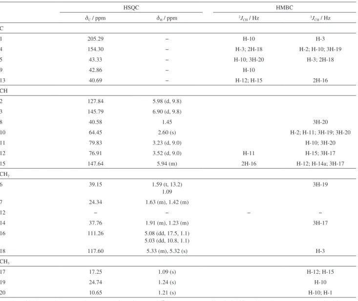
Table 1. 1H NMR (500 MHz, CDCl
3) and 13C NMR (125 MHz, CDCl3) data for 1. Chemical shifts (d, ppm) and coupling constants (J, Hz, in parenthesis)a
HSQC HMBC
dC / ppm dH / ppm 2JCH / Hz 3JCH / Hz
C
1 205.29 − H-10 H-3
4 154.30 − H-3; 2H-18 H-2; H-10; 3H-19
5 43.33 − H-10; 3H-20 H-3; 2H-18
9 42.86 − H-10
13 40.69 − H-12; H-15 2H-16
CH
2 127.84 5.98 (d, 9.8)
3 145.79 6.90 (d, 9.8)
8 40.58 1.45 3H-20
10 64.45 2.60 (s) H-2; H-11; 3H-19; 3H-20
11 79.83 3.23 (d, 9.0) H-10; 3H-20
12 76.91 3.52 (d, 9.0) H-11 H-15; 3H-17
15 147.64 5.94 (m) 2H-16 H-12; H-14a; 3H-17
CH2
6 39.15 1.59 (t, 13.2)
1.09
3H-19
7 24.34 1.63 (m), 1.42 (m)
12 − − − −
14 37.76 1.91 (m), 1.23 (m) 3H-17
16 111.26 5.08 (dd, 17.5, 1.1)
5.03 (dd, 10.8, 1.1)
18 117.60 5.33 (m), 5.32 (s) H-3
CH3
17 17.25 1.09 (s) H-12; H-15
19 24.74 1.24 (s) H-10
20 10.65 1.21 (s) H-10; H-1
aNumber of hydrogens bound to carbon atoms deduced by DEPTQ-13C NMR spectrum. Chemical shifts and coupling constants (J) obtained from 1D 1H NMR spectrum. Superimposed 1H signals are described without multiplicity and chemical shifts deduced by 1H-13C COSY 1J
CH (HMQC), 1H-13C COSY-nJ
CH-11 (dC 79.8) with the 3H-20 (dH 1.21) and of the CH-12
(dC 76.9) with H-11 (dH 3.25), H-15 (dH 5.97-5.90) and
3H-17 (dH 1.09) through the location of hydroxyl groups
at 11 and 12 positions (Table 1). Additional heteronuclear long-range couplings are summarized in Table 1.
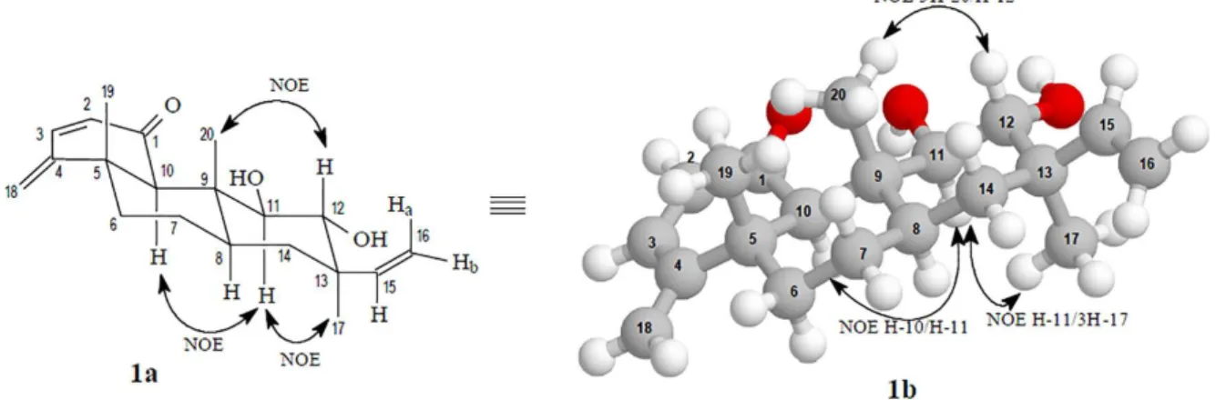
The relative stereochemistry of 1 (Figure 1) was determined by the relevant hydrogens coupling constants revealed by 1H NMR and 1H-1H-COSY spectra and from the
dipolar-dipolar interaction observed in the 1H-1H-NOESY
spectrum (Figure 2). The value corresponding to vicinal interaction (3J
H,H) between the hydrogens H-11 and H-12
(J 9.0 Hz) is consistent with axial-axial interaction7 as
appear in Figure 1, which also reveals dipolar interaction between H-12 (dH 3.52)/3H-20 (dH 1.21), H-10 (dH 2.60)/
H-11 (dH 3.23) and H-11 (dH 3.23)/3H-17 (dH 1.09)
revealed by 1H-1H nuclear Overhauser effect spectroscopy
(NOESY) spectrum through the cross-peaks assigned to corresponding dipolar interaction (special proximity) shown in Figure 2 (1a and 1b) which indicates the axial positions for these hydrogens. Furthermore, coupling values observed in the 1H NMR spectrum for these hydrogens
indicate axial-axial interaction.
Thus, the analysis of the spectral data allowed the structural characterization of this new erythroxylane diterpene 11β,12α -dihydroxy-2,4(18),15-eritroxilatrien-1-one (1), named Angelocunhol as a simple tribute to colleague and friend Angelo da Cunha Pinto, excellent researcher who died in November 7, 2015.
The results of the extensive application of 1D and 2D NMR spectral techniques were also used to confirm the structure and to establish the 1H and 13C resonance
assignments of 1 (Table 1). Proposed fragmentation mechanisms of this new diterpene 1 (only peaks classified as principals) was summarized in Scheme 1.
The known simirane B (2),6 harman (3),6,8 maxonine (4),
isomalindine (5), malindine (6),9
sitost-4-en-6-ol-3-one (7), estigmast-4,22-dien-6-ol-3-one (8),
campest-4-en-6-ol-3-one (9), sitost-4-en-3-one (10), stigmast-4,22-dien-3-one (11), campest-4-en-3-one (12),10,11 β-sitosterol (13), stigmasterol (14),12 and
stigmast-4,22-dien-3-ol (15) were identified by spectral data, involving mainly 1D and 2D 1H and 13C NMR spectra
and comparison with literature values.
Conclusions
A total of fifteen compounds were isolated from S. sampaioana (Rubiaceae), among which four alkaloids, nine steroids and two diterpenes. These compounds are in agreement with the secondary metabolites produced by plants of the Rubiaceae family and of the Simira genus. It is angelocunhol compound (1) unprecedented in the literature, and isomalindine (5), malindine (6), sitost-4-en-6-ol-3-one (7), estigmast-4,22-dien-6-ol-3-one (8), campest-4-en-6-ol-3-one (9), stigmast-4,22-dien-3-one (11), campest-4-en-3-one (12) and stigmast-4,22-dien-3-ol (15) were reported by first time in the genus. And yet the harman alkaloid (2), corroborating the proposition of this alkaloid the taxonomic marker of genus.
Experimental
General experimental procedures
Measure of IR data was on Shimadzu IRAffinity-1. NMR spectra were obtained on Bruker DRX-500 and Avance IIIH (both 500 MHz for 1H and 125 MHz for 13C), with CDCl
3 (0.1% tetramethylsilane, TMS) or
dimethylsulfoxide (DMSO-d6) as solvents,used as internal
references (dH 7.24 and dC 70.00). Low-resolution mass
analysis was done on Shimadzu GCMS-QP5050A and high-resolution mass spectrometry (HRMS) analysis on Bruker microTOF electrospray-time-of-flight mass spectrometer (ESI-TOF-MS) equipped with an ESI source in the
positive and negative modes. Column chromatography was conducted using silica gel (Merck). Precoated thin layer chromatography (TLC) sheets (Merck) of silica gel 60 GF254 (0.25 mm) were used, and visualization of plates was carried out using a lamp UV 254 and 356 nm and vanillin (1%) solution in H2SO4 (5%).
Plant material
The S. sampaioana specimen employed in this study was collected at the Companhia Vale do Rio Doce (CVRD) Atlantic Rainforest, in Linhares city, Espírito Santo State, Brazil, and was identified by Domingos A. Folli. A voucher specimen (CVRD 8796) is deposited at the company’s herbarium.
Scheme 1. Proposed fragmentation mechanisms of 1 (only peaks classified as principals).
Extraction and isolation
The dried and powdered wood (6 kg) was extracted with Hexane and MeOH at room temperature and providing 0.004 kg of Hexane crude extract and 0.5 kg of MeOH crude extract after solvent evaporation.
The Hexane extract was subjected to column chromatography (CC) (SiO2, gradient Hex/EtOAc)
furnishing eleven fractions. The fraction 9 (333.7 mg) was chromatographed over a silica gel column with a hexane/ethyl acetate (9:1, v/v) to yield pure compound 1
hexane/ethyl acetate (9:1, v/v) to yield pure compound 2
(4 mg). The fraction 7 (522.3 mg) was chromatographed on silica gel column using hexane/acetone (9:1, v/v), which furnished a solid identified as a mixture of the compounds
11 + 12 (7.6 mg) by GC-MS analysis. The mixture of the compounds 10 + 15 (55.5 mg) and the mixture of the compounds 13 + 14 (94.4 mg) identified by GC-MS analysis, were isolated from the fraction 5 (515.2 mg) by chromatography using hexane/ethyl acetate (9.5:0.5, v/v).
An aliquot of this extract (2.5 g) was dissolved in a MeOH-H2O (8:2 (v/v), 100 mL) mixture and partitioned
with CH2Cl2 (5 × 100 mL), EtOAc (5 × 100 mL) and
n-BuOH (5 × 100 mL), to give the following fractions: CH2Cl2 (1.3 g), EtOAc (0.2 g), n-BuOH (0.9 g). The
CH2Cl2 fraction was subjected to CC (SiO2, gradient
MeOH/CH2Cl2) to yield pure compounds 3 (6.4 mg) and
4 (5.4 mg), and the n-BuOH fraction was subjected to CC (SiO2, gradient MeOH/CH2Cl2) to yield pure compounds
5 (4 mg) and 6 (7.3 mg).
Supplementary Information
Supplementary information, including 1H NMR, 13C NMR, COSY, NOESY, HSQC, and HMBC spectra, as
well as mass spectra and IR (Figures S1-S9), are available free of charge at http://jbcs.sbq.org.br as a PDF file.
Acknowledgments
The authors are grateful to FAPERJ, CAPES and CNPq for the financial support and research fellowships (CNPq). To Prof Mário Geraldo de Carvalho, Departamento de Química-Instituto de Ciências Exatas, UFRRJ, for access to the NMR and GC-MS facilities through the technicians
Vitor de Almeida/Maurício Lemos and Frances Regiane dos Santos, respectively.
References
1. Barbosa, M. R. V.; Peixoto, A. L.; Acta Amaz. 1989, 19, 27. 2. Capasso, A.; Aquino, R.; de Tommasi, N.; Piacente, S.; Rastrelli,
L.; Pizza, C.; Curr. Med. Chem.: Cent. Nerv. Syst. Agents2002,
2, 1.
3. Moreira, V. F.; Vieira, I. J. C.; Braz-Filho, R.; Nat. Prod. J. 2014,
4, 290.
4. Moreira, V. F.; Vieira, I. J. C.; Braz-Filho, R.; Am. J. Plant Sci.
2015, 6, 2612
5. herbario.iac.sp.gov.br/ accessed in May 2016.
6. Araújo, M. F.; Vieira, I. J. C.; Braz-Filho, R.; Carvalho, M. G.;
Nat. Prod. Res. 2011, 25, 1713.
7. Silverstein, R. M.; Webster, F. X.; Kiemle, D.; Spectrometric Identification of Organic Compounds, 7a ed.; LTC: Rio de
Janeiro, 2007, p. 490.
8. Seki, H.; Hashimoto, A.; Hino, T.; Chem. Pharm. Bull. 1993,
41, 1169.
9. Hasbun, C. P.; Calderon, M.; Castro, O.; Gacs-Baitz, E.; Delle Monache, G.; Delle Monache, F.; Tetrahedron Lett.1989, 30, 6199.
10. Greca, M. D.; Monaco, P.; Previtera, L.; J. Nat. Prod. 1993, 53, 1430.
11. Correia, S. J.; David, J. P.; David,J. M.; Quim. Nova2003, 26, 36.
12. Chaturvedula, V. S. P.; Prakash, I.; Int. Curr. Pharm. J.2012,
1, 239.
Submitted: February 27, 2016