r e v b r a s r e u m a t o l . 2017;57(6):610–612
ww w . r e u m a t o l o g i a . c o m . b r
REVISTA
BRASILEIRA
DE
REUMATOLOGIA
Case
report
Presence
of
riziform
bodies
in
a
patient
with
juvenile
idiopathic
arthritis:
case
report
and
literature
review
夽
Presenc¸a
de
corpos
riziformes
em
paciente
com
artrite
idiopática
juvenil:
relato
de
caso
e
revisão
de
literatura
Leonardo
Rodrigues
Campos
a,
Fernanda
Cardoso
das
Neves
Sztajnbok
a,
Stélio
Galvão
b,
Marise
de
Araújo
Lessa
b,
Ierecê
Lins
Aymoré
c,
Flavio
Sztajnbok
d,∗aUniversidadeFederaldoRiodeJaneiro,RiodeJaneiro,RJ,Brazil
bUniversidadedoEstadodoRiodeJaneiro,FaculdadedeCiênciasMédicas,RiodeJaneiro,RJ,Brazil
cHospitalMárioKroeff,LaboratórioCláudioLemosAnatomiaPatológicaLtda.,RiodeJaneiro,RJ,Brazil
dUniversidadedoEstadodoRiodeJaneiro,NúcleodeEstudosdaSaúdedoAdolescente,RiodeJaneiro,RJ,Brazil
a
r
t
i
c
l
e
i
n
f
o
Articlehistory: Received17April2014 Accepted14September2014 Availableonline28November2014
Introduction
Juvenileidiopathicarthritis(JIA)isthe mostcommon form ofchronicarthritisinpediatricpatients,anditsdiagnosisis madefollowingexclusionofseveralotherconditionsthatmay presentwithprolongedmusculoskeletalmanifestations.The mostcommon form ofJIAis the oligoarticular one,which maypresentasachronicmonoarthritis.1Inthesecases,there aremultiplediagnosestobeinvestigated:tuberculosis, sar-coidosis,villonodularsynovitis,hemarthrosis,hemangioma, synovial osteochondromatosis, arborescent lipoma, malig-nanciesandsomeautoinflammatorydiseases.2–4Ourgoalis
夽
WorkcarriedoutintheDepartmentofRheumatologyoftheCenterforAdolescentHealthStudies,UniversidadedoEstadodoRiode Janeiro,RiodeJaneiro,RJ,Brazil.
∗ Correspondingauthor.
E-mail:[email protected](F.Sztajnbok).
toreportthecaseofachildwithchronicmonoarthritiswhose biopsyshowedagreatamountofriziformbodiesthatare sel-domdescribedinthisagegroup.Thisisthe9thcasereported inliteratureaboutthepresenceofriziformbodiesinpatients withJIAand,toourknowledge,thefirstcasereportedinBrazil.
Case
report
Amale8-year-oldchild,2ndtwin,borninthecityofRiode Janeiro,hadahistoryofdifficultypracticingexercises,with limitedrangeofmotionoftheleftknee,approximatelyfour monthsbeforethe1stconsultation.Afteramonth,theparents
http://dx.doi.org/10.1016/j.rbre.2014.09.004
rev bras reumatol.2017;57(6):610–612
611
noticedagreatincreaseinvolumethat remaineduntilthe dayofconsultation.Withinthisperiod,therewasnofever, skin lesions or any signs or symptoms of involvement of other organs. There was no previous history oftrauma or infectioninthethreemonthsbeforetheonsetofsymptoms. Hehasahealthy twinbrother,thereisnofamilyhistoryof spondyloarthritis,and the mother had a facialskin lesion about 25 years ago, diagnosed as sarcoidosis, and treated withintralesionalcorticosteroids.Physicalexaminationinthe 1stconsultationwasnormalexceptforthepresenceoflarge swellingoftheleftknee,withheatandslighthyperemia,piano keysignandmotionlimitation(flexionat60◦andextension
at150◦).
Withthe syndromic diagnosis ofchronic monoarthritis, testswereordered,theresultsofwhichshowedbloodcount, C-reactiveprotein,erythrocytesedimentationrate,lipid pro-file, blood glucose, calcium, urea, creatinine, complement,
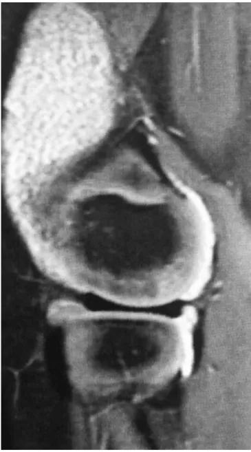
Fig.1–Magneticresonanceofleftkneeshowedbulkyjoint effusionandpresenceofmultiplesmallelongatedimages withhypotensesignalinallsequences,suggestiveof riziformbodies.
Fig.2–Macroscopicaspectofleftkneesynovium,showing moderateamountofwhitishmass,similartoricegrains.
muscle enzymes, proteinelectrophoresis, angiotensin con-verting enzyme(ACE)and urinary sedimentwithinnormal ranges. Therheumatoid factor, antinuclear antibodies and HLAB-27werenegative.SerologicmarkersforhepatitisBand C,toxoplasmosis,HIVandHTLVwerealsonegative.Serology for rubellaand cytomegalovirusshowed negative IgM.The tuberculintestwasnegativeandchestradiographwasnormal. Also,hiselectrocardiogram,echocardiogram,and ophthalmo-logicevaluationwerenormal.LeftkneeX-rayshowedonlysoft tissueswelling,andultrasonography(USG)showedjoint effu-sionwithdebrisandsynovialthickening.Magneticresonance imagingshowedmassivejoint effusionwithheterogeneous signalinthepresenceofmultiplesmallelongatedimageswith hypotensivesignalintensityonallsequences,suggesting riz-iformbodies,andpronouncedenhancementofthesynovium (Fig.1).Leftkneesynovialbiopsywasrequiredand,insurgery, there wasoutput ofminimalamount ofsynovialfluid (SF) andamoderateamountofwhitemass,similartoricegrains (Fig.2).Anatomicpathologyshowed,onmacroscopic exam-ination, the presence ofnumerous oval, friable, white and firmstructuresconsistentwiththediagnosisofriziform bod-ies.Microscopyshowedsynoviumfragmentswithhyperplasia ofliningcells,andextensivedepositsoffibrin,withedema, vascularectasiaandneogenesisseeninthestroma,and lym-phocytic inflammatory infiltrateoccasionally aggregated in follicles,andsomegranulocytes(Fig.2).Thematerial repre-sentedbyriziformbodiesconsistedoforganizedfibrinandhad pervadingmononuclearcells,andsomepolymorphonuclear cells.Thehistologicalreportwassuggestiveofchronic inflam-matory synovitis, or JIA. The patient was initially treated, beforesurgery,withnon-steroidalanti-inflammatorydrugs, with no clinical response.After surgery,methotrexate was addedtothetreatmentand,inabouttwomonths,thepatient wasasymptomatic.
Discussion
612
rev bras reumatol.2017;57(6):610–612outsarcoidosis,notconfirmedbyhistopathology.Atsurgery, a small amount of synovial fluid was found, along with the presence ofa moderate amount of whitish mass that histopathologyshowedtoberiziformbodies.
Riziform bodies are structures which can be found in synovialfluidoradheredtosynovium,andhavethis denom-ination for its similar appearance to the rice grains. They consistoffibrininvolvingmononuclearcells, polymorphonu-clearcellsand red bloodcells,and representa nonspecific responsetosynovialinflammation.5–8Theyhavealreadybeen described in several diseases suchas tuberculous arthritis (where they were originally first reported in 1895), other infectiousarthritis, osteoarthritis,rheumatoid arthritisand JIA.2–11
Itsetiopathogenesisiscontroversial.Theycanarisefrom areas of microinfarctions of inflamed synovium that are releasedinto theSFandencapsulatedbyfibrin,abundantly producedininflammatoryprocessesofthesynovium.7,9,10,12 Thereisalsoanothertheorythatsuggeststhatriziform bod-iesareformeddenovointheSF,quiteinterestingforcasesof osteoarthritisandthepresenceofapatitecrystalsandcalcium pyrophosphate.4,8 Rovenska et al. suggestedthat although thereisaphysiologicallymphoangiogenesisassociatedwith chronicinflammationinordertoimprovedrainageof exces-siveSF,theformationofriziformbodiescanbeassociatedwith thedifficultyoflymphaticdrainageoftheinflamedsynovial fluid.13 Theriziformbodiesoccur moreoften inknees and shouldersand wecould saythattheyare theendproducts ofinflammation,synovialproliferationanddegeneration.9On theotherhand,thereisadescriptionofriziformbodiesinthe pleuralfluid,tendonsheathsandbursae,suggestinga possi-bilityofnonsynovialorigin.2,8,14
ThefindingofriziformbodiesinJIAwasfirstreportedby Wynne-Robertsetal.in1979inan17-year-oldadolescent12 and, after, we found 7 more cases reported in the litera-ture,withagesrangingfrom2to17years.5–7,9–11Thisfinding appearstobeindependentfromarthritisseverityandlength ofthedisease.7,9,10,15 Inthe casesreportedinchildrenand adolescents,thetimebetweentheonsetofarthritisandthe findingofriziformbodiesrangedfrom2monthsto5years, butnotallreportscontainthisinformation.Inourcase,we observethattheappearanceoccurredafteronly3monthsof theonsetofarthritis.
Itwouldbeinterestingtoconductsynovialbiopsytocheck forthepresenceofriziformbodieswhenevertheUSGshow thefinding ofdebris.Thedifficulty ofSFsuctioninajoint containinga largeeffusion may bedue tothe presenceof riziformbodiesandthereforetheuseofathickerneedleor asurgicalapproachmaybenecessary.10Theperformanceof synovialbiopsyforcheckingthepresenceofriziformbodiesis important,especiallyincasesofarthritisthatisbulkyand/or unresponsivetoconventionaltreatment,asitswithdrawalcan causesymptomaticreliefforthepatient.7
Ourobjectivewastodescribewhatwebelieveisthe9th casereportedaboutthepresenceofriziformbodiesinJIA,and itspresenceshouldbemorefrequentthanisreportedinthe
literatureandweareusedtofind.9Inrheumatoidarthritis, theycanbefoundin72%ofjoints,whensearched.15In gen-eral, we donotorder synovialbiopsy inother typesofJIA otherthanthemonoarticularand,withthat,weareprobably under-diagnosingthepresenceofriziformbodies,whichmay beresponsibleforlargearthritisofdifficultclinicaltreatment, butthatrespondwelltosurgicalintervention.
Conflict
of
interest
Theauthorsdeclarenoconflictsofinterest.
r
e
f
e
r
e
n
c
e
s
1.OenKG,CheangM.Epidemiologyofchronicarthritisin childhood.SeminArthritisRheum.1996;26:575–91. 2.HuangG-S,LeeC-H,ChenC-Y.Clinicalimages:tuberculous
ricebodiesofthewrist.ArthritisRheum.2005;52:1950. 3.BuckiB,LansamanJ,JansonX,Billon-GallandMA,MartyC,
RuelM,etal.Osteoarthritiswithricebodiesincalcium microcrystals4caseswithultrastructuralstudy.RevRheum. 1994;61:415–20.
4.JeongYM,ChoHY,LeeS-W,HwangYM,KimY-K.Candida septicarthritiswithricebodyformation:acasereportand reviewofliterature.KoreanJRadiol.2013;14:465–9. 5.MartiniG,TregnaghiA,BordinT,VisentinMT,ZulianF.Rice
bodiesimaginginjuvenileidiopathicarthritis.JRheumatol. 2003;30:2720–1.
6.DiVitoA,KanJH.Juvenileidiopathicarthritiswithricebodies. PediatrRadiol.2008;38:1263.
7.ChungC,ColeyBD,MartinLC.Ricebodiesinjuvenile rheumatoidarthritis.AJR.1998;170:698–700.
8.ForseCL,MuchaBL,SantosMLZ,OngcapinEH.Ricebody formationwithoutrheumaticdiseaseortuberculosis infection:acasereportandliteraturereview.ClinRheumatol. 2012;31:1753–6.
9.DruschelC,FunkJF,KallinichT,LiebA,PlaczekRP. Developmentofricebodiesin2childrenyoungerthan3 years.JClinRheumatol.2013;19:35–7.
10.CoxA,AllenR,AkikusaJ.Ricebodiesinjuvenileidiopathic arthritis:aclinicalimage.JPaediatrChildHealth.
2012;48:279–80.
11.TeramotoA,WatanabeK,KiiY,KudoM,OtsuboH,WadaT, etal.Recurrentkneearthritisdiagnosedasjuvenile
idiopathicarthritiswitha10-yearasymptomaticperiodafter arthroscopicsynovectomy:acasereport.JMedCaseRep. 2013;7:166–70.
12.Wynne-RobertsCR,CassidyJT.JRAwithricebodies:lightand electricmicroscopicstudies.AnnRheumDis.1979;38:8–13. 13.RovenskaE,StvrtinaS,GreguskaO,PravdaL,RovenskyJ.
Conspicuoussynoviallymphaticcapillariesinjuvenile idiopathicarthritissynovitiswithricebodies.AnnRheum Dis.2005;64:328–9.
14.ChavanS,SableSS,TekadeS,PuniaP.Tuberculous
tenosynovitispresentingasganglionofwrist.CaseRepSurg. 2012:143921.
15.PopertAJ,ScottDL,WainwrightAC,WaltonKW,Williamson N,ChapmanJH.Frequencyofoccurrence,modeof