ASSESSMENT OF CARDIAC FUNCTION BY MAGNETIC RESONANCE
IMAGING: SEGMENTED
×
REAL-TIME STEADY-STATE FREE
PRECESSION SEQUENCES*
Leonardo Bernardo Bezerra1, Edson Marchiori2, Paulo V. Pontes3
OBJECTIVE: To compare ventricular systolic parameters on segmented and real-time steady-state free pre-cession cine-MRI sequences and ECG-gated MRI in patients presenting or not with cardiac arrhythmias. MATERIALS AND METHODS: Ejection fraction and end-diastolic/end-systolic volumes have been compared in 31 patients, 11 presenting with cardiac arrhythmias, and 20 with regular sinus rhythm, using ECG-gated segmented and real-time sequences. The statistical analysis was performed using Pearson’s correlation and Bland-Altman agreement plot, with p < 0.01. RESULTS: Real-time acquisitions demonstrated endocardial borders blurring effects, but both sequences presented a clear, positive correlation: ejection fraction r = 0.94; end-diastolic volume r = 0.93 and end-systolic volume r = 0.98. The assessment of 11 patients with arrhythmias has not demonstrated a statistically significant difference, despite the lower blood pool-myocar-dial contrast ratio. CONCLUSION: Real-time sequences may be utilized for cardiac function assessment, regardless the patient’s cardiac rhythm.
Keywords: Cine-RMI; Ventricular function; Steady-state free precession; Real-time; ECG-gated MRI.
Avaliação da função cardíaca por ressonância magnética com seqüências em equilíbrio estável: segmentadas × tempo real.
OBJETIVO: Comparar os índices de função sistólica ventricular obtidos entre as seqüências de cine-resso-nância magnética em equilíbrio estável, em tempo real e acoplada ao eletrocardiograma, em pacientes com ritmo regular ou não. MATERIAIS E MÉTODOS: Foram comparados a fração de ejeção e os volumes diastó-lico e sistódiastó-lico finais, em 31 pacientes, 11 com ritmo cardíaco irregular e 20 com ritmo cardíaco sinusal regular, utilizando-se seqüências segmentadas acopladas ao eletrocardiograma e em tempo real. O trata-mento estatístico foi feito através da correlação de Pearson e a concordância de Bland-Altman, com p < 0,01. RESULTADOS: As aquisições em tempo real demonstraram borramento dos contornos endocárdicos, mas ambas as seqüências tiveram forte correlação positiva entre os valores obtidos: fração de ejeção, r = 0,94; volume diastólico final, r = 0,93; volume sistólico final, r = 0,98. A análise dos 11 pacientes com ritmo irregular não demonstrou diferença estatisticamente significativa, apesar da menor relação de contraste sangue-miocárdio. CONCLUSÃO: Seqüências em tempo real podem ser utilizadas para a análise da função cardíaca, independente do ritmo cardíaco dos pacientes.
Unitermos: Cine-RM; Função ventricular; Equilíbrio estável; Tempo real; Acoplamento de imagens.
Absrtract
Resumo
* Study developed at Clínica Multimagem – Ipanema, Rio de Janeiro, RJ, Brazil.
1. Master in Radiology by Universidade Federal do Rio de Janeiro, Assistant Professor at Disciplina de Imagenologia da Universidade Federal do Rio Grande do Norte.
2. Titular Professor of Radiology at Universidade Federal Fluminense, Adjunct Coordinator for Course of Post-Graduation in Radiology at Universidade Federal do Rio de Janeiro.
3. Master in Cardiology by Universidade Federal Fluminense. Mailing address: Dr. Leonardo Bernardo Bezerra. Rua dos Tororós, 420, Ed. Therra Mater, ap. 1203, Bloco B, Lagoa Nova. Natal, RN, Brazil, 59054-550. E-mail: warrenhellwind@yahoo. com.br
Received January 11, 2006. Accepted after revision February 20, 2006.
INTRODUCTION
Currently, magnetic resonance imaging (MRI) is the gold-standard for assessing the ventricular function(1,2). Notwithstanding,
the complexity of the cyclic heart motion has posed a challenge to this imaging
method(3), and the long acquisition time as
well as the frequent presence of respiratory movement artifacts have restricted its wide-spread use in clinical practice(4–6). The
ECG-gated MRI(6) has been an alternative
method for acquisition of images free from motion artifacts, while respiratory control techniques have reduced diaphragmatic motion artifacts(7).
Currently, cine-MRI steady-state se-quences, TrueFISP (free steady-state pre-cession), segmented and ECG-gated se-quences are the methods of choice for car-diac function evaluation, because of the high contrast between blood pool and myo-cardium(5,6,8). These sequences utilize the
ECG R-R interval regularity to perform the images acquisition. Another significant factor in the implementation of this method
has been the introduction of true fast im-aging with steady-state precession se-quences, reducing the acquisition time within a single breath-hold(7).
Notwithstanding this reduction in ac-quisition time, there are cases where com-plex arrhythmias inherent to some ventricu-lar dysfunctions complicate the MRI evalu-ation. This difficulty only could be over-come when images acquisition could be performed independently of the cardiac rhythm. Then, real-time sequences are in-troduced(7), allowing images acquisition
independently of the cardiac cycle rhyth-micity, differently from segmented acqui-sition.
studies with ECG-gated TrueFISP (seg-mented) and real-time sequences and, also, to observe if the presence of arrhythmias may affect the analysis of such ventricular systolic function parameters.
MATERIALS AND METHODS
Thirty-one patients were retrospectively evaluated by cardiac MRI for investigating possible myocardial viability, myocarditis and arrhythmogenic morphological alter-ations, in the period between August 2003 and March 2004. Regardless the indication and the presence or not of cardiovascular dysfunctions, all the patients had their ven-tricular function studied by means of left ventricular volumetric analysis by cine-MRI.
MR images were acquired in a Magne-ton Symphony 1.5 T scanner equipped with a high-performance gradient system (40 mT/m; slew rate – 125 mT/m/s), using TrueFISP sequences. Short cardiac axis views were acquired in cine-MRI seg-mented and real-time sequences, all of them under expiratory apnea. The repeti-tion time (TR) was reduced to a minimum possible for both sequences and magneti-zation angle was the maximum possible, in compliance with the workstation predeter-mined protocol aiming at the sequence maximum effectiveness.
TrueFISP sequences (TR/TE = 1.4/1.2; flip angle = 65°and 8 mm section thick-ness) obtained the highest number possible of segments per heartbeat according to the size of R-R interval for each patient The software utilized predetermined 28 seg-ments as a basis for each heartbeat, this number being increased or reduced accord-ing the R-R variation in milliseconds (msec) (Figure 1), resulting in a mean tem-poral resolution of 39 msec. Acquisition matrix was 192 × 82, with a mean acqui-sition time of 14 heartbeats. Each apnea at the moment of the data acquisition resulted in a slab (a set o slices) with three slices (multislice technique). This slab was re-peated for two or three times up to the whole imaging of left ventricle long axis was completed without any gap between slabs. Therefore, for ventricular function data acquisition, two or three breath-hold periods were necessary.
Figure 2. Schematic diagram showing real-time TrueFISP sequences ECG-gating and TR-gating, demon-strating k-spaces and mean number of segments. Figure idealized by the author.
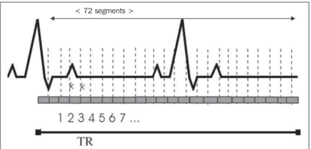
Real-time sequences(TR/TE = 3.5/0.9; flip angle = 40° and 8 mm section thick-ness) had a 128 × 72 acquisition matrix, with 72 segments per slice (Figure 2), an effective total TR of 2.300 msec.
Single-shot acquisitions were utilized in real-time sequences, optimizing the tempo-ral resolution, allowing the study of the whole heart during a single breath-hold period. The number of sections was simi-lar to the number of segmented acquisitions (six to nine sections for real-time and two or three slabs for segmented acquisitions) Total acquisition time (estimated in heart-beats) of real-time sequences ranged be-tween 12 and 18 heart-beats.
Parallel acquisition (multichannel) pro-tocols were utilized with an iPAT factor 2 in both sequences.
The initial FOV was of 350 mm, but varied among patients, aiming at offering
a better spatial resolution at each acquisi-tion, ranging between 280 and 420 mm.
For images acquisition, six leads were placed: four anterior and two on the dorsum. For real-time acquisitions, the ECG-gating served for the purpose of synchro-nizing the start signal for data acquisition, while in segmented images it was utilized for triggering pulse sequences.
The short cardiac axis was studied from the base to the apex with a 2 mm gap.
Ventricular function parameters were calculated with the Argus software (Sie-mens Medical Systems) through semi-au-tomatic endocardial cavity volume render-ing on the several sections of the short car-diac axis, with manually corrected data. All the patients had their parameters for analy-sis of the left ventricular function mea-sured: end-systolic (ESV) and end-diastolic (EDV) volumes, and ejection fraction.
Figure 1. Schematic diagram showing segmented TrueFISP sequences ECG-gating and TR-gating, dem-onstrating k-spaces and mean number of segments. Figure idealized by the author.
All the analyses were performed by a single observer in both sequences.
Pearson’s correlation and t-Student test were employed for statistical analysis. The concordance between the sequences was analyzed by means of the Bland-Altman plot with p < 0.01 as an indicator of statis-tically significant difference. The analyses were performed with the aid of the softwares Analyse-it forExcelTM, version 1.71, and MedCalc,version 7.6.0.0.
RESULTS
The epidemiological analysis of the sample included in the present study dem-onstrated a female predominance (18 women and 13 men) and an age range be-tween 7 and 77 years (mean age 47 years), with a standard deviation of ± 19 years.
The analysis of the cardiac rhythm dur-ing examinations demonstrated that 11 patients presented with irregular cardiac rhythm due the presence of frequent ven-tricular extra-systoles and other arrhyth-mias such as tachyarrhytharrhyth-mias, bigeminy and atrial fibrillation. The remaining 20 patients presented with regular sinusal rhythm.
Steady-state, real-time and segmented MRI acquisitions were performed for all the patients, from the base to the apex, as per Figures 3 and 4, with temporal resolu-tion of 39 msec and 126 msec respectively in cine-MR segmented and real-time acqui-sitions.
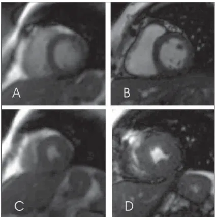
Real-time images showed lower spatial resolution than segmented images, a fact demonstrated by a slight blurring of con-tours on acquired images (Figure 5), which complicated the myocardial contour delin-eation. At the moment of the endocardial contour delineation, the papillary muscles were added to this contour if contacting the ventricular wall, otherwise, they were added to the ventricular cavity volume.
Volumetric parameters
There was no statistically significant difference (p < 0.01) between values ob-tained from both sequences. The mean val-ues obtained from real-time and segmented acquisitions were, respectively 55.7% and 53.8% for ejection fraction, 44 ml and 45 ml for ESV and 96.4 ml and 94.2 ml for
EDV. These functional parameters pre-sented a strong positive correlation r, as per Table 1.
The concordance between methods was tested by means of the Bland-Altman plot updated by NCCLS, with mean differences of respectively –2.1%, –2.2 ml and 1.7 ml for ejection fraction, EDV and ESV. The proposed graphic analysis (NCCLS guide-line EP9-A(9)) plotted the difference
be-tween the sequences values compared with values obtained from segmented acquisi-tions, demonstrating only two patients
fall-ing out of the confidence interval of agree-ment for ejection fraction and one patient for EDV and ESV (Graph 1).
Cardiac rhythm influence
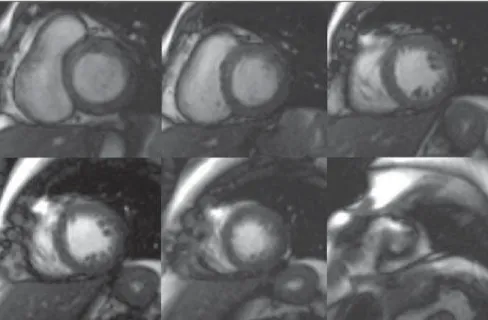
A comparison also has been performed between data obtained for both sequences — real-time and segmented — in patients with irregular cardiac rhythm due the pres-ence of arrhythmias, with a strong correla-tion and establishing the absence of statis-tically significant difference between val-ues by means of the t-Student test (Table 2) Figure 3. Short cardiac axis views acquired in segmented TrueFISP sequences, from the base to the apex.
(sinusal rhythm) is necessary for appropri-ate image acquisition in cardiac MRI. This is not always feasible due the frequent pres-ence of cardiac rhythm disorders in patients with cardiopathy. The introduction of real-time MRI sequences has allowed the acqui-sition of cardiovascular images indepen-dently from the ECG regularity. Manning and Pennell(10) consider that ECG-gating
and regular cardiac cycle are essential for an appropriate quality RM image, assertion that we consider real although, in our opin-ion the absence of these factor does not impede the performance of cardiac MRI. Real-time sequences utilize the cardiac gating as a start signal for data acquisition, however they do not depend on a normal R-R interval for images acquisition.
In this study we compared cardiac ECG-gated MRI steady-state sequences (seg-mented) with real-time sequences (multi-channel). Systolic ventricular function pa-rameters were utilized as a tool to indicate the existence of concordance between both sequences. Also, one has investigated if the presence of cardiac rhythm disorder affected the concordance between ventricular sys-tolic function parameters in both sequences. This is one of few studies to include, in the systolic parameters evaluation, patients presenting an irregular cardiac rhythm with arrhythmias (bigeminy, ventricular and supraventricular extra-systoles). These car-diac rhythm alterations affect the R-R in-terval causing irregularity and difficulting the EGC-gating. Barkhausen et al.(8) have
developed a similar study including a lower number of patients, but they have not re-ported any potential cardiac rhythm disor-der although they have discussed the pres-ence of already existent cardiac diseases.
Images quality
Systolic function parameters from two cine-MRI sequences (segmented and real-time acquisition) were utilized to determine the concordance between them. The deter-mination of such parameters required en-docardial contour delineation aiming at the quantitation of ventricular volumes. Such delineation is related to the level of clear-ness distinguishing myocardium from blood. Therefore, spatial and contrast reso-lution (images quality) was an essential attribute in the present study.
Figure 5. Short axis TrueFISP MR images of one of the patients included in the present study, demon-strating endocardial contour blurring. A: Heart base real-time image. B: Heart base segmented image. C: Heart apex real-time image. D: Heart apex segmented image.
Table 1 Result of comparison between averages of measurements for evaluation of ventricular func-tion and respective correlafunc-tion rates (r value), obtained in segmented and real-time TrueFISP sequences.
Parameter
Ejection fraction End-diastolic volume End-systolic volume
Segmented
55.7% 96.4 ml 44.0 ml
Real-time
53.8% 94.2 ml 45.0 ml
Correlation coefficient
0.94 0.93 0.98
Table 2 Values calculated according to Pearson’s correlation, t-Student test and p demonstrating the inexistence of difference between parameters for 11 patients with irregular cardiac rhythm.
Parameter
Ejection fraction End-diastolic volume End-systolic volume
Value t calculated
1.56 1.146 0.037
p
0.147 0.278 0.971
Pearson’s correlation
0.94 0.82 0.94
and the concordance between values ob-tained in both sequences of those 11 pa-tients.
Volumetric parameters acquired from patients with irregular cardiac rhythm dem-onstrated mean differences of respectively 2%, 5.2 ml and –0.1ml for ejection fraction, EDV and ESV (Graph 2), with only one pa-tient falling out of the confidence interval for EDV. This was the parameter which
presented a high confidence interval in the whole studied population.
DISCUSSION
Graph 1. Graphic representation of concordance as proposed by NCCLS EP9-A guideline updating Bland-EP9-Altman plot. EP9-A comparison was utilized between methods difference and segmented sequences. A: Concordance between ejection fraction in real-time sequences (FE – real) and segmented (FE – seg.). B: Concordance between end-diastolic volume for data obtained in real-time (EDV – real) and segmented (EDV – seg.) sequences. C. Concordance be-tween end-systolic volume for data obtained in real-time (ESV – real) and segmented (ESV – seg.) sequences.
Graph 2. Graphic representation of concordance as proposed by NCCLS EP9-A guideline updating Bland-EP9-Altman plot. EP9-A comparison was utilized between methods difference and segmented sequences. A: Concordance between ejection fraction in real-time sequences (FE – real) and segmented (FE – seg.). B: Concordance between end-diastolic volume for data obtained in real-time (EDV – real) and segmented (EDV – seg.) sequences. C. Concordance between end-systolic volume for data obtained in real-time (ESV – real) and segmented (ESV – seg.) sequences.
C
Mean = –0,1 CI = 11,2
ESV – seg.
D if fe re n c e E D V – s e g . × E D V – re a l D if fe re n c e E S V – s e g . × E S V – re a l
Mean = 5.2 CI = 29.7
B EDV – seg.
D if fe re n c e E D V – s e g . × E D V – re a l
Mean = 2 CI = 8.6
A D if fe re n c e F E – s e g . × F E – re a l
FE – seg.
C
Mean = 1.7 CI = 14.4
ESV – seg.
D if fe re n c e E S V – s e g . × E S V – re a l B
Mean = –2.2 CI = 24.4
EDV – seg.
D if fe re n c e E D V – s e g . × E D V – re a l A
Mean = –2.1 CI = 8.16
FE – seg.
The balance between spatial resolution (images quality) and temporal resolution (images acquisition time) is necessary for appropriate images acquisition.
The ability of patients to maintain breath-hold periods is directly related with his/her clinical condition, especially in cases of patients with cardiopathy. The breath-hold significance is paramount, since cardiac images acquisition with high-field MR equipment is performed within this temporal window.
Segmented acquisitions required two or three breath-hold periods to cover the whole left ventricle, while real-time acqui-sitions do it within just one apnea. This gain in the examination time resulted in loss of spatial resolution evidenced by the comparison between levels of images de-tailing in both sequences, particularly re-garding the delimitation of the blood-en-docardium interface. Such loss of spatial resolution has already been reported by other authors(4,11–13).
For comparison between parameters utilized in images segmented and real-time acquisitions, the following factors will af-fect the spatial resolution: images matrix, pixels and voxels size, acquisition time duration and images reconstruction soft-ware utilized.
In the present study, the matrix utilized in real-time sequences provided a mean pixel size of 3 mm., which is slightly larger than those reported in the study of Barkhausen et al.(4), while segmented
se-quences resulted in mean pixels size of 2.3 mm, contributing to a low contrast resolu-tion in real-time sequences. Miller et al.(12)
report that a pixel size of 1-2 mm may be obtained with high magnetic field (1.5 T) equipment with good temporal resolution and that alterations in cardiac volumetric parameters, especially the EDV, only would be observed on images with >3-mm pixels. These authors also say that section thickness >10 mm affect the values of ac-quired cardiac parameters. The slight blur-ring of real-time images has not evidenced any significant interference on the ventricu-lar function analysis, even making the de-lineation a little difficult, especially in pa-tients with extra-systoles, as a result of the difficulty in determining systole and dias-tole in the slices sequence..
The multichannel or integrated parallel imaging techniques (iPAT) have favored the temporal resolution of both sequences, reducing acquisition time, since they utilize simultaneous data acquisition because of variation of MR equipment coil sensitivity. Lee et al.(14) have evaluated cardiac
volumes with segmented and real-time TrueFISP sequences, obtaining similar spa-tial resolution, but they suggest that this similarity could inexist in case of an in-crease in the patient cardiac frequency. In the present study, real-time and segmented sequences presented different spatial reso-lutions, but with no statistical significance for volumetric evaluations, even in the 11 patients presenting with irregular cardiac rhythm.
Steady-state sequences present a better contrast resolution because they do not depend on the blood flow and their images formation is based on the relation between T1 and T2 or, more specifically, the tissues composition, differently from FLASH se-quences previously utilized to study the heart and which depended on the presence of blood flow to generate cine-MR images. The present study has utilized steady-state sequences for both for segmented and real-time acquisition methods, facilitating the endocardial contour delineation due the better contrast resolution. With steady-state (TrueFISP), real-time sequences present a lower signal intensity (signal noise)(8). This
fact has been observed in the comparison between short-axis views in both sequences included in our study.
The post-processing analysis of real-time and segmented TrueFISP sequences for endocardial contour delineation is a process that takes considerable time to be accomplished. Segmented sequences took, on average, less time than the real-time sequences, which is explained by the best contras resolution of the first ones. This analysis was extended in cases where pa-tients presented with irregular cardiac rhythm, since the presence of extra-systoles and other arrhythmias complicated the or-ganized disposition of sections for volu-metric quantitation by the Simpson’s tech-nique. Our data do not indicate a significant difference between parameters obtained, but this analysis was more prolonged, es-pecially for the patients with irregular
car-diac rhythm, in agreement with the current literature(9,15–17).
The utilization of a modified Bland-Altman graphic plotting for statistical con-cordance comparison has not demonstrated any significant variation between ventricu-lar systolic function parameters. The men-tioned plotting fitted effectively in the present study, since it compares a gold-standard method, which is closest to real values, with a new method. In the present study, the graphic analysis has shown only two patients with values falling out of the confidence interval for ejection fraction, and only one for EDV and another for ESV, with no common factor between them. This reflects an excellent concordance between sequences.
When separately analyzed, the 11 pa-tients with arrhythmias demonstrated a strong correlation and a good concordance between both sequences, even in the pres-ence of an irregular cardiac rhythm, with only one patient with EDV falling out of the confidence interval. However, a 7 ml increase has been identified in the analy-sis of EDV in these 11 patients with arrhythmias and of the mean EDV in the study population. This may represent a deterioration of the measurement accuracy due the presence of an irregular cardiac rhythm. The high confidence interval dem-onstrated by volumetric parameters graphs for EDV is compatible with this parameter variability, and previous studies have dem-onstrated absence of concordance for both sequences, as explained by Miller et al.(12).
Cine-MRI has progressed with potential to become a supplementary method for providing a higher number of data for car-diovascular evaluation. However, cardiac rhythm alterations present in patients with cardiopathy affect the images quality. The present study, comparing TrueFISP seg-mented sequences (currently the gold-stan-dard in cardiac evaluation) with real-time TrueFISP sequences, has concluded that the latest may be safely utilized for ven-tricular function assessment and determi-nation, independently from the presence of an irregular cardiac rhythm.
Acknowledgement
for the opportunity to accomplish the present study.
REFERENCES
1. Higgins CB. What standard has the gold? J Am Coll Cardiol 1992;19:1608–1609.
2. Lladó GP, Costa FC, Beiras AC, et al. Guías de práctica clínica de la Sociedad Espanõla de diología en resonancia magnética. Rev Esp Car-diol 2000;53:542–559.
3. Boxt LM. Cardiac MR imaging: a guide for the beginner. RadioGraphics 1999;19:1009–1025. 4. Barkhausen J, Ruhm SG, Goyen M, Buck T, Laub
G, Debatin JF. MR evaluation of ventricular func-tion: true fast imaging with steady-state preces-sion versus fast low-angle shot cine MR imaging – feasibility study. Radiology 2001;219:264–269. 5. Bellenger NG, Francis JM, Davies CL, Coats AJ, Pennell DJ. Establishment and performance of a magnetic resonance cardiac function clinic. J Cardiovasc Magn Reson 2000;2:15–20. 6. Lanzer P, Botvinick EH, Schiller NB, et al.
Car-diac imaging using gated magnetic resonance. Radiology1984;150:121–127.
7. Sakuma H, Fujita N, Foo TKF, et al. Evaluation of left ventricular volume and mass with breath-hold cine MR imaging. Radiology 1993;188: 377–380.
8. Barkhausen J, Goyen M, Ruhm SG, Eggebrecht H, Debatin JF, Ladd ME. Assessment of ventricu-lar function with single breath-hold real-time steady-state free precession cine MR imaging. AJR Am J Roentgenol 2001;178:731–735. 9. Lelieveldt BPF, van der Geest RJ, Lamb HJ, Kayser
HWM, Reiber JHC. Automated observer-indepen-dent acquisition of cardiac short-axis MR images: a pilot study. Radiology 2001;221:537–542. 10. Manning WJ, Pennell DJ. Cardiovascular
mag-netic resonance. 1sted. Philadelphia: Churchill Livingstone; 2002.
11. Kaji S, Yang PC, Kerr AB, et al. Rapid evalua-tion of left ventricular volume and mass without breath-holding using real-time interactive cardiac magnetic resonance imaging system. J Am Coll Cardiol 2001;38:527–533.
12. Miller S, Simonetti OP, Carr J, Kramer U, Finn JP. MR imaging of the heart with cine true fast imaging with steady-state precession: influence of spatial and temporal resolutions on left
ven-tricular functional parameters. Radiology 2002; 223:263–269.
13. Setser RM, Fischer SE, Lorenz CH. Quantifica-tion of left ventricular funcQuantifica-tion with magnetic resonance images acquired in real-time. J Magn Reson Imaging 2000;12:430–438.
14. Lee US, Resnick D, Burdy JM, Simonetti OP, Lee P, Weinreb JC. Cardiac function: MR evaluation in one breath hold with real-time true fast imag-ing with steady-state precession. Radiology 2002; 222:835–842
15. Graves MJ, Dommett DMT. Comparison of car-diac stroke volume measurement determined us-ing stereological analysis of breath-hold cine MRI and phase contrast velocity mapping. Br J Radiol 2000;73:825–832.
16. Lima JAC, Milind Y, Desai MD. Cardiovascular magnetic resonance imaging: current and emerg-ing applications. J Am Coll Cardiol 2004;44: 1164–1671.