Article
J. Braz. Chem. Soc., Vol. 26, No. 3, 498-505, 2015. Printed in Brazil - ©2015 Sociedade Brasileira de Química 0103 - 5053 $6.00+0.00
A
*e-mail: [email protected]
Rapid Preparation of (BiO)
2CO
3Nanosheets by Microwave-Assisted Hydrothermal
Method with Promising Photocatalytic Activity Under UV-Vis Light
Juliane Z. Marinho,a Lidiaine M. Santos,a Leilane R. Macario,b Elson Longo,b Antonio E. H. Machado,a,c Antonio O. T. Patrocinioa and Renata C. Lima*,a
aInstituto de Química, Universidade Federal de Uberlândia, CP 593, 38400-902 Uberlândia-MG, Brazil
bInstituto de Química, Universidade Estadual Paulista, 14800-900 Araraquara-SP, Brazil
cDepartamento de Química, Universidade Federal de Goiás, Campus Catalão, 75704-020 Catalão-GO, Brazil
Crystalline (BiO)2CO3 nanosheets were synthesized by a rapid one-step reaction via
microwave-assisted hydrothermal method using urea as a morphology mediator and carbon source. The hydrothermal method combined with microwave heating allowed to obtain sheet-like (BiO)2CO3 particles at shorter reaction times when compared to the conventional heating hydrothermal method. The photocatalytic activity of the as prepared samples was evaluated towards degradation of Ponceau 4R (C.I. 16255) under artificial UV-Vis light irradiation. The results show that good photocatalytic efficiency can be obtained for powders prepared with reaction times as low as 2 minutes.
Keywords: (BiO)2CO3, nanosheets, microwave-assisted hydrothermal method, photocatalysis
Introduction
Over the past decade, considerable efforts have been made to synthesize nanostructures for organic pollutant degradation under UV and visible-light irradiation.1-4 Bare
and doped anatase TiO2 structure, the most commonly used photocatalyst, has shown excellent results on complete mineralization of pollutants as well as in devices for effective solar energy conversion.5-12 From the first report
by Fujishima and Honda13 on water-splitting using TiO 2
and UV light, considerable attention have been deposited on the synthesis of low dimensional nanomaterials capable of realize photocatalysis.14-19 Given the interest
in semiconductor photocatalysts for environmental applications, it is necessary to develop new materials with improved visible-light-response in order to efficiently use the solar energy.
Typical Aurivillius oxides such as Bi2MO6 (M = W, Mo), BiVO4, BiOX (X = Cl, Br) and Bi2O3 have shown excellent
photocatalytic activity for both, solar energy conversion and environmental remediation.20-25 Zhang et al.26 reported
the effect of morphology and variation in local structure on Bi2MoO6 photocatalytic activities at different pHs.
Ren et al.27 investigated the different BiVO4 microstructures
and their photocatalytic activities for the degradation of rhodamine B under visible light. Yu et al.28 showed
different photocatalytic performances of Bi2WO6 powders in the degradation of organic pollutants depending on their calcination temperatures. Bismuth compounds with perovskite structure, such as BiFeO3, also present
photocatalysis applications.29
Bismuth subcarbonate ((BiO)2CO3) Aurivillius
structured was firstly reported by Grice research group.30
This oxy-carbonate has an alternate intergrown Bi2O22+
and CO32- layers in which the plane of the CO
32- ions is
orthogonal to the one of the Bi−O bond. Moreover, the internal layered structure of (BiO)2CO3 could guide the lower growth along (001) axis and thus morphologies like nanosheets or nanoplates can be obtained.31-33
(BiO)2CO3 with layered structure has presented expressive
antibacterial34 and photocatalytic35-38 properties.
and can also easily screen a wide range of experimental conditions in order to optimize the material preparation and properties.39-44
Herein, we report the (BiO)2CO3nanosheets prepared
by a rapid one-step reaction using the microwave-assisted hydrothermal (MAH) method. Their photocatalytic properties towards the degradation of the organic dye Ponceau 4R under artificial UV-Vis light irradiation were investigated and compared of (BiO)2CO3 prepared by other
methods. The azo-dye Ponceau 4R was chose due its large use in the food industry in Brazil,45 despite the fact of
several countries, including the United States, Norway and Finland, its classified as a carcinogen.46,47 Moreover, several
works in the literature have described the photocatalytic activities of new materials against azo compounds.48-50 The
synthetic methodology used allows a rapid and uniform heating of particles, leading to the production of efficient photocatalysts at short reaction times.
Experimental
Synthesis of (BiO)2CO3 nanostructures
All chemicals were analytical grade and were used without further purifications. 1.0 × 10⁻³ mol of
Bi(NO3)3·5H2O was dissolved in concentrated nitric acid
under constant stirring. 40 mL of distilled water with 5 × 10⁻³ mol of urea were added to the solution and the
pH was adjusted to 9 using a 3 mol L-1 KOH solution. The
suspension was transferred to a 50 mL Teflon autoclave and heated in 800 W microwave at 130 ºC during 2 and 8 min. The pale yellow solid obtained was washed with distilled water and ethanol and dried at 60 ºC.
Characterization
Crystalline phases were determined using a Shimadzu X-ray diffraction (XRD) 6000 equipped with CuKradiation (λ= 1.5406 Å) in the 2θ range from 10° to 60° with 0.02°
per min scan increment. Scanning electron microscopy
with a field emission gun (FE-SEM) Supra 35-VP was used to investigate particle size and morphology. The diffuse reflectance absorption spectra of the powders were recorded at room temperature on a Shimadzu UV-1650PC spectrophotometer equipped with an integrating sphere, in the region between 190 and 850 nm. Potassium bromide was used as reference in these experiments. The infrared spectra was measured from powder samples diluted in potassium bromide on a FTIR Shimadzu IR-Prestige 21 spectrometer in the 400-4000 cm-1 region. Raman spectra
was recorded on a RFS/100/S Bruker Fourier Transform
Raman (FT-Raman) spectrometer. The 1064 nm line of a Nd:YAG ion laser was used as the excitation source.
Photocatalytic studies
The photocatalytic activity of the new (BiO)2CO3
nanostructures were evaluated against the degradation of the commercial dye Ponceau 4R, P4R (acid red 18, C.I. 16255, Sigma-Aldrich, 80% dye content), under UV-Vis irradiation. The photocatalytic experiments were carried out in water cooled reactor described in detail previously.6 A 400 W high pressure mercury lamp was
used as irradiation source to provide a photonic flux of c.a. 3.3 × 10⁶ Einsteins s-1. 100 mg mL-1 of the catalyst was
added to 125 mg mL-1 P4R aqueous solution (pH = 6.1)
under magnetic stirring (4 L irradiated volume). The mixture was irradiated during 120 minutes. The system was kept at 40 ± 2 ºC. Aliquots were taken at 20 minutes intervals, filtered and analyzed spectrophotometrically, following the bleaching at 507 nm.6 The results presented
are averages of at least three different experiments. Control measurements in the dark were performed and no dye adsorption at the catalyst surface was observed.
Results and Discussion
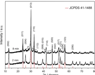
In the Figure 1 are shown the X-ray diffraction patterns of the powders obtained using the microwave-assisted hydrothermal (MAH) method with 2 or 8 minutes heating. The diffraction peaks can be indexed to the tetragonal (BiO)2CO3 structure with lattice constants, a = b = 3.865 Å,
c = 13.675 Å and space group I4/mmm, which are consistent
with the standard card, JCPDS No. 41-1488. The sharp and intense peaks indicated the highly crystalline nature of the (BiO)2CO3 samples obtained over 2 and 8 min
using the MAH conditions. Results obtained by Rietveld refinement of the XRD patterns of the samples show the presence of small amounts of secondary phases relative to polymorphs of bismuth oxides (Bi2O3) mixture and
the principal tetragonal (BiO)2CO3 phase with 99.9% and
81.3% to the samples obtained after 2 and 8 min by MAH method (Supplementary Information). In addition, the intensity ratio of the (110) peak to the (013) is about 0.42, higher than the correspondent standard card of bismutite, indicating that the crystals have anisotropic growth along the (110) plane.37
The morphology of the samples observed by FE-SEM is shown in Figure 2. The 2 min heated (BiO)2CO3 powders
two-dimensional (2D) nanosheets of fairly homogeneous thickness of about 10 nm, as shown in Figure 2b (inset).
During the heating into autoclave the prolonged urea hydrolysis results in either CO2 in acidic medium or
to CO32− ions in basic medium.51 Under hydrothermal
conditions the concentration of urea and the reaction time are important factors influencing the particles growth and the morphology of the products.52 The hydrothermal
method combined with microwave heating can accelerate the kinetics of crystallization by one to two orders of magnitude compared to the conventional hydrothermal method,39,53 which leads to an anisotropic crystal growth
and crystallization of (BiO)2CO3 under mild time and
temperature conditions.
In the MAH process at high pH,54 Bi(NO
3)2 and urea are
hydrolyzed to yield BiO+ and CO
32-, respectively, that react
between themselves to produce (BiO)2CO3 agglomerates.
At short reaction times, the intense and homogeneous microwave heating leads to the production of crystalline but non-ordered agglomerates, Figure 2a. As the reaction time increases, a series of redissolution and recrystalyzation
steps take place, and the resulting powder exhibits regular and ordered shape as well as crystallinity. Thus, the nucleation and growth of the regular nanosheets observed in Figure 2b is likely dependent on the reaction time. In Figure 3 the effect of reaction time on the morphology of MAH synthesized (BiO)2CO3 is illustrated.
The MAH (BiO)2CO3 particles exhibit broad absorbance
bands in the UV-Vis region (Figure 4). The abrupt decay of the absorbance between 380 and 420 nm is attributed to the band gap transition. The Kubelka-Munk function was used to estimate the optical absorption edge energy55
(Figure 4, inset). The as determined band gap energy are 2.9 and 2.7 eV for (BiO)2CO3 samples obtained at 2 and 8 min
heating, respectively and can be considered experimentally similar. These gapvalues are slightly smaller than the reported by Liu et al.32 using first-principle calculations, but
higher than the value reported by Zhang et al.56(2.23 eV).
Such difference can probably be related to the presence of defects in the crystalline structure of the synthesized samples and also to the influence of Bi2O3 phases that
exhibit lower band gaps57 and could explain the low lying
absorption features observed.
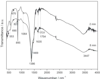
The infrared spectra of (BiO)2CO3 samples are shown
in Figure 5. Strong absorption bands of vibrational modes of the CO32− group are observed at 1386 and 1469 cm−1
assigned to anti-symmetric vibration ν3 and 823 and 845 cm−1 relative to out of plane bending modeν
2, being
at 845 cm-1 band characteristic of (BiO)
2CO3.58 In addition,
two weak absorption bands at 1064 and 670-690 cm−1
are ascribed to the symmetric stretching ν1andin-plane deformation ν
4modes of the carbonate ion; vibration
peaks at 1754 and 1731 cm−1 are observed for both
samples.59 Two sharp absorption bands at around 3447
and 1635 cm−1 can be attributed to the O−H stretching
mode and bending modes of adsorbed water molecules, respectively.58The medium strong band at 553 cm-1 is
assigned to the metal-oxygen bonds, Bi−O of (BiO)2CO3.
10 20 30 40 50 60 70 80
α β α α β β
αβ (20
6 ) JCPDS 41-1488 (1 2 3 ) (0 2 4 ) (1 2 1 ) (0 1 7 ) (0 2 2 ) (0 2 0 ) (1 1 4 ) (0 1 5 ) (0 0 6 ) (1 1 2 ) (1 1 0 ) (0 1 3 ) (0 0 4 ) (0 1 1 ) 2 min Intensit y / a .u .
2θ/ degree
8 min
(0
0
2
)
Figure 1. X-ray diffraction patterns of (BiO)2CO3 obtained by MAH
method.
Figure 2. FE-SEM images of (BiO)2CO3 obtained by MAH method with 2 min (a) and 8 min (b) heating time.
10 20 30 40 50 60 70 80
α β α α β β
αβ (20
6 ) JCPDS 41-1488 (1 2 3 ) (0 2 4 ) (1 2 1 ) (0 1 7 ) (0 2 2 ) (0 2 0 ) (1 1 4 ) (0 1 5 ) (0 0 6 ) (1 1 2 ) (1 1 0 ) (0 1 3 ) (0 0 4 ) (0 1 1 ) 2 min Intensit y / a .u .
2θ/ degree
8 min
(0
0
2
The bands ascribed to the Bi−O−Bi stretching vibrations relative to Bi2O3 impurities are covered by the band
centered at 553 cm-1. The peaks at low frequencies, at 420
and 474 are characteristic to the vibration of Bi−O bonds of monoclinic α-Bi2O3.60,61
Figure 6 shows the Raman spectra of (BiO)2CO3
samples obtained by MAH method. A shoulder at
162 cm-1 and bands around of 360 and 668 cm-1 assigned
to the lattice vibrations were observed. The weak band at 360 cm-1 is probably arise from the motion of oxygen
atoms in the polymeric (BiO)nn+ cation.62,63 The (BiO)2CO3
tetragonal lattice, as shown by X-ray diffraction, could only contained the CO32- ions in orientations parallel to the
(110) planes, since other dispositions result in exceedingly close inter-anionic contacts.The band at 229 and 307 cm-1
attributed to internal stretching modes of Bi−O bonds of β-Bi2O3 tetragonal structure61 is covered by the broad
band centered at 162 cm-1 referring to the presence of the
secondary phases identified by Rietveld refinement. Bands
characteristic of monoclinic α-Bi2O3 at 89 and 122 cm-1 are
more pronounced for the sample obtained for 8 min heating, which contains a higher concentration of Bi2O3 impurities
when compared to the sample prepared in 2 min.64
The photocatalytic activities of the MAH (BiO)2CO3
were evaluate against the degradation of the commercial dye Ponceau 4R (P4R). Continuous irradiation of P4R aqueous solutions in the presence of the catalysts lead to a progressive bleaching of the dye absorption band (Figure 7). As can be observed in Figure 7 (inset),no other bands appear in the spectral region monitored, indicating that no new chromophore groups have being formed during the dye degradation.
After 120 min irradiation, 43% discoloration was reached for the 2 minutes heated sample, and 40% for the sample heated during 8 minutes, which is lower than the observed for commercial TiO2 P25 under the same
conditions, but considerably higher in relation to the photo-discoloration of the dye in the absence of a catalyst.6
The discoloration follows a pseudo first-order kinetics in relation to the organic substrate (Figure 8) which agree with the Langmuir-Hinschelwood model.6,7,65 The rate constants
Figure 3. Scheme of rapid (BiO)2CO3 nanosheets formation by the
microwave-assisted hydrothermal (MAH) method.
300 400 500 600 700 800
8 min
A
b
s
o
rb
a
n
c
e
/
a
.u
.
Wavelength / nm
2 min 2.0 2.5 3.0 3.5 4.0 4.5
8 min
(
α
h
ν
)
1
/2
hν/ eV
2 min
Figure 4. UV-Vis spectra of the (BiO)2CO3 nanostructures. The inset shows
plots of (αhν)1/2versus photon energy (hν) for the (BiO)
2CO3 samples.
500 1000 1500 2000 2500 3000 3500 4000
670
1754 823
690
8 min 2 min
1469 1635
T
ra
n
s
m
it
ta
n
c
e
/
a
.u
.
Wavenumber / cm-1
3447
1386 1064
845 553
1731
Figure 5. FT-IR spectra of (BiO)2CO3 obtained by MAH method.
100 200 300 400 500 600 700
2 min
8 min 668 162
In
te
n
s
it
y
/
a
.u
.
Raman shift / cm-1
360
are 4.1 × 10⁻³ min-1 and 5.0 × 10⁻³ min-1, respectively for the
2 and 8 min heated MAH (BiO)2CO3 samples.
The results show that the photocatalytic activities of both samples are very similar, despite the different morphologies observed by electron microscopy. Moreover, the results are in the same order of previous reported photocatalytic activities for (BiO)2CO3 nanosheets
(Table 1). It is worthwhile to note the efficacy of the MAH method in produce this material in softer conditions and at shorter reaction times than the conventional hydrothermal method.
The small influence of the (BiO)2CO3 morphology
on the photocatalytic behavior, for the samples prepared by the MAH method at 2 or 8 minutes of heating time, can be related to the nature of the dye employed in this study. It is well known that the catalytic process involves dye adsorption/desorption on catalyst surface. Thus, high specific surface area as well as an open porous structure should enhance the catalytic activity. The 8 min MAH (BiO)2CO3 is constituted by regular nanosheets well
separated from each other, while the 2 min heated sample exhibit large agglomerates. Thus, the adsorption sites should be more available in the samples heated by 8 min. However, such a change in the morphology did not result in an increase in the P4R degradation, probably due to the size of the dye that inhibits the interaction with part of the catalytic sites. Probably, the photoprocess is limited by the charge separation efficiency of the semiconductor
0 20 40 60 80 100 120
0.0 0.2 0.4 0.6 0.8 1.0
200 300 400 500 600 0.0
0.5 1.0 1.5 2.0 2.5
A
b
so
rb
a
n
c
e
/a.u.
Wavelength / nm
Ab
s
/
Ab
s0
time / min
Figure 7. Absorption changes during the photocatalytic degradation of P4R, mediated by (BiO)2CO3 nanostructures heated by 2 min () and 8 min (), obtained by the MAH method. The photocatalytic activities were compared to that for the commercial TiO2 P25 () under the same conditions and also to the direct photolysis of the dye in the absence of catalyst (). Inset: electronic spectra of P4R at different irradiation times (∆t = 20 min).
0 20 40 60 80 100 120
0.0 0.1 0.2 0.3 0.4 0.5 0.6 0.7
-l
n
(
Abs/Ab
s0
)
time / min (a)
0 20 40 60 80 100 120
0.0 0.1 0.2 0.3 0.4 0.5 0.6 0.7
-ln
(
a
b
s
/a
b
s0
)
time / min (b)
Figure 8. P4R discoloration mediated by (BiO)2CO3 catalysts prepared
by microwave heating for 2 min (a) and 8 min (b), fitted according to a pseudo-first order kinetics.
Table 1. Bismuth subcarbonate (BiO)2CO3 sheet-like nanostructures obtained under different synthesis conditions and pollutants studied
Method Synthesis conditions Dye Discoloration Rate constant k / min-1 Ref.
Conventional hydrothermal 180 °C / 24 h RhB 47% in 60 min - 31
180 °C / 24 h RhB 60% in 50 min - 66
200 °C / 2 h RhB 40% in 100 min - 32
180 °C / 24 h RhB 35% in 120 min 0.0030 18
Microwave-hydrothermal 130 °C / 2 min P4R 43% in 120 min 0.0044 This work
130 °C / 8 min P4R 40% in 120 min 0.0035
and, the dye adsorption is controlled by physicochemical parameters, such as pH. Nevertheless, the higher rate constant observed for the 8 min heated sample evidences the effect of the more porous structure.
It is also important to consider the influence of secondary phases on the photocatalytic activity. These impurities can act as electron/hole traps, changing the charge recombination rates and, consequently, the photocatalytic activities. Cai et al.67 have shown enhanced
photocatalytic activities for β-Bi2O3/Bi2O2CO3 composites
in relation to pure Bi2O2CO3, which were attributed to
formation of efficient heterojunctions. As the Rietveld refinement have shown the presence of secondary phases on the MAH Bi2O2CO3 nanosheets, similar behavior can
also be occurring here and can be matter of future studies. The absence of dye adsorption at the experimental conditions employed evidences that the negatively charged sulfonate groups present in P4R did not favor an effective adsorption of dye on the catalyst surface. Hence, better photocatalytic activity can be obtained for smaller molecules and/or at different pH. Nevertheless, the MAH (BiO)2CO3 particles can be prepared faster and in softer
conditions than other catalysts reported in literature. Further studies are in progress to enhance the photocatalytic activity.
Conclusions
The microwave-assisted hydrothermal (MAH) is an appropriated, facile and environmentally friendly method for preparing rapidly (BiO)2CO3 nanosheets with
controllable shape and size. The hydrothermal conditions under microwave heating accelerates the kinetics of (BiO)2CO3 crystallization and this material can be obtained
at short reaction times when compared with the conventional hydrothermal method. Furthermore, the MAH conditions associated to the urea concentration play an important role on the morphology of the samples obtained. The microwave-hydrothermal time is a key factor to produce pure (BiO)2CO3 nanosheets. The as-prepared (BiO)2CO3
nanosheets obtained after 2 min and 8 min under MAH presented good photocatalytic activity for degradation of P4R under UV-Vis light, which motivates us to develop photocatalytic bismuth oxides and nanocomposites using the MAH method for degrading organic pollutants under solar light irradiation.
Supplementary Information
Supplementary data are available free of charge at http://jbcs.sbq.org.br as PDF file.
Acknowledgements
The authors are thankful to Coordenação de Aperfeiçoamento de Pessoal de Nível Superior (CAPES), Conselho Nacional de Desenvolvimento Científico e Tecnológico (CNPq, 477150/2008-0), Fundação de Amparo à Pesquisa do Estado de Minas Gerais (FAPEMIG, APQ-01842-09/APQ-00425-12) and Rede Mineira de Química (RQ-MG) by the financial support.
References
1. Hoffmann, M. R.; Martin, S. T.; Choi, W.Y.; Bahnemann, D. W.;
Chem. Rev.1995, 95, 69.
2. Mills, A.; LeHunte, S.; J. Photochem. Photobiol., A1997,
108, 1.
3. Papoulis, D.; Komarneni, S.; Panagiotaras, D.; Stathatos, E.; Toli, D.; Christoforidis, K. C.; Fernandez-Garcia, M.; Li, H.; Yin, S.; Sato, T.; Katsuki, H.; Appl. Catal. B: Environ. 2013, 132, 416.
4. Li, W. J.; Li, D. Z.; Meng, S. G.; Chen, W.; Fu, X. Z.; Shao, Y.;
Environ. Sci. Technol.2011, 45, 2987.
5. Stylidi, M.; Kondarides, D. I.; Verykios, X. E.; Appl. Catal. B:
Environ. 2004, 47, 189.
6. Oliveira, D. F. M.; Batista, P. S.; Muller Jr., P. S.; Velani, V.; França, M. D.; de Souza, D. R.; Machado, A. E. H.; Dyes
Pigments2012, 92, 563.
7. Machado, A. E. H.; Franca, M. D.; Velani, V.; Magnino, G. A.; Velani, H. M. M.; Freitas, F. S.; Muller, P. S.; Sattler, C.; Schmucker, A.; Inter. J. Photoenergy2008, 2008, 1.
8. Patrocinio, A. O. T.; El-Bacha, A. S.; Paniago, E. B.; Paniago, R. M.; Iha, N. Y. M.; Inter. J. Photoenergy2012,
2012, 1.
9. Brennaman, M. K.; Patrocinio, A. O. T.; Song, W. J.; Jurss, J. W.; Concepcion, J. J.; Hoertz, P. G.; Traub, M. C.; Iha, N. Y. M.; Meyer, T. J.; ChemSusChem2011, 4, 216.
10. Zhang, H.; Zong, R. L.; Zhao, J. C.; Zhu, Y. F.; Environ. Sci.
Technol.2008, 42, 3803.
11. Patrocinio, A. O. T.; Paniago, E. B.; Paniago, R. M.; Iha, N. Y. M.; Appl. Surf. Sci.2008, 254, 1874.
12. Feil, A. F.; Migowski, P.; Scheffer, F. R.; Pierozan, M. D.; Corsetti, R. R.; Rodrigues, M.; Pezzi, R. P.; Machado, G.; Amaral, L.; Teixeira, S. R.; Weibel, D. E.; Dupont, J.; J. Braz.
Chem. Soc.2010, 21, 1359.
13. Fujishima, A.; Honda, K.; Nature1972, 238, 37.
14. Chamjangali, M. A.; Boroumand, S.; J. Braz. Chem. Soc.2013,
24, 1329.
15. Chen, Y.; Dionysiou, D. D.; Appl. Catal. B: Environ. 2008, 80, 147.
17. Feng, B.; Yang, J. H.; Cao, J.; Yang, L. L.; Gao, M.; Wei, M. B.; Zhai, H. J.; Sun, Y. F.; Song, H.; J. Alloys Compd.2013, 555, 241.
18. Liang, H. Y.; Yang, Y. X.; Tang, J. C.; Ge, M.; Mater. Sci.
Semicond. Process.2013, 16, 1650.
19. Zhang, L.; Yan, J. H.; Zhou, M. J.; Yang, Y. H.; Liu, Y. N.; Appl.
Surf. Sci.2013, 268, 237.
20. Gui, M. S.; Zhang, W. D.; Su, Q. X.; Chen, C. H.; J. Solid State
Chem.2011, 184, 1977.
21. Dong, L.; Zhang, X.; Dong, X.; Zhang, X.; Ma, C.; Ma, H.; Xue, M.; Shi, F.; J. Colloid Interface Sci.2013, 393, 126. 22. Pare, B.; Sarwan, B.; Jonnalagadda, S. B.; J. Mol. Struct.2012,
1007, 196.
23. Xu, J.; Meng, W.; Zhang, Y.; Li, L.; Guo, C.; Appl. Catal. B:
Environ. 2011, 107, 355.
24. Ai, Z.; Huang, Y.; Lee, S.; Zhang, L.; J. Alloys Compd.2011,
509, 2044.
25. Zhang, L. S.; Wang, H. L.; Chen, Z. G.; Wong, P. K.; Liu, J. S.;
Appl. Catal. B: Environ. 2011, 106, 1.
26. Zhang, L. W.; Xu, T. G.; Zhao, X.; Zhu, Y. F.; Appl. Catal. B:
Environ. 2010, 98, 138.
27. Ren, L.; Ma, L. L.; Jin, L.; Wang, J. B.; Qiu, M. Q.; Yu, Y.;
Nanotech.2009, 20, 405602.
28. Yu, J. G.; Xiong, J. F.; Cheng, B.; Yu, Y.; Wang, J. B.; J. Solid
State Chem.2005, 178, 1968.
29. Ponzoni, C.; Rosa, R.; Cannio, M.; Buscaglia, V.; Finocchio, E.; Nanni, P.; Leonelli, C.; J. Eur. Ceram. Soc.2013, 33, 1325. 30. Grice, J. D.; Can. Mineral.2002, 40, 693.
31. Madhusudan, P.; Ran, J.; Zhang, J.; Yu, J.; Liu, G.; Appl.
Catal. B: Environ. 2011, 110, 286.
32. Liu, Y. Y.; Wang, Z. Y.; Huang, B. B.; Yang, K. S.; Zhang, X. Y.; Qin, X. Y.; Dai, Y.; Appl. Surf. Sci.2010, 257, 172.
33. Marinho, J. Z.; Silva, R. A. B.; Barbosa, T. G. G.; Richter, E. M.; Munoz, R. A. A.; Lima, R. C.; Electroanalysis2013, 25, 765.
34. Chen, R.; So, M. H.; Yang, J.; Deng, F.; Che, C. M.; Sun, H. Z.;
Chem. Commun.2006, 21, 2265.
35. Zhao, T.; Zai, J.; Xu, M.; Zou, Q.; Su, Y.; Wang, K.; Qian, X.;
CrystEngComm2011, 13, 4010.
36. Madhusudan, P.; Zhang, J.; Cheng, B.; Liu, G.; CrystEngComm
2013, 15, 231.
37. Chen, L.; Huang, R.; Yin, S.-F.; Luo, S.-L.; Au, C.-T.; Chem.
Eng. J.2012, 193, 123.
38. Cheng, H.; Huang, B.; Yang, K.; Wang, Z.; Qin, X.; Zhang, X.; Dai, Y.; ChemPhysChem2010, 11, 2167.
39. Komarneni, S.; Roy, R.; Li, Q. H.; Mater. Res. Bull.1992, 27, 1393.
40. de Moura, A. P.; Lima, R. C.; Paris, E. C.; Li, M. S.; Varela, J. A.; Longo, E.; J. Solid State Chem.2011, 184, 2818. 41. Hu, X. L.; Yu, J. C.; Adv. Funct. Mater.2008, 18, 880. 42. de Moura, A. P.; Lima, R. C.; Moreira, M. L.; Volanti, D. P.;
Espinosa, J. W. M.; Orlandi, M. O.; Pizani, P. S.; Varela, J. A.; Longo, E.; Solid State Ionics 2010, 181, 775.
43. Komarneni, S.; Katsuki, H.; Ceram. Int.2010, 36, 1165. 44. Volanti, D. P.; Keyson, D.; Cavalcante, L. S.; Simoes, A. Z.;
Joya, M. R.; Longo, E.; Varela, J. A.; Pizani, P. S.; Souza, A. G.;
J. Alloys Compd.2008, 459, 537.
45. Comissão Nacional de Normas e Padrões para Alimentos (CNNPA); Resolução No. 44: Brasília, Brasil,1977.
46. U.S. Food and Drug Administration (FDA); Compliance Program Guidance Manual. In http://www.fda.gov/ downloads/Food/GuidanceComplianceRegulatoryInformation/ ComplianceEnforcement/ucm073305.pdf accessed in December 2014.
47. European Food Safety Authority (EFSA); Scientific Opinion on
the Re-evaluation of Ponceau 4R (E 124) as a Food Additive. In
http://www.efsa.europa.eu/en/scdocs/scdoc/1328.htm accessed in December 2014.
48. Hernandez-Martinez, A. R.; Estevez, M.; Vargas, S.; Rodriguez, R.; Compos. Part. B-Eng.2013, 44, 686.
49. Salem, M. A.; Abdel-Halim, S. T.; El-Sawy, A.; Zaki, A. B.;
Chemosphere2009, 76, 1088.
50. Palfi, T.; Wojnarovits, L.; Takacs, E.; Radiat. Phys. Chem.2011,
80, 462.
51. Kieke, M. L.; Schoppelrei, J. W.; Brill, T. B.; J. Phys. Chem.
1996, 100, 7455.
52. Marinho, J. Z.; Romeiro, F. C.; Lemos, S. C. S.; Motta, F. V.; Riccardi, C. S.; Li, M. S.; Longo, E.; Lima, R. C.; J. Nanomater.
2012, 2012, 1.
53. Riccardi, C. S.; Lima, R. C.; dos Santos, M. L.; Bueno, P. R.; Varela, J. A.; Longo, E.; Solid State Ionics2009, 180, 288. 54. Wang, H. C.; Lu, C. H.; Mater. Res. Bull.2002, 37, 783. 55. Patterson, E. M.; Shelden, C. E.; Stockton, B. H.; Appl. Opt.
1977, 16, 729.
56. Zhang, X. C.; Guo, T. Y.; Wang, X. W.; Wang, Y. W.; Fan, C. M.; Zhang, H.; Appl. Catal. B: Environ. 2014, 150, 486.
57. Hou, J.; Yang, C.; Wang, Z.; Zhou, W.; Jiao, S.; Zhu, H.; Appl.
Catal. B: Environ. 2013, 142, 504.
58. Tsuji, M.; Ikeda, Y.; Sazarashi, M.; Yamaguchi, M.; Matsunami, J.; Tamaura, Y.; Mater. Res. Bull.2000, 35, 2109. 59. Wang, C. J.; Zhao, Z. W.; Luo, B.; Fu, M.; Dong, F.;
J. Nanomater.2014, 2014, 1.
60. Sun, Y. Y.; Wang, W. Z.; Zhang, L.; Zhang, Z. J.; Chem. Eng. J.
2012, 211, 161.
61. Wang, Y.; Wen, Y. Y.; Ding, H. M.; Shan, Y. K.; J. Mater. Sci.
2010, 45, 1385.
62. Taylor, P.; Sunder, S.; Lopata, V. J.; Can. J. Chem.1984, 62, 2863.
63. Dong, F.; Lee, S. C.; Wu, Z. B.; Huang, Y.; Fu, M.; Ho, W. K.; Zou, S. C.; Wang, B.; J. Hazard. Mater.2011, 195, 346. 64. Narang, S. N.; Patel, N. D.; Kartha, V. B.; J. Mol. Struct.1994,
65. Yang, H.; Li, G. Y.; An, T. C.; Gao, Y. P.; Fu, J. M.; Catal. Today
2010, 153, 200.
66. Zheng, Y.; Duan, F.; Chen, M.; Xie, Y.; J. Mol. Catal. A: Chem.
2010, 317, 34.
67. Cai, G. Y.; Xu, L. L.; Wei, B.; Che, J. X.; Gao, H.; Sun, W. J.;
Mater. Lett.2014, 120, 1.
Submitted: March 15, 2014
Published online: January 20, 2015