*e-mail: [email protected]
Hongyan Su, Jianwei Li, Hai Lin and Huakuan Lin*
aDepartment of Chemistry, Nankai University, Tianjin 300071, China
bEducation Ministry Key Laboratory of Functional Polymer Materials, Nankai University, Tianjin
300071, China
Um receptor para reconhecimento de ânions derivado da tiouréia foi sintetizado combinando-se indolina-2,3-diona e 1,3-diaminotiouréia. A interação pode combinando-ser facilmente monitorada acompanhando-se as mudanças no espectro de absorção na região do UV-vis induzidas pela complexação de um dado ânion. Por exemplo, veriicou-se que o luoreto liga-se ao receptor na proporção de 1:2, mas esta muda para 1:1 no caso dos ânions di-hidrogenofosfato e acetato. Este último apresenta a maior constante de ainidade dentre eles indicando um melhor reconhecimento molecular pelo novo receptor. Finalmente, a natureza da interação entre o novo receptor e o ânion acetato foram explorados utilizando-se espectroscopia de 1H-RMN.
An anion recognition receptor, a kind of thiourea derivative, was designed and synthesized by combining indoline-2,3-dione and 1,3-diaminothiourea. The anion recognition can be easily monitored by anion complexation induced changes in UV-vis absorption spectra. In particular, the binding ratio between the receptor and luoride is 1:2 but in the case of dihydrogenphosphate and acetate are 1:1. Moreover, the afinity constants revealed that the receptor can recognize acetate well. Finally, 1H NMR experiments were carried out to explore the nature of interaction between
this new receptor and acetate.
Keywords: thiourea; anion receptor; optical recognition; acetate
Introduction
Anions play signiicant roles in life processes and in the environment, such that the development of new anion receptors is of great interest and signiicance in host-guest chemistry.1 Anion artiicial receptor has represented an unique application prospect in anion sensor,2 membrane transmit carrier3 and simulation, enzyme catalyst synthesis, etc.4,5 Also their effects as environmental pollutants have only recently been realized and several types of synthetic chemosensors have been developed to date. For the molecular design of the chemosensors, how to achieve the speciic recognition of a certain anion and how to convert the recognition event into a signal are the key points.6
Among the various important anionic analytes, biologically important acetate anion is one of the most signiicant due to its speciic biochemical functions in the enzymes and antibodies.7 Acetate has unique chemical properties and can form the strongest hydrogen-bond
interaction with hydrogen-bond donors because of the trigonal geometry and the high basicity. Many examples are available on selective receptor molecules for acetate anion.8-10 However, there is paucity of reports that describe the change in color in the visible region of the spectrum, and thereby allowing the visual detection of the acetate anion using a receptor. Accordingly, we designed and synthesized the new receptor 1 (Scheme 1) containing indoline-2,3-dione and 1,3-diaminothiourea subunits. UV-Vis spectral and 1H NMR titration experiments were employed to investigate
the interaction between receptor 1 and several anions.
Experiments
Reagents
from Sigma-Aldrich Chemical Co., stored under vacuum in a dessiccator containing self-indicating silica-gel and fully dried before use. DMSO (dimethyl sulfoxide) was dried over CaH2 and then distilled at reduced pressure.
Apparatus
1H NMR spectra were obtained in a Varian UNITY
Plus-400 MHz Spectrometer using tetramethylsilane (TMS) as an internal reference. SI-MS experiments were performed in a MARINER apparatus. C, H, N elemental analyses were carried out in an Elementar Vario EL. UV-vis spectra were recorded in a Shimadzu UV-2450 Spectrophotometer using a quartz cuvette (path length = 1 cm).
General method
All experiments were carried out at 298 K, unless otherwise mentioned. A 2.0 × 10⁻⁴ mol L-1 solution of the compound 1 in DMSO was prepared and stored in dry atmosphere. This freshly prepared stock solution was used in all spectroscopic studies after appropriate dilution. 1.0 × 10⁻² mol L-1 solutions of tetra-n-butylammonium (TBA) salts of the respective anions were prepared using dried and distilled DMSO and were appropriately stored in dry atmosphere.
1H NMR titration experiments were carried out in
DMSO-d6 solution (TMS as internal reference). Receptor 1 (0.91 mg) was dissolved in DMSO-d6 to obtain a 5 × 10⁻³ mol L-1 solution. Then, increasing amounts of acetate anion (in DMSO-d6) were added into that solution, and the host-guest interaction was monitored by 1H NMR spectroscopy.
Synthesis of N, N’-di-(indolino-2-one-3-yl)-1,3-diiminothiourea (1)11
A solution of indoline-2,3-dione (isatin) (588 mg, 4 mmol) in ethanol (20 mL) was added dropwise to a hot solution of 1,3-diaminothiourea (212 mg, 2 mmol) in ethanol/water (20 mL, v/v = 1/1) with stirring at relux. After being stirred for 5 h, the solvent was removed in a lash evaporator. Recrystallization from ethanol yielded 664 mg of yellow crystals (83%). 1H NMR (400 MHz, DMSO-d6, Me4Si): 14.95 (s, 2H, N–H), 11.34 (s, 2H,
S=C-N–H), 7.58 (d, 2H, Ar-H, J 7.8 Hz), 7.43 (d, 2H, Ar-H, J 7.6 Hz), 7.25 (d, 2H, Ar-H, J 7.6 Hz), 6.97 (d, 2H, Ar-H, J 8.2 Hz). Elemental analysis: Calc. for C17H12N6O2S: C, 56.04; H, 3.32; N, 23.06; Found: C, 56.25; H, 3.37; N, 23.23. ESI-MS (m/z): 400.10 (M+H, calcd. 400).
Results and Discussion
UV-vis spectroscopic measurement
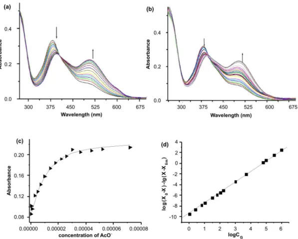
Firstly, to evaluate the binding ability, UV-vis titration experiments were carried out using a solution of the receptor 1 in dry DMSO tetrabutylammonium salts of AcO–, F–, H
2PO4 –,
Cl–, Br– and I–, at 298.2 ± 0.1K. The set of UV-vis spectra of 1 (1.0 × 10⁻⁵ mol L-1) recorded upon the addition of increasing concentration of AcO– is shown in Figure 1a. Two bands, one at 380 and another at 490 nm can be easily identiied in the spectrum of the pure solution of 1. Upon addition of AcO–, there is an decrease of the 380 nm band intensity, while the lower energy band intensity increased gradually as a function of acetate concentration, generating an isosbestic point at 404 nm. As a consequence, the color of the solution changed from yellow to red, which could provide a convenient method for naked-eye detection of AcO–, as it was coordinated to 1 and the negative charge was dispersed to the π-conjugated system through intramolecular charge transfer (ICT) effect. A similar spectral change was observed upon the addition of H2PO4– but the change was smaller. However, as Cl–, Br- and I– were titrated into 1, the spectra hardly changed even after an excess of the anions were added. In the case of F–, spectra changed without clear isosbestic points (see Figure 1b) implying formation of higher order complexes such as 1/ F– = 1/2,12 in analogy to the formation of [HF
2]- species. 13-14
This should be the result of the too small size of F– anion. Continuous variation method was used to determine the stoichiometric ratios of the receptors to the anion guests. In Figure 2 Job plot15,16 of receptor 1 and AcO−
in DMSO shows the maximum at a mole fraction of 0.5. This result indicates that the receptor 1 binds AcO−
guest with a 1:1 ratio. Moreover, similar results can also be obtained for H2PO4−
. On the other hand, the Job plot reveals that 1 coordinated F−
with a 1:2 ratio, showing the maximum at a molar fraction of 0.67.
For a complex of 1:1 stoichiometry, the relation in Equation (1) could be derived easily, where X is the absorption intensity, and CHor CG is the concentration of the host or the anion guest correspondingly.17
X=X0+ (Xlim-X0) {CH + CG+1/Kass-[CH + CG +1/Kass)2 -4 C
H CG] 1/2 }/2 C
H (1)
For a complex of 1:2 stoichiometry, the relation in Equation (2) could be derived easily, where X is the absorption intensity, and CHor CG is the concentration of the host or the anion guest correspondingly.18
(2)
The afinity constants of receptor 1 for anionic species were calculated and listed in Table 1 below.
Obviously, the recognition function of 1 for AcO− is more remarkable than H2PO4−
. Because AcO−
is a trigonal planar species and the angle of O-C-O is about 120º while the angle of O-P-O is about 108º,19 the distance between two oxygen atoms in AcO−
might it better to the recognition sites forming stronger hydrogen bonds. Furthermore, AcO−
is a stronger base than H2PO4− and F−
. The afinity constants of Cl− , Br−
and I−
for 1 were so small probably due to the much lower basicity of those species.
As a validation to the titration experiments, selective recognition experiments were performed by UV-vis spectral investigation. One equivalent of the AcO−
, H2PO4− , F−
,
Figure 1. (a). Family of spectra taken in the course of the titration of a 1.0 × 10⁻⁵ molL-1 solution of 1 with a standard solution of AcO− at 298.2 ± 0.1K. (b) Family of spectra taken in the course of the titration of a 1.0 × 10⁻⁵ molL−1 solution of 1 with a standard solution of F− at 298.2 ± 0.1K. (c) Fitting curve of the titration of a 1.0 × 10⁻⁵ molL-1 solution of 1 with a standard solution of AcO− at 298.2 ± 0.1K. (d) Fitting curve of the titration of a 1.0 × 10⁻⁵ mol L-1
solution of 1 with a standard solution of F− at 298.2 ± 0.1K.
Cl− , Br−
and I−
anions, were added to the solution of 1 (1.0 × 10⁻⁵ mol L-1), respectively. The UV-vis spectra were recorded and the results presented that 1 could recognize AcO−
well (Figure 3).
1H NMR spectroscopic analyses
Further supporting for the notion that hydrogen bond formed between 1 and anions came from 1H NMR spectroscopic analyses. They were carried out in DMSO-d6 under normal conditions of so-called NMR titration where the spectra of the receptor 1 were recorded in the presence of increasing concentrations of anions (Figure 4). Upon the addition of AcO– to the solution of 1, the original peak of H
a (marked in Scheme 1) at 14.95 ppm disappeared, but that at 11.34 ppm assigned to Hb (marked in Scheme 1) shifted downield. When 1.5 equivalents of AcO– were added to
1, the peaks corresponding to Hb broadened and moved downield to 12.64 ppm (∆d = 1.30 ppm), which suggests that AcO– was being combined with the two H
b of receptor
1 by hydrogen bonding. However, on the one hand, as to the signals of Ha, they were still not observed in the whole titration process of AcO– anions. On the other hand, the signals of the phenyl group shifted upield, which indicated the increase of the electron density on the phenyl ring owing to the deprotonation taking place. As a function of the titration experiments and spectra analysis, the proposed binding mode of receptor 1 and AcO– is given in Scheme 2.12
Conclusion
In conclusion, 1 has been developed as a novel colorimetric receptor for AcO– anion. It has been demonstrated that the receptor can bind anions in a 1:1 stoichiometry (except for F–) with obvious colorimetric changes, where the complex had been formed between 1 and AcO– by hydrogen bonding as proved by 1H NMR titration.
Table 1. Afinity constants of receptor 1 with anions at 298.2 ± 0.1 K in DMSO
Anion AcO– F– H
2PO4
– Cl– Br– I–
logKass 5.38 ± 0.27 4.73 ± 0.16 4.46 ± 0.12 ND ND ND
ND = can not be determined
Figure 3. UV-vis spectral changes of 1 in DMSO solution (1.0 × 10⁻⁵ mol L−1) after the addition of 1 equivalent of AcO−, H
2PO4
−, F−, Cl−, Br− and I−.
Figure 4.1H NMR spectra of 1 in DMSO-d
6 in the absence (a) and in the
presence (b) of 0.5, (c) 1.0, (d) 1.5 equivalent of AcO−.
References
1. Miao, R.; Zheng, Q. Y.; Chen, C. F.; Huang, Z. T.; Tetrahedron Lett. 2005,46, 2155.
2. Buhlmann, P.; Pretsch, E.; Bakker, E.; Chem. Rev.1998,98, 1593.
3. Kral, V.; Sessier, J. L.; Tetrahedron1995, 51, 539.
4. Kavauierators, K.; Carbtree, R. H.; Chem. Commun. 1999, 20, 2109.
5. Hubner, G. M.; Glaser, J.; Sell, C.; Angew Chem. Int. Ed. Engl.
1999,38, 383.
6. Yamagchi, S.; Akiyama, S.; Tamao, K.; J. Am. Chem. Soc.1999, 121, 10438.
7. Gunnlaugsson, T.; Davis, A. P.; O’Brien, J. E.; Glynn, M.; Org. Lett. 2002, 4, 2449.
8. Hossain, M. A.; Llinares, J. M.; Powell, D.; Bowman-James, K.; Inorg. Chem. 2001, 40, 2936.
Commun. 2006, 624.
13. Amendola, V.; Gomez, D. E.; Fabbrizzi,L.; Licchelli, M.; Acc. Chem. Res. 2006, 343.
14. Bonizzoni, M.; Fabbrizzi, L.; Taglietti, A.; Tiengo, F.; Eur. J. Org. Chem. 2006, 3567.
15. Job, A.; LiebigsAnn. Chem. 1928, 9, 113.
16. Liu, Y.; You, C. C.; Zhang, H. Y.; Supramolecular Chemistry, Nankai University Press, Tianjin. 2001.
17. Valeur, B.; Pouget, J.; Bourson, J.; Kaschke, M.; Ernsting, N. P.; J. Phys. Chem. 1992, 96, 6545.
18. Pan, B.; Gao, F.; He, R.; Cui, D.; Zhang, Y.; J. Colloid Interface Sci. 2006, 297, 151.
19. Sessler, J. L.; Gale, P.; Cho, W. S.; Anion Receptor Chemistry, The Royal Society of Chemistry. 2006.
Received: October 12, 2008