Short Report
S
J. Braz. Chem. Soc., Vol. 22, No. 5, 993-996, 2011. Printed in Brazil - ©2011 Sociedade Brasileira de Química 0103 - 5053 $6.00+0.00
*e-mail: [email protected]
Novel Anthraquinone Derivatives Produced by Pestalotiopsis guepinii,
an Endophytic of the Medicinal Plant Virola michelii (Myristicaceae)
Marilene N. Oliveira,a Lourivaldo S. Santos,*,a Giselle M. S. P. Guilhon,a Alberdan S. Santos,a Isabel C. S. Ferreira,a Manoel L. Lopes-Junior,a
Mara Silvia P. Arruda,a Andrey M. R. Marinho,a Milton N. da Silva,a Edson Rodrigues-Filhob and Maria C. F. Oliveirac
a Programa de Pós-Graduação em Química, Instituto de Ciências Exatas e Naturais,
Universidade Federal do Pará, 66075-110Belém-PA, Brazil
bDepartamento de Química, Universidade Federal de São Carlos, CP 676,
13565-905 São Carlos-SP, Brazil
cDepartamento de Química Orgânica e Inorgânica, Universidade Federal do Ceará,
60455-970 Fortaleza-CE, Brazil
Um novo derivado de antraquinona, denominado guepinone (1), juntamente com as conhecidas substâncias isossulocrina (2) e cloroissosulocrina (3) foram isolados de uma cultura em arroz de
Pestalotiopsis guepinii, um fungo endoitico de Virola michelii. Os compostos foram identiicados pela análise de seus dados espectrométricos de RMN 1D e 2D e EM. A atividade antimicrobiana dos compostos isolados foi avaliada e a cloroisossulcrina (3) foi a mais ativa.
A new anthraquinone derivative, named guepinone (1), along with the known substances isosulochrin (2) and chloroisosulochrin (3), were isolated from a rice culture of Pestalotiopsis guepinii, an endophytic fungus of Virola michelii. The compounds were identiied by analysis of 1D and 2D NMR and MS spectral data. The antimicrobial activity of these compounds was evaluated and chloroisosulchrin (3) was the most active.
Keywords: Virola michelii, Pestalotiopsis guepinii, anthraquinone derivatives, endophytic fungus
Introduction
Endophytic fungi have been collected from nearly all plants studied. They colonize the plants without causing visible disease symptoms. The function of the host plant is not yet clear and is supposed to depend on the organ-fungus interaction of each plant indicating that there are no neutral interactions but a balanced antagonism between the plant and the endophytic fungi.1
Endophytic fungi are very useful in agriculture and industry. They can be used as a tool for introduction of genes of interest into the plants,2,3 like plague and
pathogen inhibitors.4,5 They are also a source of primary
and secondary metabolites of interest.
In spite of the several reports concerning the production of substances by endophytic fungi that show biological
activities, such as fungicide, herbicide and algicide, among others,6 there are not many studies concerning the chemical
composition of endophytic microorganisms, especially when we analize the wide fungic biodiversity and the speciicity in the colonization of plants by fungi.
This paper deals with the study of the metabolites produced by the fungus Pestalotiopsis guepinii isolated from Virola michelii.
V. michelii belongs to the family Myristicaceae and it is used in folk medicine in the treatement of diseases caused by fungi and in the treatment of infections of the skin.7 Phytochemical study, anti-inlammatory and
allelopathic activities of the leaves of the V. michelii were reported.8-11
There have been described 205 species of Pestalotiopsis,12
Novel Anthraquinone Derivatives Produced by Pestalotiopsis guepinii J. Braz. Chem. Soc. 994
to the isolation of the diterpenoid taxol, an important anticancer drug.14
Pestalotiopsis also produce sesquiterpenes, other diterpenes, phenolic derivatives and anthraquinone derivatives.14-16
This paper reports the isolation and structural elucidation of three anthraquinone derivatives, a new lactone (1) named guepinone, isosulochrin (2) and chloroisosulochrin (3),17
Figure 1. In addition, this is the irst report of P. guepinii as an endophyte from V. michelii and its production of anthraquinones derivatives.
Experimental
General experimental procedures
UV spectra were obtained in MeOH solution on a Hewlett Packard 8452-A spectrophotometer, and IR spectra were measured with a Bomem MB-102 spectrophotometer in KBr pellets. High-resolution mass spectra were measured in a QTOF I (quadrupole-hexapole-TOF) mass spectrometer with an orthogonal Z-spray-electrospray interface (Micromass, Manchester, UK). Low resolution ESI-MS data were acquired in the negative ion mode, using a Quattro-LC instrument (Micromass, Manchester, UK) equipped with an ESI/APCI “Z-spray” ion source. HPLC was carried out in a semi-preparative LC-8A Shimadzu system with SPD-10AV Shimadzu UV detector (Tokyo, Japan) using a RP sinergy fusion column (250 mm × 10 mm, 5 µm), isocratic systems of H2O:MeCN
(40:60) and H2O:MeCN:MeOH (55:20:25), and a low rate
of 4.7 mL min−1. Detection was performed at 270 nm. All
solvents were iltered through a 0.45 mm membrane ilter
prior to use. Absorbance measurements were recorded on a Spectrum UV SPD-20A spectrophotometer. 1H NMR
(300 MHz) and 13C NMR (75 MHz) spectra were recorded
on a Varian Mercury 300, in CDCl3 and CD3OD, with
residual solvent peak as internal reference.
Plant material
Leaves of Virola michelii were collected in Belém, state of Pará, Brazil and a voucher specimen (No. 180621) has been deposited at the herbarium of the Empresa Brasileira de Pesquisa Agropecuária - Embrapa - (Botanic Division in Belém, State of Pará, Brazil).
Microorganism
The general procedures adopted for isolation of the microorganism followed the methodology described by Pereira.18 After collected, healthy leaves of Virola michelii
were washed with water and the surface sterilized by immersion in 70% aqueous ethanol (1 min), followed by 5% aqueous sodium hypochlorite (4 min), and inally with 70% aqueous ethanol (30 s). After these procedures, the leaves were rinsed with sterilized water. This latter water was incubated in Petri dishes to guarantee the elimination of all epiphytic microorganisms. Small pieces of the leaves were excised and placed in Petri dishes containing PDA medium at 30 °C. Individual hyphal tips of the emerging fungi were removed and replaced on PDA.
The pgfvm-04 strain was identiied as Pestalotiopsis guepinii by Francisco das Chagas de Oliveira Freire, Embrapa, State of Ceará, Brazil. A voucher specimen (LISB 68) has been deposited at the Laboratório de Investigação Sistemática em Biotecnologia, LABISISBIO, UFPA, Brazil.
Rice culture of Pestalotiopsis guepinii and isolation of the anthraquinones
The fungus was statically cultured in 45 erlenmeyer lasks (500 mL) containing 100 g of rice (“Uncle Ben’s”-parboiled) and 30 mL of distilled water per lask, and autoclaved at 121 °C for 45 min. A small disc of the PDA medium from the Petri dish containing mycelium of P. guepinii was transferred under sterile conditions to each erlenmeyer lasks. Three lasks were kept for control purposes. After 30 days of growth at 28 °C, methanol (300 mL) was added to each lask and allowed to stand for 5 h, and then it was iltered by gravity. The methanol was evaporated under reduced pressure to afford 157.3 g of a dark residue. This residue was suspended in 500 mL MeOH:H2O (1:3) solution. The suspension was extracted
Oliveira et al. 995 Vol. 22, No. 5, 2011
successively with hexane (3 × 1000 mL), dichlorometane (3 × 1000 mL) and EtOAc (3 × 1000 mL) and was further concentrated under vacuum. The hexane fraction (1.03 g) was separated by column chromatography on silica gel using as eluents mixtures of hexane, ethyl acetate and methanol of increasing polarities. After analysis by NMR of the sub-fractions, the hexane:EtOAc (5:1) fraction was submitted to semi-preparative reversed phase HPLC (250/10 synergi fusion C18, H2O:MeCN 40:60, low rate 4.7 mL min−1, 215 nm) to
yield 1 (32 mg). The dichloromethane fraction (1.04 g) was submitted to the same procedure and two sub-fractions were obtained (hexane:EtOAc 40% andhexane:EtOAc 60%). These fractions were submitted to semi-preparative reversed phase HPLC (250/10 synergi fusion C18, H2O:MeCN:MeOH
55:20:25, low rate 4.7 mL min−1, 215 nm) to yield 2 (32 mg)
and 3 (16 mg).
Guepinone (1)
White amorphous solid; mp 162.0-167.0 °C; UV lmax/nm (MeOH): 216, 255 and 313; IR (KBr) νmax/cm−
1
3000, 1712, 1653, 1618-1600; HRESIMS [M+Na]+ Found:
337.0691. Calc. for C17H14O6Na: 337.0688;
1H NMR and
13C NMR spectral data: see Table 1.
Isosulochrin (2)
Yellowish amorphous solid; mp 180-185 °C (MeOH); lit.17 182-189 °C; C
17H16O7; 1H NMR (300 MHz, CDCl3)
d10.2 (1H, br s, OH), 6.89 (1H, d, J 2.1 Hz, H-4), 6.54 (1H, d, J 2.1 Hz, H-6), 6.13 (2H, s, H-5’, H-3’), 3.52, 3.64
(each 3H, s, CH3O), 2.15 (3H, s, CH3); 13C NMR (75 MHz,
CDCl3) d199.5 (CO), 167.3 (COOCH3), 161.0 (C-5), 160.2
(C-6’, C-2`), 156.2 (C-1), 147.9 (C-4’), 131.2 (C-3), 122.4 (C-2), 109.8 (C-1’), 108.5 (C-5’, C-3’), 107.1 (C-4), 105.5 (C-6), 55.4 (CH3O), 52.2 (CH3O), 21.8 (CH3).
Chloroisosulochrin (3)
Yellowish amorphous solid; mp 166-169 oC (MeOH);
lit.17 170-173 oC. C
17H15O6Cl, ESIMS m/z 367.2 [M-H]−, 1H NMR (300 MHz; CD
3OD) d 7.15 (1H, s, H-4); 6.13 (2H,
s, H-5’, H-3’); 3.68, 3.92 (each 3H, s, CH3O), 2.19 (3H, s,
CH3);
13C NMR (75 MHz; CD
3OD) d200.6 (CO), 167.5
(COOCH3), 163.3 (C-6’, C-2’), 156.5 (C-5), 151.8 (C-1),
149.5 (C-4’), 129.5 (C-3), 127.2 (C-6), 115.7 (C-2), 110.5 (C-1’), 108.8 (C-5’, C-3’),104.8 (C-4), 55.4 (CH3O), 52.2
(CH3O), 21.8 (CH3).
Antimicrobial bioassay
The antimicrobial properties of compounds 1, 2 and 3
were evaluated by the disc diffusion method.19,20 The study
was carried out with the following microorganisms species: Escherichia coli (ATCC 8739), Pseudomonas aeruginosas (ATCC 9027), Staphylococcus aureus (ATCC 6538) and Candida albicans.
The compounds 1, 2 and 3 were weighed under aseptic conditions in sterile lasks and dissolved with DMSO to obtain 5 mg mL−1 solutions. These solutions were
impregnated on sterile paper discs of 6 mm diameter (20 µL per disc) and the discs were let to dry overnight to remove any residual solvent. The solvent control (DMSO) did not show any antimicrobial activity.
Seeded agar plates were prepared and inoculated with 0.1 mL of inoculum. Discs were then placed on the seeded agar plates. The zones of growth inhibition around the discs were measured after 24 h of incubation at 37 °C. All determinations were made in triplicate.
Results and Discussion
The MeOH extract obtained from the cultivation of Pestalotiopsis guepinii afforded after chromatographic separation, three anthraquinones derivatives (Figure 1).
The compounds 2 and 3 in conformitywith literature refer to isosulochrin and chloroisosulochrin.17 The
molecular formula of compound 1 was established as C17H14O6 (11 degrees of insaturation) on the basis of
HRESIMS (m/z 337.0691 [M+Na]+; ∆−0.3 mmu] analysis
and the NMR data (Table 1). The UV spectrum showed absorption maxima at 255 and 313 nm. The IR spectrum showed characteristic absorption bands of a hydroxy group (3000 cm−1), carbonyl of a lactone (1712 cm−1), carbonyl
Table 1. 1H and 13C NMR chemical shift (d in ppm) assignments for compound 1 in CDCl3a
Number 1H NMR dH 13C NMR dC
1 161.4
2 6.60 (dd, J 1.5 and 0.6)b 111.7
3 148.6
4 6.68 (dd, J 1.5 and 0.6) 107.1
5 6.88 (d, 2.4) 101.4
6 164.7
7 6.86 (d, 2.4) 112.1
8 169.3 9 179.7 10 158.0 4a 155.7 5a 135.0 8a 111.3 9a 106.6
Me 2.41 (s) 22.5
OMe-8 4.01 (s) 53.1
OMe-6 3.93 (s) 56.1
OH 12.27 (s)
Novel Anthraquinone Derivatives Produced by Pestalotiopsis guepinii J. Braz. Chem. Soc. 996
of a α, β-unsaturated ketone (1653 cm−1) and characteristic
absorption bands of aromatic ring (1618-1600 cm−1). The 13C NMR spectrum of 1 showed 17 signals attributed to:
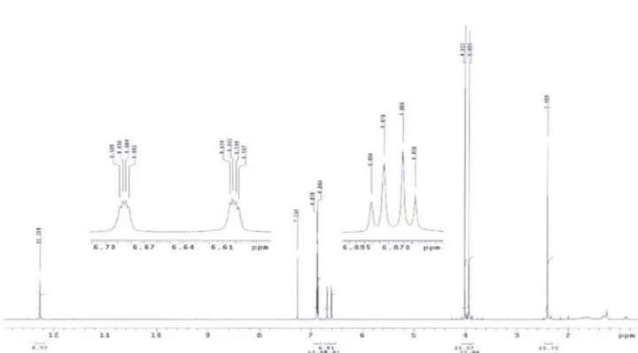
two carbonyls carbons (d158.0 and d 179.7 ppm), eight non-hydrogenated carbons, four aromatics CH carbons, one aromatic methyl and two methoxyl groups (Table 1), identiied by DEPT. The 1H NMR spectrum of compound 1
(Table 1) showed a singlet at d 12.27 ppm from a hydroxyl group H-bonded to a carbonyl, two doublets attributed to meta-coupled aromatic hydrogens, H-5 (d 6.88 ppm) and H-7 (d 6.86 ppm) and two double doublets at d 6.60 (J 1.5 and 0.6 Hz) andd6.68 (J 1.5 and 0.6 Hz) from H-2 and H-4, respectively. The COSY spectrum conirmed the coupling between one hydrogen from the methyl group (d 2.41 ppm) and the aromatic hydrogens (H-2 and H-4). The signals at d 3.93 and 4.01 ppm are characteristic of OCH3 groups. A
nOe difference experiment with irradiation at d 6.60 ppm (H-2) resulted in the enhancement of the signal of the CH3
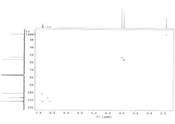
and also of phenolic OH and conirmed the position of the hydroxyl group. The conirmation of the position of the other substituents on the rings was obtained from HMBC data shown in Figure 2.
The antimicrobial activity of guepinone (1), isosulochrin (2) and cloroisosulochrin (3) were evaluated in the presence of Escherichia coli, Pseudomonas aeruginosa, Staphylococcus aureus and Candida albicans. Guepinone (1) and isosulochrin (2) were completely inactive against the microorganisms tested and cloroisosulochrin (3) was toxic only to Staphylococcus aureus (13 mm inhibition zone).
Supplementary Information
Supplementary information data (Figure S1-S33) are available free of charge at http://jbcs.sbq.org.br as PDF ile.
Acknowledgements
We thank CAPES/PROCAD for inancial support and a fellowship (M. N. O.), to CNPq and Fundação de Amparo a Pesquisa do Estado do Pará (FAPESPA) for inancial support.
References
1. Gotz, M.; Nirenberg, H.; Krause, S.; Wolters, H.; Draeger, S.; Buchner, A.; Lottmann, J.; Berg, G.; Smalla, K.; FEMS Microbiol. Ecol. 2006, 58, 404.
2. Fahey, J. W.; J. Am. Chem. Soc. 1988, 380, 120.
3. Murray, F. R.; Latch, G. C. M.; Scott, D. B.; Mol. Gen. Genet.
1992, 233, 1.
4. Hallmann, J.; Sikora, R. A.; Eur. J. Plant Pathol.1996, 102, 155.
5. Volksch, B.; Ullrich, M.; Frytsche, W.; Mycrobial Ecol. 1992, 24, 305.
6. Strobel, G.; Daisy, B.; Castillo, U.; Harper, J.; J. Nat. Prod.
2004, 67, 257.
7. Schultes, R. E.; Holmstedt, B.; Rhodora 1968, 70, 113. 8. Santos, L. S.; Corrêa, M. J. C.; Campos, L. M. O.; Andrade, M.
A.; Fitoterapia1996, 67, 555.
9. Carvalho, J. C. T.; Corrêa, M. J. C.; Campos, L. M. O.; Santos, L. S.; Bastos, J. K.; Sarti, S. J.; J. Ethnopharmacol. 1999, 64, 173.
10. Souza Filho, A. P. S.; Borges, F. C.; Santos, L. S.; Planta Daninha2006, 24, 205.
11. Santos, L. S.; Borges, F. C.; Oliveira, M. N.; Ferreira, I. C. S.; Guilhon, G. M. S. P.; Souza Filho, A. P. S.; Santos, A. S.; Arruda, M. S. P.; Muller, A. H.; Arruda, A. C.; Allelopathy J.
2007, 20, 235.
12. Jeewon, R.; Liew, C.Y. E.; Simpson, A. J.; Hodgkiss, I. J.; Hyde, D. K.; Mol. Phylogenet. Evol.2003, 27, 372.
13. Strobel, G.; Ford, E.; Worapong, J.; Harper, K. J.; Arif, M. A.; Grant, M. D.; Fung, W. C.; Chau, M. W. R.; Phytochemistry
2002, 60, 179.
14. Strobel, G.; Hess, W. M.; Ford, E.; Sears, J.; Sidhu, R. S.; Summerell, B.; Aust. J. Bot.1997, 45, 1073.
15. Li, J. Y.; Harper, J. K.; Grant, D. M.; Tomb, B. O.; Bashyal, B.; Hess, W. M.; Strobel, G. A.; Phytochemistry2001, 56, 463. 16. Magnani, R. F.; Rodrigues-Filho, E.; Souza, A. Q. L.; Ferreira,
A. G.; Daolio, C.; J. Biosci.2003, 58, 319.
17. Shimada, A.; Takahashi, I.; Kawano, T.; Kimura, Y.; Z. Naturforsch., B 2001, 56, 797.
18. Pereira, J. O.; Vieira, M. L. C.; Azevedo, J. L.; World J. Microbiol. Biotechnol.1999, 15, 43.
19. Bauer, A. W.; Kirby, M. D. K.; Sherries, J. C.; Truck, M.; Am. J. Clin. Pathol.1966, 45, 493.
20. Kartal, M.; Yildiz, S.; Kaya, S.; Kurucu, S.; Topçu, G.; J. Ethnopharmacol.2003, 86, 69.
Submitted: November 26, 2009
Published online: February 8, 2011
FAPESP has sponsored the publication of this article. Figure 2. Key HMBC (H → C) correlations of compound 1.
O O O HO CH3 OCH3
Supplementary Information
J. Braz. Chem. Soc., Vol. 22, No. 5, S1-S17, 2011. Printed in Brazil - ©2011 Sociedade Brasileira de Química
0103 - 5053 $6.00+0.00
S
I
*e-mail: [email protected]
Novel Anthraquinone Derivatives Produced by Pestalotiopsis guepinii,
an Endophytic of the Medicinal Plant Virola michelii (Myristicaceae)
Marilene N. Oliveira,a Lourivaldo S. Santos,*,a Giselle M. S. P. Guilhon,a Alberdan S.
Santos,a Isabel C. S. Ferreira,a Manoel L. Lopes-Junior,a Mara Silvia P. Arruda,a
Andrey M. R. Marinho,a Milton N. da Silva,a Edson Rodrigues-Filhob and
Maria C. F. Oliveirac
a Programa de Pós-Graduação em Química, Instituto de Ciências Exatas e Naturais,
Universidade Federal do Pará, 66075-110Belém-PA, Brazil
bDepartamento de Química, Universidade Federal de São Carlos, CP 676,
13565-905 São Carlos-SP, Brazil
cDepartamento de Química Orgânica e Inorgânica, Universidade Federal do Ceará,
60455-970 Fortaleza-CE, Brazil
Figure S1. 1H NMR spectrum (300 MHz, CDCl
Novel Anthraquinone Derivatives Produced by Pestalotiopsis guepinii J. Braz. Chem. Soc.
S2
Figure S2. 13C NMR spectrum (75 MHz, CDCl
3) of compound 1.
Figure S3. 13C NMR spectrum (75 MHz, CDCl
Oliveira et al. S3 Vol. 22, No. 5, 2011
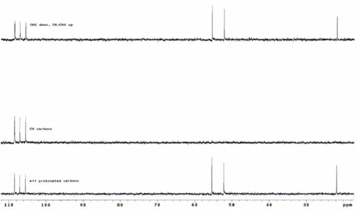
Figure S4. DEPT spectrum (75 MHz, CDCl3) of the compound 1.
Novel Anthraquinone Derivatives Produced by Pestalotiopsis guepinii J. Braz. Chem. Soc.
S4
Figure S6. HETCOR spectrum (CDCl3) of compound 1.
Oliveira et al. S5 Vol. 22, No. 5, 2011
Figure S8. HMBC spectrum of compound 1 (expansion).
Novel Anthraquinone Derivatives Produced by Pestalotiopsis guepinii J. Braz. Chem. Soc.
S6
Figure S10. HMBC spectrum (CDCl3) of compound 1 (expansion).
Oliveira et al. S7 Vol. 22, No. 5, 2011
Figure S12. NOEDIF spectrum (irradiation at 2.41 ppm) of compound 1.
Novel Anthraquinone Derivatives Produced by Pestalotiopsis guepinii J. Braz. Chem. Soc.
S8
Figure S14. NOEDIF spectrum (irradiation at 12.0 ppm) of compound 1.
Oliveira et al. S9 Vol. 22, No. 5, 2011
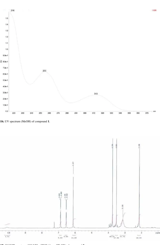
Figure S16. UV spectrum (MeOH) of compound 1.
Figure S17. 1H NMR spectrum (300 MHz, CDCl
Novel Anthraquinone Derivatives Produced by Pestalotiopsis guepinii J. Braz. Chem. Soc.
S10
Figure S18. 13C NMR spectrum (75 MHz, CDCl
3/drops CD3OD) of compound 2.
Oliveira et al. S11 Vol. 22, No. 5, 2011
Figure S20. HETCOR spectrum (CDCl3/drops CD3OD) of compound 2.
Novel Anthraquinone Derivatives Produced by Pestalotiopsis guepinii J. Braz. Chem. Soc.
S12
Figure S22. HMBC spectrum (CDCl3 /drops CD3OD) of compound 2 (expansion).
Oliveira et al. S13 Vol. 22, No. 5, 2011
Figure S24. NOEDIF spectrum (irradiation at 3.76 ppm) of compound 2.
Figure S25. 1H NMR spectrum (300 MHz, CD
Novel Anthraquinone Derivatives Produced by Pestalotiopsis guepinii J. Braz. Chem. Soc.
S14
Figure S26. 13C NMR spectrum (75 MHz, CD
3OD) of compound 3.
Oliveira et al. S15 Vol. 22, No. 5, 2011
Figure S28. HETCOR spectrum (CD3OD) of compound 3 (expansion).
Novel Anthraquinone Derivatives Produced by Pestalotiopsis guepinii J. Braz. Chem. Soc.
S16
Figure S30. HMBC spectrum (CD3OD) of compound 3.
Oliveira et al. S17 Vol. 22, No. 5, 2011
Figure S32. NOEDIF spectrum (irradiation at 7.15 ppm) of compound 3.