337
Fibrosing mediastinitis: a case report
Radiol Bras. 2009 Set/Out;42(5):337–339
Case Report • Relato de Caso
Fibrosing mediastinitis: a case report*
Mediastinite fibrosante: relato de casoThales de Carvalho Lima1, Edson Marchiori2, Domenico Capone3, Miriam Menna Barreto4, Rosana Souza Rodrigues4, Gláucia Zanetti5
The present study reports the case of a 51-year-old woman with chest pain, dyspnea and upper chest vessels engorgement, with no clinical evidence of granulomatous disease. Chest imaging study revealed a mediastinal mass with calcifications that was approached by mediastinoscopy with biopsy. Histopathological study demonstrated an etiologically undefined chronic inflammatory pattern compatible with fibrosing mediastinitis. Keywords: Fibrosing mediastinitis; Mediastinal mass; Fibrosis; Chest radiology.
Neste trabalho é relatado o caso de uma mulher de 51 anos de idade, com dor torácica, dispneia e engurgi-tamento de vasos do tórax superior, sem evidências clínicas de doença granulomatosa. O estudo por ima-gem do tórax revelou massa mediastinal com calcificações, abordada por mediastinoscopia com biópsia. A análise histopatológica mostrou padrão inflamatório crônico sem etiologia definida, compatível com medias-tinite fibrosante.
Unitermos: Mediastinite fibrosante; Massa mediastinal; Fibrose; Radiologia torácica.
Abstract
Resumo
* Study developed at Hospital Universitário Clementino Fraga Filho (HUCFF) – Universidade Federal do Rio de Janeiro (UFRJ), Rio de Janeiro, RJ, Brazil.
1. MD, Resident in Radiology, Universidade Federal do Rio de Janeiro (UFRJ), Rio de Janeiro, RJ, Brazil.
2. Full Professor, Department of Radiology, Universidade Fe-deral Fluminense (UFF), Niterói, RJ, Adjunct Coordinator for the Course of Post-Graduation in Radiology, Universidade Federal do Rio de Janeiro (UFRJ), Rio de Janeiro, RJ, Brazil.
3. PhD, Associate Professor at Universidade do Estado do Rio de Janeiro (UERJ), MD, Unit of Radiodiagnosis, Hospital Univer-sitário Clementino Fraga Filho (HUCFF) – Universidade Federal do Rio de Janeiro (UFRJ), Rio de Janeiro, RJ, Brazil.
4. PhDs, MDs, Radiologists, Unit of Radiodiagnosis, Hospital Universitário Clementino Fraga Filho (HUCFF) – Universidade Federal do Rio de Janeiro (UFRJ), Rio de Janeiro, RJ, Brazil.
5. PhD, Professor of Medical Practice, Faculdade de Medicina de Petrópolis, Petrópolis, RJ, Brazil.
Mailing address: Dr. Thales de Carvalho Lima. Rua Pedro de Carvalho, 658, ap. 202, Lins de Vasconcelos. Rio de Janeiro, RJ, Brazil, 20725-233. E-mail: [email protected]
Received May 15, 2008. Accepted after revision June 9, 2008.
Lima TC, Marchiori E, Capone D, Barreto MM, Rodrigues RS, Zanetti G. Fibrosing mediastinitis: a case report. Radiol Bras. 2009;42(5):337–339.
0100-3984 © Colégio Brasileiro de Radiologia e Diagnóstico por Imagem INTRODUCTION
Fibrosing mediastinitis, also known as sclerosing mediastinitis, is an uncommon condition characterized by proliferation of dense fibrous tissue in the mediastinum. In spite of its benignity, it is associated to a significant morbidity due to its fibrotic obstructive nature, and less commonly, to mortality(1). Its pathogenesis is unknown in most cases, also being possibly related to histoplasmosis, tuberculosis, and other granulomatous diseases, such as sarcoido-sis, autoimmune and fungal diseases,
radio-therapy and other fibro-inflammatory pro-cesses, for example, retroperitoneal fibro-sis(2–4). Affected patients are typically young and present signs and symptoms related to obstruction of vital mediastinal structures, such as large vessels, esophagus and airways(5).
CASE REPORT
A 51-year-old female patient presenting a history of chest pain, dyspnea on exertion over the last four months, and finally, vas-cular engorgement on the chest wall and
neck. There was no epidemiological history of histoplasmosis or tuberculosis; even so, three induced sputum tests were per-formed, all of them negative for tubercu-losis or fungi, besides serology of histo-plasmosis, which was also negative. Clini-cal and laboratory investigations for other less frequent causes of the disease were also negative.
Chest radiography demonstrated supe-rior mediastinal enlargement, notably to the right, with no other noticeable finding (Fig-ure 1). Chest computed tomography dem-onstrated a mass with soft tissue density
338
Lima TC et al.
Radiol Bras. 2009 Set/Out;42(5):337–339
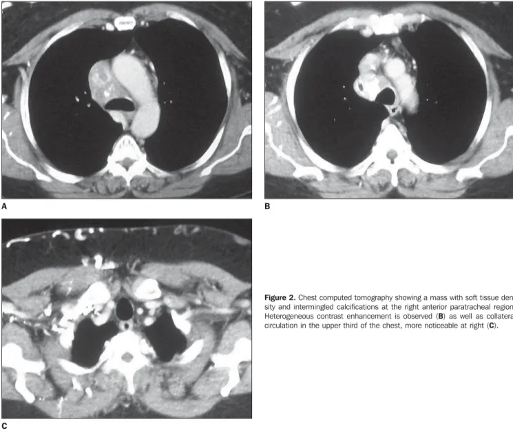
and intermingled calcifications at the ante-rior right paratracheal region, with hetero-geneous contrast enhancement, without cleavage plane with the superior vena cava, extending to the carina, besides the pres-ence of collateral circulation in the chest (Figure 2).
Finally, a mediastinoscopy was per-formed, with a biopsy of the mediastinal mass, whose histopathological study dem-onstrated a chronic inflammatory fibrotic-necrotic chronic process, negative for mi-croorganisms at silver, PAS and Ziehl-Nielsen stain. The findings were compat-ible with the fibrosing mediastinitis hy-pothesis. The patient progressed satisfac-torily, with stabilization of clinical signs, and undergoing clinical follow-up.
DISCUSSION
Fibrosing mediastinitis is caused by an extensive, infiltrating, and frequently inva-sive mediastinal fibrotic process. The in-flammatory process usually sets in the up-per half of the mediastinum, in the paratracheal region. It is commonly located to the right, anteriorly to the trachea and close to the pulmonary hilum; however it may also develop as a diffuse fibrosis in the mediastinum, extending from the brachio-cephalic veins to the pulmonary bases(1,6). The symptoms are generally caused by obstruction of the superior vena cava, esophagus, trachea, bronchi or pulmonary veins, also causing pulmonary arterial hy-pertension by direct compression of
pulmo-nary arteries, or secondary to pulmopulmo-nary venous compression(4,7).
Chest radiography is nonspecific, dem-onstrating superior mediastinal enlarge-ment by a paratracheal lobular mass(8).
Cal-cifications occur in most cases, requiring a computed tomography scan to character-ize them and ruling out other causes of mediastinal involvement such as me-tastases, lymphomas and other tumors(1,9).
Secondary lung alterations such as atelecta-sis, nodules, consolidation and interstitial infiltration are also observed(6,9). Magnetic resonance imaging is useful in the determi-nation of the disease extent and in the pre-operative planning, usually showing an image with intermediate signal intensity on T1-weighted sequences and heterogeneous
Figure 2. Chest computed tomography showing a mass with soft tissue den-sity and intermingled calcifications at the right anterior paratracheal region. Heterogeneous contrast enhancement is observed (B) as well as collateral circulation in the upper third of the chest, more noticeable at right (C).
C
339
Fibrosing mediastinitis: a case report
Radiol Bras. 2009 Set/Out;42(5):337–339
signal intensity on T2-weighted sequences because of the fibrotic nature of the disease and presence of calcifications, sometimes presenting heterogeneous gadolinium en-hancement(1,6,10).
The course of the disease is variable, sometimes with spontaneous remission, and other times with symptoms exacerba-tion. Approximately 30% of patients die from complications resulting from obstruc-tion and fibrosis. The worst prognosis is re-lated to bilateral or carinal involvement(4,6). The definite diagnosis is obtained by means of biopsy of the affected lymph nodes or mediastinal tissue(6). The therapy includes the use of corticosteroids, surgical ap-proach for excision of tissue and local man-agement of obstructive complications such as the case with the use of stents(11)
.
Finally, in spite of its uncommon char-acter, fibrosing mediastinitis should be considered in cases of mediastinal enlarge-ment at chest radiography, primarily for
being related to granulomatous diseases such as tuberculosis that is endemic in Bra-zil and also for its significant morbidity, as the affected patients are typically young. The investigation must include contrast-enhanced computed tomography of chest associated with serology of tuberculosis and histoplasmosis. Biopsy with histo-pathological study confirms the diagnosis. Regular clinical follow-up must be per-formed with these patients for monitoring of possible complications resulting from the disease.
REFERENCES
1. Devaraj A, Griffin N, Nicholson AG, et al. Com-puted tomography findings in fibrosing medias-tinitis. Clin Radiol. 2007;62:781–6.
2. Lee JY, Kim Y, Lee KS, et al. Tuberculous fibrosing mediastinitis: radiologic findings. AJR Am J Roentgenol. 1996;167:1598–9.
3. Loyd JE, Tillman BF, Atkinson JB, et al. Medi-astinal fibrosis complicating histoplasmosis. Medicine (Baltimore). 1988;67:295–310. 4. Dechambre S, Dorzee J, Fastrez J, et al. Bronchial
stenosis and sclerosing mediastinitis: an uncom-mon complication of external thoracic radio-therapy. Eur Respir J. 1998;11:1188–90. 5. Kalweit G, Huwer H, Straub U, et al. Mediastinal
compression syndromes due to idiopathic fibrosing mediastinitis – report of three cases and review of the literature. Thorac Cardiovasc Surg. 1996; 44:105–9.
6. Rossi SE, McAdams HP, Rosado-de-Christenson ML, et al. Fibrosing mediastinitis. Radiographics. 2001;21:737–57.
7. Mathisen DJ, Grillo HC. Clinical manifestation of mediastinal fibrosis and histoplasmosis. Ann Thorac Surg. 1992;54:1053–8.
8. Sherrick AD, Brown LR, Harms GF, et al. The ra-diographic findings of fibrosing mediastinitis. Chest. 1994;106:484–9.
9. Weinstein JB, Aronberg DJ, Sagel SS. CT of fibrosing mediastinitis: findings and their utility. AJR Am J Roentgenol. 1983;141:247–51. 10. Erasmus JJ, McAdams HP, Donnelly LF, et al. MR
imaging of mediastinal masses. Magn Reson Imaging Clin N Am. 2000;8:59–89.