Article
J. Braz. Chem. Soc., Vol. 26, No. 9, 1956-1966, 2015. Printed in Brazil - ©2015 Sociedade Brasileira de Química 0103 - 5053 $6.00+0.00
A
*e-mail: [email protected]
Coupling DLLME-CE for the Stereoselective Analysis of Venlafaxine and Its Main
Metabolites after Biotransformation by Fungi
Marcela A. Bortoleto,a Mariana Z. Bocato,b Mônica T. Pupo,b Cristiane M. Gaitanib and
Anderson R. M. Oliveira*,a
aDepartamento de Química, Faculdade de Filosofia, Ciências e Letras de Ribeirão Preto,
Universidade de São Paulo, 14040-901 Ribeirão Preto-SP, Brazil
bFaculdade de Ciências Farmacêuticas de Ribeirão Preto, Universidade de São Paulo,
14040-903 Ribeirão Preto-SP, Brazil
Fungal biotransformations have become very important in the study of chiral drugs because the reactions performed by these microorganisms may be enantioselective. However, analyses of analytes present in liquid culture medium have proved to be very difficult due to the complexity of this matrix. The aim of this work was to couple dispersive liquid-liquid microextraction (DLLME) with capillary electrophoresis to evaluate the biotransformation of the antidepressant drug venlafaxine (Vx) into its chiral metabolites, N-desmethylvenlafaxine (NDV) and O-desmethylvenlafaxine (ODV) by fungi. The chiral separation was carried out in 50 mmol L-1 sodium phosphate buffer pH 2.0 containing 8 mmol L-1α-cyclodextrin and 1.0% (m/v) carboxymethyl-β-cyclodextrin. The temperature of the capillary was set at 20 °C. A voltage of +20 kV was applied during analysis. The DLLME was accomplished using 300 µL of isopropanol (disperser solvent) and 200 µL of chloroform (extraction solvent). The method was completely validated and showed to be linear over the concentration range of 75-938 ng mL-1 for ODV and NDV enantiomers and of 500-15000 ng mL-1 for venlafaxine enantiomers with a correlation coefficient higher than 0.99. The selectivity of the method was evaluated and no interference peaks were detected in the migration time of the analytes. The limit of quantification was 75 ng mL-1 for metabolite enantiomers and 500 ng mL-1 for venlafaxine enantiomers. The study showed a stereoselective biotransformation of venlafaxine into (+)-(S)-N-desmethylvenlafaxine by the fungus Cunninghamella elegans ATCC 10028B with an enantiomeric excess of 100%.
Keywords: venlafaxine, dispersive liquid-liquid microextraction, fungal biotransformation, chiral separation
Introduction
Venlafaxine (Vx) hydrochloride ((R/S
)-1-[(2-dimethylamine)-1-(4-methoxyphenyl) ethyl] cyclohexanol) is a second-generation antidepressant used in the treatment of depression and anxiety associated with depression.1
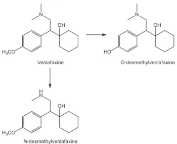
After oral administration, Vx is metabolized by the hepatic cytochrome P450 enzymes (CYP2D6 and CYP3A4) producing two main metabolites: N-desmethylvenlafaxine
(NDV) and O-desmethylvenlafaxine (ODV) which are
also chiral molecules (Figure 1). ODV is the metabolite produced in higher proportion, and it has similar pharmacological activity when compared to venlafaxine.1
Therapeutic study conducted with the enantiomers of Vx
proved a stereoselective activity. The (R)-venlafaxine inhibits the reuptake of norepinephrine and serotonin, while (S)-venlafaxine, exclusively, inhibits serotonin reuptake.2
In February 2008, the Food and Drug Administration (FDA) approved the metabolite ODV for the treatment of major depressive disorder in adults.3 Numerous patent
applications for synthesis of ODV have been deposited which confirm the trend to obtain this drug.
In vitro biotransformation has become an important tool to perform in vitro metabolism studies. These studies have several advantages over the use of in vivo models or chemical processes due to their lower complexity. The use of microorganisms is an inexpensive procedure and allows the control of conditions strictly. In addition, the biotransformation processes may be enantioselective.4,5
may produce a chiral compound (from a prochiral molecule) and/or enantiomers of a racemic mixture may be biotransformed at different rates leading to an enantiomeric excess of an enantiomer. The biotransformation carried out by fungi is feasible because these microorganisms may mimic the mammalian enzymatic reactions providing the same (and also some different) metabolites that are found in mammalians.4 In addition, they can resist to high amount of
substrates which may lead to a production of high amount of the target metabolite(s).4,5 There are several studies
showing the ability of fungi in promoting stereoselective biotransformation,6-11 however, there are no reports using
the drug Vx as substrate.
Some methods have been reported the stereoselective analysis of Vx and its metabolites in biological fluids mainly by chromatography systems12-14 using classical sample
preparation techniques such as liquid-liquid extraction12
or solid phase extraction.14 Moreover, only one report of
the use of a miniaturized sample preparation technique has been described for the analysis of Vx and its metabolites.13
There are, until now, only three methods describing the enantioselective analysis of Vx and its metabolite ODV by capillary electrophoresis (CE);15-17 and none of them
describe a simultaneous enantioseparation of NDV, ODV and Vx in a single run. Rudaz et al.,15 reported the analysis
of Vx and ODV in serum samples by CE using phosphated-gamma-cyclodextrin as chiral selector. The resolution reported by the authors for Vx and ODV was higher than 1.5. Fanali et al.16 described the analysis of these same
molecules in human plasma by electrochromatography using a vancomycin packed capillary. The mobile phase was composed of 100 mmol L-1 ammonium acetate buffer
pH 6:water:acetonitrile (5:5:90, v/v/v). The resolution achieved by the authors was 1.68 for Vx enantiomers and
1.57 for ODV enantiomers. Finally, Rudaz et al.,17 described
the chiral analysis of Vx and ODV by CE employing a mixture of two cyclodextrins (CD): carboxymethyl-β-CD (CM-β-CD) and α-CD.
In 2006, Rezaee et al.18 developed a novel liquid-phase
microextraction technique named dispersive liquid-liquid microextraction (DLLME). DLLME is based on a cloudy solution, which is formed when an appropriate mixture of extraction and disperser solvents is quickly injected into an aqueous sample containing the target analytes. The surface areas between the extraction solvent and sample solution are very large. Therefore, the extraction equilibrium can be achieved easily and quickly. At the end of the extraction procedure, the sample is centrifuged and the sedimented phase enriched with the analyte of interest is determined by a suitable analytical technique. The main advantage of DLLME is the simplicity, speed, low cost and high extraction efficiency.18-23 Recently, some reports have been
proving the usefulness of DLLME in extracting drugs and their metabolites from different biological matrices.24-28
Based on that, the aim of this work was to develop a new CE-DLLME method for the analysis of Vx and its metabolites after enantioselective fungal biotransformation of venlafaxine.
Experimental
Materials and reagents
Ve n l a f a x i n e , N- d e s m e t h y l v e n l a f a x i n e a n d
O-desmethylvenlafaxine were obtained from Toronto
Research Chemicals Inc. (Toronto, Canada). Standard solutions were prepared in methanol at a concentration of 100 µg mL-1. These solutions were stored in amber glass
tubes at –20 oC.
were obtained from Acumedia (Lansing, Michigan, USA). Water used was purified by a Milli-Q plus system (Millipore, Bedford, MA, USA).
The reagents used for CE analyses were: monosodium phosphate acquired from Synth (SP, Brazil), phosphoric acid supplied by VETEC (RJ, Brazil) and α-CD acquired from Thermo Separation Products (Santa, NM, USA). CM-β-CD, degree of substitution (DS) ca. 3, was obtained from Sigma Aldrich (St. Louis, MO, USA).
Electrophoretic conditions
The analyses were performed on a CE equipment from Beckman Coulter, model P/ACE MDQ (Fullerton, CA, USA), consisting of an analyzer, an automatic sampler with temperature control and a diode array detector. The software 32 Karat® was used to control the instrument and the data acquisition. An uncoated fused-silica capillary (Beckman Coulter, USA) with a 75 µm i.d., 30 cm effective length and 40 cm total length was used. All the solutions employed in CE analyses were filtered through a Millex-HA 0.45 µm disk filter (Millipore Corporation, Bedford, MA, USA). Before the first use, the capillary was conditioned with aqueous 1 mol L-1 NaOH (30 min) and water (30 min). On
a daily basis, before using, the capillary was washed with aqueous 0.1 mol L-1 NaOH (10 min) and water (10 min).
Prior to each analysis, the capillary was rinsed with aqueous 0.1 mol L-1 NaOH (2 min), water (4 min), and
the background buffer (BGE) (2 min). At the end of day, the capillary was washed with aqueous 0.1 mol L-1 NaOH
(10 min) and water (10 min).
The electrophoretic separations were carried out in 50 mmol L-1 sodium phosphate buffer pH 2.0 containing
8 mmol L-1 α-CD and CM-β-CD 1.0% (m/v). The
temperature of the capillary was set at 20 °C. A constant voltage of +20 kV was applied during analysis. All samples were injected by using the hydrodynamic mode employing a pressure of 0.5 psi for 8 s. The wavelength used to monitor the analytes was 204 nm.
Migration order of venlafaxine enantiomers and its metabolites
To establish the migration order of the enantiomers, pure enantiomers were firstly obtained by high performance liquid chromatography (HPLC) employing the chromatographic conditions previously described in the literature.13 Briefly,
the enantiomers of Vx and its metabolites were separated on a Chiralpak AD column (250 mm × 4.6 mm, 10 µm particle size, Chiral Technologies, Exton, PA, USA) under isocratic conditions using n-hexane-2-propanol (95:5, v/v)
plus diethylamine (0.025%) as mobile phase. After HPLC separation, each enantiomer peak was collected at the end of the column and the mobile phase was evaporated. The remaining residue, containing each enantiomer, was solubilized in 120 µL of water and analyzed by CE under the conditions described in section Electrophoretic separation. Then, in order to determine the migration order by CE, the migration times of the pure enantiomers analyzed under the electrophoretic conditions established in this present work were compared with retention times of the enantiomers described before.13
Dispersive liquid-liquid microextraction (DLLME)
Aliquots of 2 mL of Czapek liquid culture medium spiked with 25 µL Vx, ODV and NDV (at a concentration of 100 µg mL-1) or samples obtained in the biotransformation
process were transferred to 10 mL conical glass tubes and buffered with 2 mL of 100 mmol L-1 borate buffer solution,
pH 10. Next, 300 µL of isopropanol (disperser solvent) and 200 µL of chloroform (extraction solvent) were injected rapidly into the sample by using a 1 mL microsyringe (Hamilton Bonaduz, Swiss). A cloudy solution was formed in the tube and, immediately after that the samples were vigorously shaken by vortex agitation during 30 s. After that, the samples were centrifuged (Hitachi CF16RXII, Himac®, Tokyo, Japan) for 5 min at 3000 rpm at 20 ± 2 °C
and the extraction solvent was sedimented in the bottom of the conical tube. Next, 200 µL of the sedimented phase was transferred to another conical glass and the extract was evaporated to dryness under a gently compressed air stream. Finally, the dried residue was solubilized in 120 µL of water and injected into the CE system.
Method validation
Because there is not a specific guide specifying a standard procedure for analysis of drugs and metabolites in liquid culture medium, the European Medicines Agency (EMA) guideline for analysis of drugs in biological matrices was followed as closely as possible.29
The linearity of the calibration curve was performed in fivefold replicate and the results were weighted (1/χ²). Risperidone was used as internal standard (IS). Vx and its metabolites were added in 2 mL Czapek liquid culture medium in the concentration range of 500-15000 ng mL-1
and 75-938 ng mL-1, respectively. The correlation
The limits of quantification were determined by analyzing 2 mL Czapek liquid culture medium samples spiked at the concentration of 500 ng mL-1 and 75 ng mL-1
of Vx and its metabolites, respectively, in quintuplicate. The accuracy, expressed by the relative error (RE, %) was set at ± 20% and the precision, expressed by the relative standard deviation, was set at 20%.29
The absolute recoveries were performed by spiking Vx at concentrations of 1500, 7500 and 12000 ng mL-1 and its
metabolites at concentration of 150, 300 and 700 ng mL-1
in 2 mL Czapek liquid culture medium (n = 5). Next, the areas obtained were compared with the areas obtained from the analysis of standard pure solutions, which were directly injected into the CE system at the same concentrations. The absolute recovery was expressed by the percentage of the extracted amount and the relative standard deviation was calculated (RSD, %).
Within-day and between-day precision and accuracy (n = 5) was determined by spiking Vx and its metabolites at concentrations of 1500, 7500, 12000 ng mL-1 and 150,
300 and 700 ng mL-1, respectively, in 2 mL Czapek liquid
culture medium. The accuracy, expressed by the relative error (RE, %) was set at ± 15% and the precision, expressed by the relative standard deviation was set at 15%.29
The selectivity of the method was evaluated by analyzing the liquid culture medium in the absence of the analytes, but with the fungus, under the CE conditions previously established.
In order to evaluate the stability of the analytes, freeze-thaw cycles and short-term room temperature stability (n = 5) were carried out. The Czapek liquid culture medium was spiked with Vx and its metabolites in the following concentrations: 1500, 7500, 12000 ng mL-1 and 150, 300
and 700 ng mL-1, respectively. To perform freeze thaw
cycle stability, the samples were stored at −20 °C for 24 h and thawing at room temperature. Then, the samples were refrozen for 12 h and after the third freeze-thaw cycle the samples were analyzed. The accuracy, expressed by the relative error (RE, %) was set at ± 15% and the precision, expressed by the relative standard deviation was set at 15%. Short-term room temperature stability was performed after keeping the samples at room temperature (22 ± 2 ºC) for 8 h. The acceptability criterion was the same adopted for the freeze-thaw cycle stability.
Fungus isolation and maintenance
The selected strains of endophytic fungi were
Penicillium crustosum (VR4)and Aspergillus fumigatus
(VR12) isolated from Viguiera robusta; Papulaspora immerse, Hotson SS13, Nigrospora sphaerica (Sacc.),
E.W. Mason SS67 and Fusarium oxysporum SS50 isolated from Smallanthus sonchifolius.30These fungi have been
maintained in agar plugs in sterile glycerol:water (8:2, v/v) solution at −20 °C. The strains have been deposited in the Laboratório de Química de Microorganismos, Faculdade de Ciências Farmacêuticas (Universidade de São Paulo, Ribeirão Preto, Brazil). Cunninghamella echinulata var. elegans ATCC 8688A and Cunninghamella elegans: NRRL 1393 ATCC 10028B strains were purchased from ATCC (University Boulevard, Manassas, VA, USA).
Minimal inhibitory concentration (MIC)
The effect of Vx on fungal growth was evaluated by monitoring the minimal inhibitory concentration (MIC). The MIC test was carried out in a 96-well plate, where each well was loaded with the liquid culture medium (200 µL), the evaluated fungus and Vx. The Vx concentration was varied from 200 µg mL-1 to 97.7 ng mL-1. The plate was
incubated for 7 days at 30 °C and the evaluation of the results was performed visually by comparing the fungal growth in the presence of an inhibitory agent and in the presence of Vx. The MIC is the lowest concentration capable of inhibiting the fungal growth.
Enantioselective biotransformation of venlafaxine
The biotransformation procedure was carried as described by our group.8-11 Three disks of 0.5 cm of diameter containing
the fungal mycelia were aseptically transferred to 9.0 cm diameter Petri dishes containing potato dextrose agar and allowed to grow for 6 days at 22 ± 2 °C. Then, three uniform disks of 0.5 cm diameter of the fungus mycelia were cut with a transfer tube (Fischer, Scientific, Pittsburgh, PA, USA) and then inoculated in 50 mL Falcon tubes containing 10 mL of pre fermentative medium (named as Malt medium) (10 g malt extract, 10 g dextrose, 5 g triptone, and 3 g yeast extract and deionized water to 1 L and pH adjusted to 6.2 with a solution of 0.5 mol L-1 HCl) that was used for the appropriate
growth of microorganism for 96 h, 120 rpm at 30 °C. After that, the mycelium was completely transferred to 125 mL erlenmeyer flask containing 80 mL of modified Czapek liquid medium (25 g glucose, 2 g NaNO3, 1 g KH2PO4,
0.5 g MgSO4.7H2O, 0.5 g KCl, 0.01 g FeSO4.7H2O, and
deionized water to 1 L, pH adjusted to 5 with a solution of 1.0 mol L-1 HCl). At this point, 3.0 mg Vx was added to
culture medium with venlafaxine but without the fungus and culture medium with the fungal mycelium of the studied fungi without Vx. All these control samples were performed at the same time. The enantiomeric excess (ee) was given by the equation: ee = (A − B/A + B) × 100; where: A is the enantiomer with higher concentration and B is the enantiomer with lower concentration.
Results and Discussion
Electrophoretic separation
Venlafaxine and its metabolites present pKa values above 9.0.15 In addition, they exhibit the amine functional
group with a nitrogen atom that contains a lone pair of electrons giving to these molecules a basic character. One of the strategies for chiral separation of basic analytes by CE is the use of neutral chiral selectors in acid run buffer. Based on the work developed by Rudaz et al.,17 CM-β-CD
and α-CD were chosen as chiral selectors for the first set of experiments. Therefore, different electrophoretic conditions were evaluated in order to obtain a new and fast chiral separation method for Vx and its metabolites, simultaneously. Our focus was to develop a more efficient chiral separation in a shorter migration time. The first evaluated parameter was the concentration of CM-β-CD and α-CD. The chiral resolution employing cyclodextrins occurs due to a diastereoisomer complex formation between the analytes and CDs leading to different migration times.31 The α-CD and CM-β-CD were evaluated in the
concentration range of 5, 8, 10 and 15 mmol L-1 and
0.5, 0.8 and 1.0% (m/v), respectively, by their addition into 50 mmol L-1 phosphate buffer pH 2.0. The applied
voltage and the temperature of analysis were kept +20 kV and 20 °C, respectively. The best resolution values were obtained by using 8 mmol L-1 of α-CD and CM-β-CD 1%
(m/v). Next, pH of the background electrolyte (BGE) was optimized. In order to keep the analytes ionized and the CM-β-CD non-ionized, the pH of the BGE was lightly varied (from 2.0 to 3.5) by using an acid run buffer. At this condition, the analytes are positively charged, and in a pH value until, approximately 4, CM-β-CD behaves itself as a neutral CD. This phenomenon may improve the resolution between the enantiomers and decreases the migration time variability, since the migration of the analytes will be, mainly, due to its charge/mass ratio.32 The
best resolution between the enantiomers was achieved by adjusting the BGE pH to 2.0. The BGE concentration is of great importance in CE analysis because it controls the electroosmotic flow and the generated current.32 The BGE
concentration was evaluated in the following range: 50,
60, 70, 80, 90 and 100 mmol L-1. The increase in the BGE
concentration led to an increase in the generated current. A decrease in resolution values was observed at higher concentration of BGE which may be attributed to the Joule heating.31,32 Therefore, 50 mmol L-1 was chosen to further
experiments. Finally, the applied voltage and capillary temperature were varied from +10 to +20 kV and from 15 to 20 °C, respectively. The best resolution values within a short migration time were obtained using +20 kV of applied voltage and keeping the capillary temperature at 20 °C.
After the whole optimization, the best condition for the chiral separation of venlafaxine and its metabolites was: phosphate buffer 50 mmol L-1 pH 2.0 plus 8 mmol L-1α-CD
and of CM-β-CD 1% as BGE. The applied voltage and
temperature of analysis was +20 kV and 20 °C, respectively. The analytes were injected hidrodynamically by applying a pressure of 0.5 psi during 8 s. The migration time for all analytes was below 19 min with resolution values above 1.5. The migration order was determined and the first peak was (+)-(S)-venlafaxine, the second (−)-(R)-venlafaxine, the third (+)-(S)-N-desmethylvenlafaxine, the forth (−)-(R)-N-desmethylvenlafaxine, the fifth (+)-(S)-O -desmethylvenlafaxine and the sixth peak was (−)-(R)-O -desmethylvenlafaxine.
Among the methods described in the literature, this work proved to be advantageous due to the simultaneous enantioselective separation of Vx and its two metabolites, ODV and NDV, in a single run with the shortest run time described for CE analysis.
DLLME Optimization
DLLME is based on a dispersion of an extracting and disperser solvent in an aqueous phase quickly done by using a microsyringe. After solvent injection a cloud solution consisting of the analyte in the extracting solvent is formed. A centrifugation is done and the organic phase is sedimented and further collected and analyzed.18-23 The
analytes should be in their non-ionized form to allow their partition into organic phase. Thus, the control of sample pH is necessary. Since venlafaxine and its metabolites present a pKa value higher than 9.015 the control of sample pH
was performed by the addition of borate buffer solution 100 mmol L-1 pH 10. Higher pH values were tried with
phosphate buffer however the results were not reproducible. The following parameters were optimized: (i) volume and type of disperser solvent, (ii) volume and type of extracting solvent and (iii) agitation before centrifugation (assisted DLLME).18 All optimization procedure was performed
of 100 µg mL-1. All experiments were performed at room
temperature (24 ± 2 oC) in triplicate.
Optimization of the disperser solvent
The disperser solvent type was optimized by setting a volume of 100 µL of chloroform (extracting solvent) and 500 µL of different disperser solvents (acetone, isopropanol, acetonitrile, methanol and ethanol). Isopropanol and methanol (Figure 2) showed higher efficiency to extract the metabolites. Considering that the metabolites will probably be in low concentration in the sample after biotransformation and the ODV metabolite is the active one, isopropanol was chosen for further experiments.
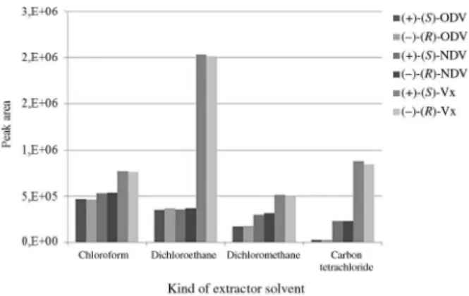
Optimization of the extractor solvent
In order to optimize the type of extractor solvent, the same strategy was done as described before by fixing a disperser solvent (500 µL isopropanol) and varying the extractor solvent (chloroform, dichloromethane, dichloroethane and carbon tetrachloride). Based on the results showed in Figure 3, chloroform showed better extraction efficiency for the extraction of the metabolites. Therefore, this solvent was employed for further optimization procedure.
Optimization of the volumes of the extractor and disperser solvent
Next, the volume of extractor and disperser solvents was optimized. The disperser solvent volume influences in the cloudy solution formation and also in the sedimented phase volume after centrifugation.18-23 To perform that,
100 µL of chloroform was employed and the volume of isopropanol was varied as follows: 300, 400, 500, 600 and 700 µL. Higher recovery values (expressed by peak areas) were obtained by using 300 µL isopropanol (Figure 4). Higher volumes led to a decrease in the extraction of the analytes. This effect can be explained by the disperser
solvent polarity. The increase of isopropanol volume leads to a polarity reduction of the aqueous phase resulting in a lower analyte recovery. This effect is more pronounced for Vx due to its lower polarity when compared to its metabolites. Next, it was optimized the volume of the extracting solvent. The choice of the extracting solvent volume was carried out using 300 µL of isopropanol and the volume of chloroform was varied as following: 70, 100, 200 300 and 400 µL. Higher recovery value was obtained with 200 µL chloroform (data not shown). Therefore, for further experiments, 300 µL isopropanol and 200 µL chloroform were used as disperser and extraction solvents, respectively.
Optimization of the agitation before centrifugation (assisted DLLME)
Sample agitation after cloudy solution formation just before the centrifugation step is very important. This step may promote an increase of the extractor solvent surface area in the aqueous phase, thus allowing a higher analyte Figure 2. Optimization of the type of disperser solvent in DLLME
procedure. Extraction conditions: 2 mL Czapek liquid culture medium; 2 mL buffer borate 100 mmol L-1 pH 10. Extractor solvent: chloroform;
sample concentration: 1.25 µg mL-1; extraction temperature (24 ± 2 oC),
n = 3.
Figure 3. Optimization of the type of extractor solvent in DLLME procedure. Extraction conditions: 2 mL Czapek liquid culture medium; 2 mL buffer borate 100 mmol L-1 pH 10. Sample concentration:
1.25 µg mL-1; disperser solvent: isopropanol; extraction temperature
(24 ± 2 oC), n = 3.
Figure 4. Optimization of the disperser solvent volume in DLLME procedure. Extraction conditions: 2 mL Czapek liquid culture medium; 2 mL buffer borate 100 mmol L-1 pH 10. Sample concentration:
1.25 µg mL-1; disperser and extractor solvents: isopropanol and
recovery.19 Therefore; a manual vortex device was employed
to evaluate this parameter. After the formation of the cloud point, the sample was stirred for: 0, 5, 10, 20 e 30 s and then analyzed. There was a significant increase in the analytes recovery after 30 s of agitation (data not shown). Longer times were not optimized in order to obtain a faster sample preparation step. Therefore, 30 s was chosen as agitation time. Accordingly, after the DLLME optimization, the following condition was set: extracting solvent: chloroform (200 µL); disperser solvent: isopropanol (300 µL); agitation in vortex device for 30 s after the cloud point formation. Figure 5 shows an electropherogram after DLLME optimization for the extraction of Vx and its metabolites from Czapek liquid culture medium.
Although the use of univariated optimization may not guarantee the best extraction condition since the factors involved in DLLME optimization cannot be evaluated together, the absolute recovery obtained was higher than 70%, thus adequate to detect the analytes in the biotransformation process.
Validation of the method
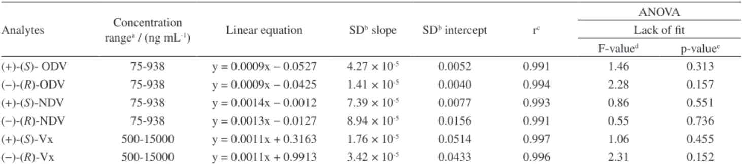
The calibration curve was linear over the concentration range of 500-15000 ng mL-1 and 75-938 ng mL-1 for Vx and
its metabolites, respectively. The correlation coefficients (r) were above 0.990 and relative errors were below 15%
for each point of the analytical curve. Submission of the analytical curves to the F test for lack-of-fit (FLOF) proved the validity of the linearity (Table 1).
The mean recoveries were 90, 83 and 75% for Vx, ODV and NDV enantiomers respectively, with relative standard deviation below 15% (data not shown). The lower limit of quantification was 90 ng mL-1 and 50 ng mL-1 for each Vx
enantiomer and its metabolite enantiomers, respectively, with RSD and RE less than 13% (Table 2).
Table 2. Limit of quantification of the method for analysis of the analytes in Czapek liquid culture medium employing DLLME-CE
Analytes Nominal concentration / (ng mL-1)
Obtained concentration /
(ng mL-1) Accuracy REa / % Precision RSDb / %
(+)-(S)-ODV 75 76 2 5
(−)-(R)-ODV 75 77 3 7
(+)-(S)-NDV 75 70 −6 6
(−)-(R)-NDV 75 71 −5 12
(+)-(S)-Vx 500 512 2 7
(−)-(R)-Vx 500 513 2 5
aExpressed as relative error, RE; bexpressed as relative standard deviation, RSD.
Table 1. Linearity of the calibration curve for analysis of the analytes in Czapek liquid culture medium employing DLLME-CE
Analytes Concentration
rangea / (ng mL-1) Linear equation SD
b slope SDb intercept rc
ANOVA Lack of fit F-valued p-valuee
(+)-(S)- ODV 75-938 y = 0.0009x − 0.0527 4.27 × 10⁻⁵ 0.0052 0.991 1.46 0.313
(−)-(R)-ODV 75-938 y = 0.0009x − 0.0425 1.41 × 10⁻⁵ 0.0040 0.994 2.28 0.157
(+)-(S)-NDV 75-938 y = 0.0014x − 0.0012 7.39 × 10⁻⁵ 0.0077 0.993 0.86 0.551
(−)-(R)-NDV 75-938 y = 0.0013x − 0.0127 8.94 × 10⁻⁵ 0.0156 0.991 0.55 0.736
(+)-(S)-Vx 500-15000 y = 0.0011x + 0.3163 1.76 × 10⁻⁵ 0.0514 0.997 1.06 0.455
(−)-(R)-Vx 500-15000 y = 0.0011x + 0.9913 3.42 × 10⁻⁵ 0.0433 0.996 2.31 0.152 aQuintuplicate replicates (n = 5) for each concentration; bSD: standard deviation; ccorrelation coefficient; dF
value < Ftabled, Ftabled = 2.85 (6.14; 0.05); ep-value > 0.05.
Figure 5. Electropherogram after DLLME optimization of (a) sample spiked with Vx and its metabolites at concentration of 1.25 µg mL-1;
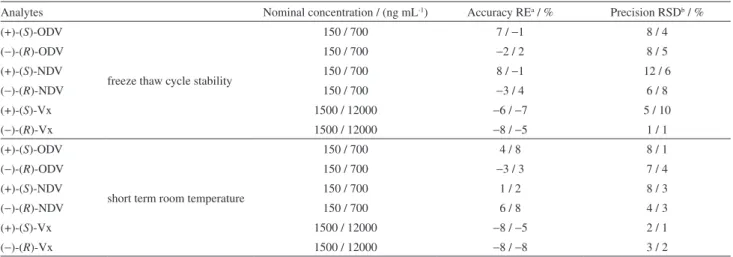
Precision and accuracy assays met the criteria defined in the EMA guidelines. The between-day (Table 3) and within-day (Table 4) precision and accuracy presented RSD values below 10% and relative errors below 12%. The samples showed to be stable under freeze-thaw cycles and short-term room temperature assays with RSD values below 13% and relative errors below 9% (Table 5).
Taking into account the possibility of secondary metabolites from the fungi, the selectivity test showed no interference peaks in the migration time of the analytes
or the internal standard (Figure 5). Therefore, the sample preparation used in this study showed to be very selective for the target analytes. In capillary electrophoresis, any change in ionic strength of the sample may lead to a variation in the migration time; therefore, the sample preparation process as well as injection sample solvent should be strictly controlled. A little variation in the migration time of the internal standard was observed during the analyses. Based on that, the repeatability of the analyte migration times was determined by analyzing 10 samples from the
Table 5. Freeze-thaw and short-term room temperature stability for analysis of the analytes in Czapek liquid culture medium employing DLLME-CE
Analytes Nominal concentration / (ng mL-1) Accuracy REa / % Precision RSDb / %
(+)-(S)-ODV
freeze thaw cycle stability
150 / 700 7 / −1 8 / 4
(−)-(R)-ODV 150 / 700 −2 / 2 8 / 5
(+)-(S)-NDV 150 / 700 8 / −1 12 / 6
(−)-(R)-NDV 150 / 700 −3 / 4 6 / 8
(+)-(S)-Vx 1500 / 12000 −6 / −7 5 / 10
(−)-(R)-Vx 1500 / 12000 −8 / −5 1 / 1
(+)-(S)-ODV
short term room temperature
150 / 700 4 / 8 8 / 1
(−)-(R)-ODV 150 / 700 −3 / 3 7 / 4
(+)-(S)-NDV 150 / 700 1 / 2 8 / 3
(−)-(R)-NDV 150 / 700 6 / 8 4 / 3
(+)-(S)-Vx 1500 / 12000 −8 / −5 2 / 1
(−)-(R)-Vx 1500 / 12000 −8 / −8 3 / 2
Table 4. Within-day accuracy and precision of the method for analysis of the analytes in Czapek liquid culture medium employing DLLME-CE
Analytes Nominal concentration / (ng mL-1)
Obtained concentration /
(ng mL-1) Accuracy REa / % Precision RSDb / %
(+)-(S)-ODV 150 300 700 135 328 701 −9 9 4 6 5 5
(−)-(R)-ODV 150 300 700 133 332 699 −10 11 4 2 2 4
(+)-(S)-NDV 150 300 700 165 315 665 10 5 −1 5 6 6
(−)-(R)-NDV 150 300 700 157 281 628 5 9 −7 7 8 2
(+)-(S)-Vx 1500 7500 12000 1398 7168 13351 −7 −4 11 2 2 1
(−)-(R)-Vx 1500 7500 12000 1402 7524 12313 −6 0.3 3 2 4 7
aExpressed as relative error, RE;bexpressed as relative standard deviation, RSD.
Table 3. Between-day accuracy and precision of the method for analysis of the analytes in Czapek liquid culture medium employing DLLME-CE
Analytes Nominal concentration / (ng mL-1)
Obtained concentration /
(ng mL-1) Accuracy RE
a / % Precision RSDb / %
(+)-(S)-ODV 150 300 700 143 332 653 −5 10 −7 5 1 3
(−)-(R)-ODV 150 300 700 148 311 683 4 −2 −2 2 4 6
(+)-(S)-NDV 150 300 700 147 316 675 −2 5 −3 6 1 9
(−)-(R)-NDV 150 300 700 144 333 650 −4 11 −7 2 1 2
(+)-(S)-Vx 1500 7500 12000 1502 7404 12140 0 −1 1 2 4 5
(−)-(R)-Vx 1500 7500 12000 1510 6859 13316 1 −8 10 2 4 1
precision/accuracy assays. The RSD of the migration time of the (+)-(S)-Vx, (−)-(R)-Vx, (+)-(S)-NDV, (−)-(R)-NDV, (+)-(S)-ODV, (−)-(R)-ODV and the IS was, respectively, 2.4, 2.5, 1.9, 1.9, 1.1, 1.0 and 2.7%.
Stereoselective fungal biotransformation study
A f t e r m e t h o d va l i d a t i o n , t h e v e n l a f a x i n e biotransformation study was performed employing the optimized parameters obtained for electrophoretic chiral separation and DLLME procedure. However, before the biotransformation study, the MIC test was carried out in order to evaluate if Vx could affect the fungal growth. The MIC test showed Vx did not inhibit the fungal growth in the concentration used for the biotransformation studies.
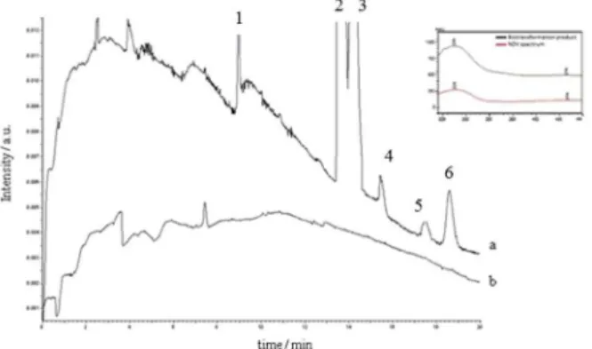
During 480 h, the fungal biotransformation was evaluated by collecting sample aliquots every 120 hours. Under the described conditions, only the fungus
Cunninghamella elegans ATCC 10028B was able to perform an enantioselective biotransformation of Vx into its metabolite (+)-(S)-N-desmethylvenlafaxine (Figure 6) (peak 4) with an enantiomeric excess of 100% during all biotransformation study (191 ng mL-1). The outset
formation of the enantiomer occurred in 120 h and remained almost constant until the end of study.
The analysis of the UV absorption spectrum of the peak eluted in the same retention time of (−)-(R)-O -desmethylvenlafaxine showed to be different of the required analyte. Therefore, it is probably an elicitation product produced by the interaction of the fungi with the drugs.10
Based on the little efficiency in the biotransformation process initially proposed, some changes in the incubation conditions were evaluated in order to improve the biotransformation and to achieve higher rate of metabolite production.
Evaluation of different conditions in the biotransformation process
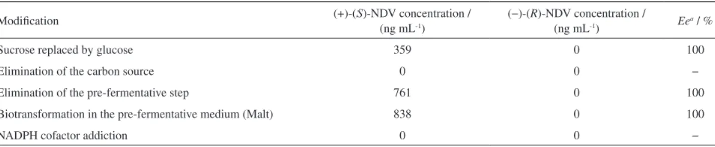
In order to attempt to improve the Vx biotransformation, some changes in the liquid culture medium were performed and the samples were analyzed only after 480 h of incubation.
Firstly, the carbon source in Czapek medium was modified (sucrose was replaced by glucose) in order to change the reaction routes. This modification led to an increase of 53% in production rate of (+)-(S)-NDV metabolite. Sucrose is commonly used because it provides less secondary fungal metabolism products and therefore, little interferences in the analysis. On the other hand, glucose is readily used by the microorganism, which might lead to an improvement in biotransformation.
Another modification was the elimination of the carbon source from Czapek liquid culture medium. This strategy aims to force the biotransformation of Vx into its metabolites since the only available carbon source would be venlafaxine. Interestingly, there was no production of metabolites, however, the concentration of Vx was dramatically reduced from the Czapek medium, about, 95% of Vx was consumed by the fungus after this modification.
Changing in the biotransformation medium was also performed. Firstly, the biotransformation of Vx was performed in the pre-fermentative medium (Malt medium) and not in the Czapek medium. Secondly, the growing of the fungus was carried out only in the Czapek medium, thereby eliminating, the pre-fermentative step (see section Enantioselective biotransformation of venlafaxine). Both modifications influence the log phase of microbial growth. This phase represents the higher stage in cellular development of the fungus (exponential growth) where essentials enzymes for the biotransformation process are produced.4 By performing the biotransformation without
the pre-fermentative step, the concentration reached of (+)-(S)-NDV was 761 ng mL-1. On the other hand, when the
biotransformation was carried out in the pre-fermentative medium (Malt medium), the concentration observed of (+)-(S)-NDV was the highest one, 838 ng mL-1.
Finally, the addition of NADPH cofactor in the Czapek liquid culture medium was evaluated. This cofactor is an important component in oxidation reactions acting as a reductor agent at the enzymes heme group. However, no biotransformation was observed. Probably, the medium was not favorable for the action of this cofactor (ionic strength and pH), and therefore there was no improvement in the biotransformation process. Table 6 summarizes the evaluated conditions in the biotransformation processes. Figure 6. (a) Electropherogram after venlafaxine biotransformation by
Table 6. Evaluation of different conditions in the fungal biotransformation process
Modification (+)-(S)-NDV concentration /
(ng mL-1)
(−)-(R)-NDV concentration /
(ng mL-1) Ee
a/ %
Sucrose replaced by glucose 359 0 100
Elimination of the carbon source 0 0 −
Elimination of the pre-fermentative step 761 0 100
Biotransformation in the pre-fermentative medium (Malt) 838 0 100
NADPH cofactor addiction 0 0 −
aEe: enantiomeric excess.
Conclusions
This paper presents, for the first time, a stereoselective fungal biotransformation study of venlafaxine by DLLME-CE. The procedure for analysis and sample preparation showed to be simple, efficient and selective for the target analytes. In addition, the sample preparation used provides a low consumption of organic solvent and high recovery efficiency. The biotransformation study showed a stereoselective biotransformation of venlafaxine into (+)-S-N-desmethylvenlafaxine metabolite by the fungus
Cunninghamella elegans ATCC 10028B with 100% of enantiomeric excess.
Acknowledgements
The authors are grateful to São Paulo Research Foundation (FAPESP, grant numbers: 2011/17508-1, 2013/17658-9), Conselho Nacional de Desenvolvimento Científico e Tecnológico (CNPq) and Coordenação de Aperfeiçoamento de Pessoal de Nível Superior (CAPES) for financial support and for granting research fellowships.
References
1. Spina, E.; Santoro, V.; D’Arrigo, C.; Clin. Ther. 2008, 30, 1206. 2. Holliday, S. M.; Benfield, P.; Drugs1995, 49, 280.
3. Perry, R.; Cassagnol, M.; Clin. Ther. 2009, 31, 1374. 4. Pupo, M. T.; Borges, K. B.; Borges, W. S.; Bonato, P. S.
In Fungal Biotransformations: a Powerful Tool in Drug Metabolism Studies; Saikai, R.; Bezbaruah, R. L.; Bora, T. C., eds.; New Delhi Publishing Agency: New Delhi, India, 2008, ch. 3.
5. Borges, K. B.; Borges, W. D.; Pupo, M. T.; Bonato, P. S.; Curr. Org. Chem. 2009, 13, 1137.
6. Borges, K. B.; Borges, W. D.; Pupo, M. T.; Bonato, P. S.; J. Pharm. Biomed. Anal. 2008, 46, 945.
7. Barth, T.; Pupo, M. T.; Borges, K. B.; Okano, L. T.; Bonato, P. S.; Electrophoresis 2010, 31, 1521.
8. Hilario, V. C.; Carrão, D. B.; Barth, T.; Borges, K. B.; Furtado, N. A.; Pupo M. T.; J. Pharm. Biomed. Anal.2012, 61, 100. 9. Carrão, D. B.; Borges, K. B.; Barth, T.; Pupo, M. T.; Bonato,
P. S.; de Oliveira, A. R. M.; Electrophoresis2011, 32, 2746. 10. de Jesus, L. I.; Alburquerque, N. C.; Borges, K. B.; Simões,
R. A.; Calixto, L. A.; Furtado, N. A.; Electrophoresis2011, 32, 2765.
11. Bocato, M. Z.; Simões, R. A.; Calixto, L. A.; Gaitani, C. M.; Pupo, M. T.; de Oliveira, A. R. M.; Anal. Chim. Acta2012, 742, 80.
12. Liu, W.; Cai, H.; Li, H.; J. Chromatogr. B2007, 850, 405. 13. da Fonseca, P.; Bonato, P. S.; Anal. Bioanal. Chem. 2010, 396,
817.
14. Kingbäck, M.; Josefsson, M.; Kalrsson, L.; Ahlner, J.; Bengtsson, F.; Kugelberg, F. C.; Carlsson, B.; J. Pharm. Biomed. Anal. 2010, 53, 583.
15. Rudaz, S.; Stella, C.; Balant-gorgia, A. E.; Fanali, S.; Veuthey, J. L.; J. Pharm. Biomed. Anal. 2000, 23, 107.
16. Fanali, S.; Rudaz, S. J. L.; Veuthey, C. D.; J. Chromatogr. A
2001, 919, 195.
17. Rudaz, J. L. S., Veuthey, C. D.; Fanali, S.; Chromatographia
1999, 50, 369.
18. Rezaee, M.; Assadi, Y.; Hosseini, M. M.; Aghaee, E.; Ahmadi, F.; Berijani, S.; J. Chromatogr. A2006, 1116, 1. 19. Caldas, S. S.; Gonçalvez, F. F.; Primel, E. G.; Quim. Nova2011,
34, 1604.
20. Meng, L.; Wang, B.; Luo, F.; Shen, G.; Wang, Z.; Guo, M.; Forensic Sci. Int.2011, 209, 42.
21. Zgota-Grzeskowiak, A.; Grzeskowiak, T.; Trends Anal. Chem.
2011, 30, 1382.
22. Rezaee, M.; Yamini, Y.; Faraji, M.; J. Chromatogr. A2010, 1217, 2342.
23. Xiao-Huan, Z.; Qiu-Hua, W.; Mei-Yue, Z.; Guo-Hong, X.; Zhi, W.; Chin. J. Anal. Chem. 2009, 37, 161.
24. Suh, J. H.; Lee, Y. Y.; Lee, H. J.; Kanga, M.; Hura, Y.; Leea, S. N.; Yanga, D.; Hana, S. B.; J. Pharm. Biomed. Anal. 2013, 75, 214.
26. Ghambari, H.; Hadjmohammadi, M.; J. Chromatogr. B2012, 899, 66.
27. Kohler, I.; Schapplera, J.; Sierroa, T.; Rudaz, A.; J. Pharm. Biomed. Anal.2013, 73, 82.
28. Fortes, S. S.; Barth, T.; Furtado, N. A. J. C.; Pupo, M. T.; de Gaitani, C. M.; de Oliveira, A. R. M.; Talanta2013, 116, 743. 29. European Medicines Agency (EMA), Guideline on Bioanalytical Method 554 Validation, 2011.http://www.ema.europa.eu/docs/ en_GB/document_library/Scientific_guideline/2011/08/ WC500109686.pdf accessed in July 2015.
30. Gallo, M. B. C.; Chagas, F. O.; Almeida, M. O.; Macedo, C. C.; Cavalcanti, B. C.; Barros, F. W. A.; Moraes, M. O.; Costa-Lotufo, L. V.; Pessoa, C.; Bastos, J. K.; Pupo, M. T.; J. Basic Microbiol. 2009, 49, 142.
31. Bonato, P. S.; Jabor, V. A. P.; Gaitani, C. M.; Quim. Nova2005, 28, 683.
32. de Gaitani, C. M.; de Oliveira, A. R. M.; Bonato, P. S. In Capillary Electromigration Techniques for the Analysis of Drugs
and Metabolites in Biological Matrices: a Critical Appraisal; García, C. D.; Chumbimuni-Torres, K. Y.; Carrilho, E., eds.; Wiley: New Jersey, 2013, ch. 12.
Submitted: April 13, 2015
Published online: July 17, 2015