Article
Printed in Brazil - ©2016 Sociedade Brasileira de Química0103 - 5053 $6.00+0.00
*e-mail: [email protected]
Cholinesterases Inhibition by Novel
cis-
and
trans-
3
-
Arylaminocyclohexyl
N,N
-Dimethylcarbamates: Biological Evaluation and Molecular Modeling
Diego A. S. Yamazaki,a Augusto A. Cândido,a Mariane C. Bagatin,a Miguel Machinski Jr.,b
Simone A. G. Mossini,b Rodrigo M. Pontes,a Fernanda A. Rosa,a Ernani A. Bassoa and
Gisele F. Gauze*,a
aDepartamento de Química and bDepartamento de Ciências Básicas da Saúde, Universidade
Estadual de Maringá, Av. Colombo, 5790, 87020-900 Maringá-PR, Brazil
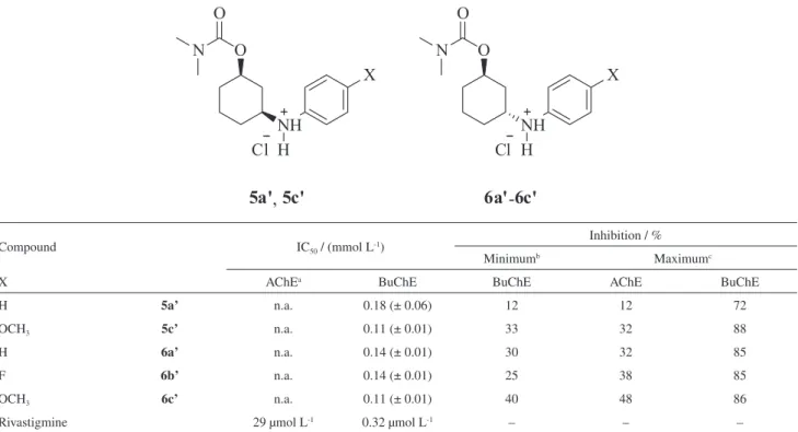
The present study describes the synthesis, assessment of the anticholinesterase activity and the inhibition type of novel cis- and trans-3-arylaminocyclohexyl N,N-dimethylcarbamates. In vitro inhibition assay by Ellman’s method with human blood samples showed that carbamates were selective for butyrylcholinesterase (BuChE) with compound concentration that inhibits 50% of enzyme activity (IC50) between 0.11 and 0.18 mmol L-1. cis- and trans-3-(4-Methoxyphenylamino)
cyclohexyl N,N-dimethylcarbamate hydrochloride were the most active for BuChE, showing that the presence of methoxyl group enhanced the anticholinesterase activity. The enzyme kinetics studies indicate a noncompetitive inhibition against acetylcholinesterase (AChE) and mixed type inhibition for BuChE. Molecular modeling studies confirm the ability of carbamates to bind both the active and peripheral sites of the BuChE.
Keywords: carbamate derivatives, butyrylcholinesterase inhibitors, molecular docking, non-covalent interactions
Introduction
Acetylcholinesterase (AChE) is a serine hydrolase that is mainly present in red blood cells, nerve endings and striated muscles.1,2 The principal biological role
of AChE is regulating the transmission of impulses at cholinergic synapses, catalyzing rapid hydrolysis of the neurotransmitter acetylcholine (ACh).3,4 Like AChE, the
butyrylcholinesterase (BuChE) has an important role in the nervous system as coregulator of ACh. Studies have shown that BuChE is able to compensate for the lack of AChE, allowing the continued regulation of cholinergic neurotransmission.5-7 Alzheimer’s disease (AD), glaucoma
and myasthenia gravis are diseases which are related to the cholinergic system and their symptomatic treatment is by administration of drugs that act as cholinesterase inhibitors (ChEI).2,8,9 Depending on the AD stage, there
is a decline in AChE levels in the brain and a progressive increase of BuChE which becomes responsible for the hydrolysis of acetylcoline.10-12 Thus, the use of specific
BuChE inhibitors may be indicated for the symptomatic treatment of AD.13
The attribute of carbamates as cholinesterase inhibitors has been known for decades. Physostigmine, an alkaloid present in Calabar bean, was the first carbamate used clinically as a cholinesterase inhibitor, in the treatment of glaucoma and later in AD. However, due to their high doses and side effects the use of physostigmine has been discontinued.14 Neostigmine and pyridostigmine are used
in the treatment of myasthenia gravis, and neostigmine is also used in the treatment of glaucoma. Rivastigmine has a dual inhibitory action on AChE and BuChE enzymes and is used in the treatment of AD.15
Since carbamates have been successfully used to treat a variety of diseases involving cholinergic dysfunction, innumerous studies have been performed to find new ChEI that can be used clinically.16-19
Recently, in vitro inhibition tests performed in human
blood samples for cis- and trans-2-arylaminocyclohexyl N,N-dimethylcarbamates showed that carbamates of
both series were selective for BuChE.20 Moreover,
carbamates tested exhibited a higher activity when compared with 2-N,N-dimethylaminecyclohexyl 1-N’,N’
-dimethylcarbamates analogues,21 indicating that the
This work describes the synthesis, biological evaluation and molecular modeling of novel cis- and trans-3
-arylaminocyclohexyl N,N-dimethylcarbamates 5a-5c and 6a-6c as potential cholinesterase inhibitors. AChE and
BuChE inhibition mode was accessed by kinetics studies, molecular docking and non-covalent interactions analysis.
Results and Discussion
Synthesis
New carbamates derivatives (5a-5c and 6a-6c) were
synthesized in three steps starting from the 2-cyclohex-1-enone (1) according to route outlined in Scheme 1.
In the first step, we obtained the 3-arylaminocyclo-hexanones (2a-2c) via aza-Michael addition of arylamines
with 2-cyclohex-1-enone (1). The more common
protocols of aza-Michael reaction use strong bases and acids as catalyst, and milder Lewis acidic catalysts. However, no conventional methodology was effective, since the arylamines exhibit a low nucleofilicity. Ying et al.22 have successfully used the basic ionic
liquid 1,8-diazabicyclo[5.4.0]-undec-7-en-8-ium acetate ([DBU][Ac]) as promoter for aza-Michael addition under solvent free conditions. The authors propose that the ionic liquid increases the nucleophilicity of the arylamines. We modified the methodology proposed by Ying et al.22 using
equimolar amounts of the Michael acceptor and arylamine and keeping the reaction time and temperature. The authors purified the compounds by column chromatography; to reduce the use of solvents we purified the products by recrystallization with good yields (75-82%). In the next step, the ketones 2a-2c were reduced by two ways to
obtain the cis- and trans-3-arylaminocyclohexanols (3a-3c; 4a-4c), Using N-selectride as reducer23 at low temperature
(–80 oC) the trans-3-arylaminocyclohexanols 4a-4c were
obtained with high diastereoselectivity (ca. 100%) in good yields (72-85%). However, when ketones 3a-3c were
reduced with NaBH4,24 we observed that the product was
obtained as the mixture of the isomers cis/trans in a ratio
of 75/25. The identification of the isomers was conducted by comparative analysis of the chemical shifts of geminal hydrogen to hydroxyl group (H1). The chemical shifts of the
H1 for the products obtained by reduction with N-selectride
were 4.10 to 4.20 ppm, while the chemical shifts of the H1
for the products obtained using NaBH4 as reducer were
3.60 to 3.81 ppm. According to literature, the hydrogens oriented in the equatorial position are more deshielded than the axial hydrogens.25,26
The mixture of isomers was used in the next step. Finally, the alcohols (3a-3c; 4a-4c) were carbamoylated with N,N
-dimethylcarbamoyl chloride to give compounds 5a-5c and 6a-6c. Further purification by column chromatography
afforded the desired carbamates in moderate yields (30-46%), except the compound 5b that was obtained as a
mixture with the isomer 6b.
Inhibition assays of AChE and BuChE
Inhibitory activities of the novel synthesized carbamates against cholinesterases (ChEs) from fresh human blood were evaluated by Ellman’s modified27 spectroscopic
method. Since this method requires water soluble compounds, the respective hydrochlorides 5a’, 5c’ and 6a’-6c’ were prepared. The assays were performed at
five different concentrations (0.067, 0.13, 0.27, 0.67 and
Scheme 1. Synthesis of cis- and trans-3-arylaminocyclohexyl N,N-dimethylcarbamates. Reagents and conditions: (i) arylamine, 1,8-diazabicyclo[5.4.0]-undec-7-en-8-ium acetate ([DBU][Ac]), r.t., 5 h; (ii) NaBH4, tetrahydrofuran (THF), r.t., 48 h; (ii’) N-selectride, THF, –80 oC, 4 h; (iii) NaH/THF, 80 oC,
8 h; N,N-dimethylcarbamoyl chloride, 16 h. O
N O
X
H
N OH
X
H N OH
X
H
N O
X O
N
H
(i) (ii)
(ii')
(iii)
(iii)
1
2a-2c
4a-4c 3a-3c
6a-6c 5a-5c
N O
X O
N
H
1.3 mmol L-1) for compounds 5c’, 6a’-6c’ and at four
different concentration (0.05, 0.1, 0.2, and 0.5 mmol L-1) for
compound 5a’. In these experiments, we could determine
the inhibition potential of each compound tested against AChE (found in red cells) and BuChE (found in plasma). The inhibitory activity was expressed as the compound concentration that inhibits 50% of enzyme activity (IC50).
The results of the AChE and BuChE inhibition and IC50
values, which were obtained from the curves of percent inhibition vs. concentration, are summarized in Table 1.
First, we observed that all carbamate derivatives exhibited a dose-dependent inhibitory activity for both cholinesterases and showed selectivity for the BuChE inhibition. The compounds 5c’ and 6a’-6c’ displayed maximum inhibitory
activity for BuChE of 85 to 88% at 1.3 mmol L-1. Due to
low solubility of compound 5a’, the maximum inhibition
for BuChE was 72% at 0.5 mmol L-1. Derivatives 5c’ and 6c’ were the more active compounds, with an IC50 of
0.11 mmol L-1. The results suggest that there is no relevant
difference in inhibition of BuChE activity between the cis
and trans isomers and that the presence of the methoxyl
group potentialized the anticholinesterase activity. The effect of methoxyl group is mainly noted in the lowest concentration tested (0.067 mmol L-1). The compound 6c’ showed an inhibition of 40% against 30 and 25%
for compounds 6a’ and 6b’, respectively. Relative to the
AChE inhibition no tested compound inhibited 50% of the enzyme activity in any of the investigated concentrations. All compounds were weakly active against AChE with an inhibition between 12 and 48% in the maximum concentration measured.
Bagatin et al.20 studied inhibitory properties of cis- and trans-2-arylaminocyclohexyl N,N-dimethylcarbamates.
Comparatively, the carbamates tested in this present study showed a significant improvement in the inhibitory activity against BuChE. The synthesized compounds showed IC50 of
0.11 to 0.18 mmol L-1 and all 1,3-disubstituted derivatives
were more active than correlated 1,2-disubstituted compounds, displaying an improvement in the inhibitory activity of 30 to 50 times. These data indicate that the arylamine group in position 3 of cyclohexyl ring enhanced the inhibitory potential of the new carbamates.
Kinetic studies of AChE and BuChE inhibition
The type of AChE and BuChE inhibition was investigated from the kinetics studies using the modified Ellman’s method27 and compound 6c’, the most potent
inhibitor of both enzymes. To assess the kinetic parameters, we measured the initial rate of enzyme activity at
Table 1. ChEs inhibitory activity of compounds 5a’, 5c’ and 6a’-6c
Compound IC50 / (mmol L-1)
Inhibition / %
Minimumb Maximumc
X AChEa BuChE BuChE AChE BuChE
H 5a’ n.a. 0.18 (± 0.06) 12 12 72
OCH3 5c’ n.a. 0.11 (± 0.01) 33 32 88
H 6a’ n.a. 0.14 (± 0.01) 30 32 85
F 6b’ n.a. 0.14 (± 0.01) 25 38 85
OCH3 6c’ n.a. 0.11 (± 0.01) 40 48 86
Rivastigmine 29 µmol L-1 0.32 µmol L-1 – – –
an.a.: Not active (IC
50 > 1.3 mmol L-1); bthe minimum concentration measured was 0.067 mmol L-1 for 5c’ and 6a’-6c’ and 0.05 mmol L-1 for 5a’; cthe
maximum concentration measured was 1.3 mmol L-1 for 5c’ and 6a’-6c’ and 0.5 mmol L-1 for 5a’. IC
50: Compound concentration that inhibits 50% of
different concentrations of substrate acetylthiocholine (0.05 to 1.0 mmol L-1) or butyrylthiocholine (0.75
to 1.0 mmol L-1) in the absence and presence of the
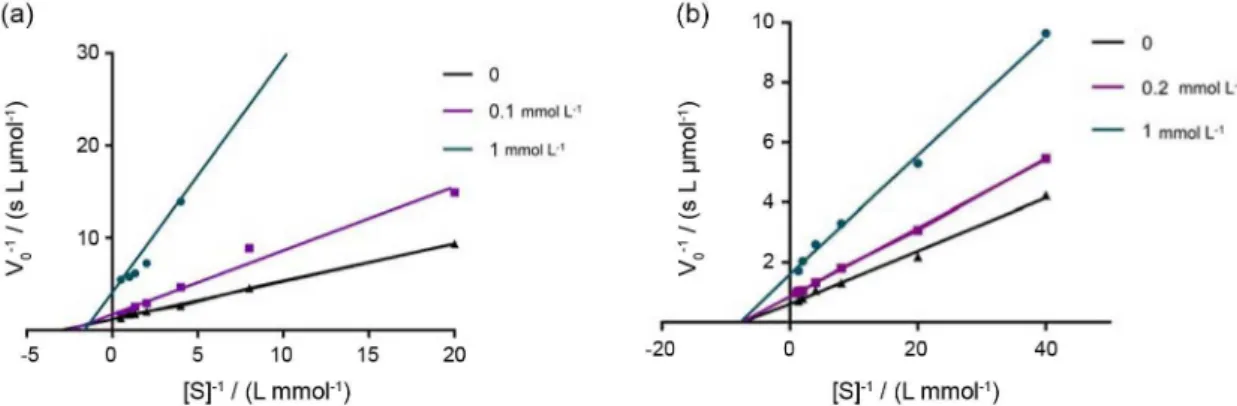
inhibitor 6c’. Lineweaver-Burk plots (Figure 1) were
built by the reciprocal of the initial rate (V0-1) against
the reciprocal of substrate concentrations ([S]-1) for the
different inhibitor concentrations resulting from the substrate-velocity curves for AChE and BuChE.
Graphical analysis of Lineweaver-Burk plots and the kinetic parameters of AChE activity (Figure 1a) showed a practically unchanged Km value with increasing inhibitor
concentration and increasing slopes with increasing inhibitor concentration, i.e., Vmax is reduced. This pattern
indicates a non-competitive-type inhibition.28 However,
Lineweaver-Burk plots and the kinetic parameters of BuChE activity (Figure 1b) showed both increasing slopes (lower Vmax) and intercepts (higher Km) with higher inhibitor
concentration, i.e., the increasing inhibitor concentration reduces Vmax and increases Km value. This pattern indicates
a mixed-type inhibition of BuChE.28 It is shown that the
compound 6c’ could interact with both the free enzyme
and the complex enzyme substrate, which could explain the higher activity against BuChE than AChE. These results also suggest that although carbamates occupy a significant fraction of the catalytic gorge, they do not compete for the same binding site as the substrate. The dissociation constant (Ki) values obtained are consistent with inhibitory activity.
Compound 6c’ was found to be more potent inhibitor of
BuChE (Ki 0.21 mmol L-1) than AChE (Ki 0.69 mmol L-1).
Molecular modeling studies
The molecular docking calculations were performed using AutoDock 4.2.329 program implemented at the
PyRx 0.930 interface in order to evaluate the
enzyme-inhibitor interactions of the newly synthesized compounds. The compound 6c’ was docked in the active site of AChE
(Protein Data Bank (PDB) 1ACJ) and BuChE (PDB 4BDS) derived from complex of the enzymes with tacrine obtained from the PDB. The best docked poses, i.e., the lowest energy conformer in the most populated cluster of conformers were subjected to energy minimization by NAMD31 program
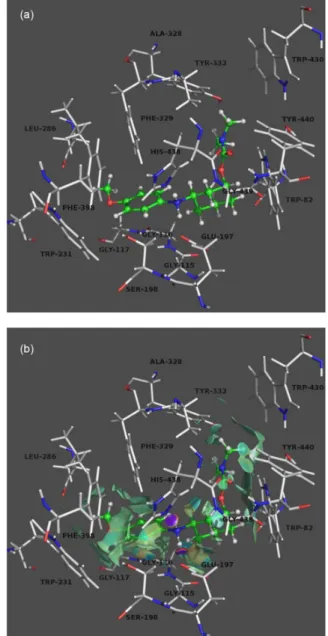
and analyzed to explain interactions between ligands and the target enzyme. Figure 2a shows the compound 6c’
complexed with BuChE. In active site gorge, both hydrogen atoms of the protonated amino group are likely to form an H-bond with an imidazolyl nitrogen atom of His438 and an oxygen atom at carbon 5 of Glu197 with a distance of 1.6 and 1.8 Å, respectively. The positively-charged nitrogen of ligand made a cation-π interaction with the imidazole ring of His438. Carbamate group is stabilized by H-bond between carbonyl oxygen and indole NH of Trp82 with a distance of 3.0 Å. Also, cyclohexyl ring is stabilized by van der Waals interaction with aromatic rings of Trp82. It is possible to observe that the aromatic ring of 6c’ is
surrounded by the aromatic rings of Trp231 and Phe398 forming T-stacking type interaction. In addition, N-methyl
group of ligand interacts with Tyr332 and the aromatic ring of compound 6c’ forms T-stacking type interaction with
Phe329. Studies performed by Macdonald et al.32 with
inhibitor competition and BuChE mutant species indicate that Tyr332 and Phe329 are amino acid residues important for binding inhibitors at the peripheral anionic site (PAS) of BuChE. This binding mode is in agreement with mixed-type inhibition of 6c’, in which the ligand is able to interact
with both active gorge site and PAS.
The new non-covalent interaction (NCI) approach developed by Yang and co-workers33 enables simultaneous
analysis and graphical visualization of a wide range of these interactions in real space surface. Thus, the network of interactions was identified using the NCI index, which defines the regions of attractive or repulsive interactions, as well as their strength.33 In what follows, the
enzyme-ligand interactions revealed by the NCI analysis are color
Figure 1. Lineweaver-Burk plot for the inhibition of (a) AChE with different acetylthiocholine concentrations (0.05 to 1.0 mmol L-1) in the absence and
presence of compound 6c’ in concentrations of 0.2 and 1 mmol L-1; and (b) BuChE with different butyrylthiocholine concentrations (0.75 to 2.0 mmol L-1)
coded from black for strongly attractive weak interactions (hydrogen bonds) to dark gray for repulsive ones (steric clashes). In between, the very weak van der Waals interactions appear in light gray. The gradient isosurface (Figure 2b) indicates two attractive interactions (black disks) corresponding to strong H-bonds and several intermolecular van der Waals interactions (in light gray) that are not atom-specific and occupy broader regions in space.33 Strongly repulsive interactions, which would appear, were not observed, showing that the ligand is well stabilized in active site of BuChE.
The orientation of the compound 6c’ in AChE
(Supplementary Information Figure S2) is quite different
from the orientation in BuChE, ergo, the interactions between the ligand and AChE are also different. The carbamate group does not exhibit any significant interaction with residues. Both hydrogen atoms of the protonated amino group form an H-bond with the indole nitrogen atom of Trp84 and oxygen atom of Gly441 with a distance of 2.3 and 1.8 Å, respectively. The cyclohexyl ring is stabilized by van der Waals interaction with aromatic rings of Trp84. Finally, the aromatic ring of 6c’ can interact with Tyr442
and Phe330 via T-stacking and π-π stacking interaction, respectively. The isosurfaces of AChE (Figure S3) are in agreement with the molecular docking.
Conclusions
This study has resulted in a series of new selective BuChE inhibitors, with IC50 values of 0.11 to 0.18 mmol L-1. The
compounds 5c’ and 6c’ were the most active in each series,
showing that the cis-trans isomerism does not influence the
activity of the compounds significantly and that the methoxyl group potentialized the anticholinesterase activity.
In general, the newly synthesized carbamates showed a significant improvement in the inhibition of BuChE activity when compared to their 1,2-substituted analogs, indicating that the presence of arylamine group in position 3 of cyclohexyl ring enhanced the activity against BuChE. Preliminar kinetics studies for the most active inhibitor (6c’) indicated a non-competitive-type inhibition for
AChE and mixed-type inhibition against BuChE which is compatible with our molecular modeling studies. Molecular docking combined with NCI approach was shown to be quite efficient in analyzing the inhibition mode of the new carbamates against AChE and BuChE.
Experimental
Chemicals
Starting materials and reagents were purchased from Sigma-Aldrich and Acros. For column chromatography, silica gel 60, 230-400 mesh (Merck) was used. Melting points were determined with a Micro-Química apparatus model MQAPF-301 and are uncorrected. Nuclear magnetic resonance (NMR) spectra were acquired with Varian Mercury Plus BB 300 MHz and Bruker Avance III HD 300 and 500 MHz The spectra were recorded in 20 mg cm-3
solutions of CDCl3, with a probe temperature of ca. 300 K
and tetramethylsilane (TMS) as reference. High-resolution mass spectrometry (HRMS) analyses were performed for new 3-arylaminocyclohexyl N,N-dimethylcarbamates.
The products were dissolved in a solution of 50% (v/v)
Figure 2. (a) Binding mode of 6c’ and BuChE. The compounds are rendered in ball-and-stick models, and the residues are rendered in sticks. (b) Non-covalent interaction (NCI) isosurfaces (dark gray to black: strongly attractive; light gray: weakly attractive)
Figure 2. (a) Binding mode of 6c’ and BuChE. The compounds are rendered in green ball-and-stick models, and the residues are rendered in grey colored sticks. (b) Non-covalent interaction (NCI) isosurfaces (blue to violet: strongly attractive; green: weakly attractive; orange to red: repulsive).
coded from violet for strongly attractive weak interactions (hydrogen bonds) to red for repulsive ones (steric clashes). In between, the very weak van der Waals interactions appear in green. The gradient isosurface (Figure 2b) indicates two attractive interactions (violet disks) corresponding to strong H-bonds and several intermolecular van der Waals interactions (in green) that are not atom-specific and occupy broader regions in space.33 Strongly repulsive
interactions, which would appear in red, were not observed, showing that the ligand is well stabilized in active site of BuChE.
The orientation of the compound 6c’ in AChE
chromatographic grade acetonitrile (Tedia), 50% (v/v) deionized water and 0.1% formic acid. The solutions were infused directly individually into the electrospray ionization (ESI) source by means of a syringe pump (Harvard Apparatus) at a flow rate of 150 µL min-1. ESI(+)-MS
and tandem ESI(+)-MS/MS were acquired using a hybrid high-resolution and high accuracy (5 µL L-1) microTof
(quadrupole time-of-flight (Q-TOF)) mass spectrometer (Bruker Scientific) under the following conditions: capillary and cone voltages were set to +3500 and +40 V, respectively, with a de-solvation temperature of 100 oC.
For ESI(+)-MS/MS, the energy for the collision induced dissociations (CID) was optimized for each component. For data acquisition and processing in QTOF-control data analysis software (Bruker Scientific) was used. The data were collected in the m/z range of 70-800 at the speed of
two scans per second, providing the resolution of 50,000
full width at half maximum (FWHM) at m/z 200.
General procedure for the synthesis
3-Arylaminocyclohexanones (2a-2c)
To a mixture of 2-cyclohexen-1-one (20 mmol) and arylamine (20 mmol) was added ionic liquid [DBU][Ac] (0.3 eq). The reaction was stirred at room temperature for 5 h. After completion of the reaction, 5 mL of water and 20 mL of ethyl acetate were added to the reaction mixture. The aqueous phase was separated and stored for the recovery of ionic liquid. To the organic phase were added 10 mL of water and a solution of NaOH (0.1 mol L-1)
until pH 12. The organic phase was washed with NaCl solution, dried over anhydrous Na2SO4 and the solvent
removed under vacuum evaporation. The crude product was purified by recrystallization from hexane/ethyl acetate. The recrystallized solid was washed with cooled hexane and dried under vacuum at room temperature.
cis-3-Arylaminocyclohexanols (3a-3c)
To a solution of NaBH4 (26 mmol) in anhydrous
tetrahydrofuran (THF) (30 mL) was slowly added 3-arylaminocyclohexanone 2a, 2b or 2c (10.5 mmol). The
reaction was stirred at room temperature under nitrogen atmosphere for 48 h. A solution of 1% HCl was added for the formation of a white salt. The mixture was extracted with ethyl acetate (3 × 30 mL), the organic phase was dried over anhydrous Na2SO4 and the solvent removed under
reduced pressure.
trans-3-Arylaminocyclohexanols (4a-4c)
A solution of 3-arylaminocyclohexanone 2a, 2b or 2c (8 mmol) in anhydrous THF (30 mL) was cooled to
–80 oC and 1.0 mol L-1N-selectride (16 mL, 16 mmol) was
added. The mixture was stirred at low temperature (–80 oC)
under nitrogen atmosphere for 4 h. The reaction mixture was allowed to attain room temperature and then was hydrolyzed with cooled water (3 mL) and ethanol (9 mL). The organoborane was oxidized with 6 mol L-1 NaOH
(6 mL) and 30% H2O2 (9 mL). The mixture was extracted
several times with ethyl acetate. The organic phase was dried with anhydrous Na2SO4 and the solvent was removed
under reduced pressure. The resultant crude product was purified by recrystallization from hexane/ethyl acetate. The recrystallized product was washed with cooled hexane and dried under vacuum at room temperature.
Spectral data of compounds 2a-2c, 3a-3c and 4a-4c are
available in Supplementary Information.
3-Arylaminocyclohexyl N,N-dimethylcarbamates (5a-5c;
6a-6c)
To a solution of 3-arylaminocyclohexanol 3a, 3c, 4a, 4b or 4c(3.3 mmol) in anhydrous THF (30 mL) was
added sodium hydride (6.7 mmol). The resulting mixture was stirred to 80 oC under nitrogen atmosphere for 8 h.
After this time, dimethylcarbamyl chloride (5.4 mmol) was slowly added and the reaction was stirred under reflux for 16 h. The reaction mixture was allowed to attain room temperature, poured into a cold solution of 1% NaHCO3
and extracted with ethyl acetate (3 × 30 mL). The organic phase was dried with anhydrous Na2SO4 and the solvent
removed under vacuum evaporation. The residue was purified by column chromatography (hexane/ethyl acetate at increasing gradient polarity) to give the pure product.
cis-3-(Phenylamino)cyclohexyl N,N-dimethylcarbamate (5a)
Crystal solid; m.p. 101.2-101.5 oC; yield: 46%; 1H NMR
(300 MHz, CDCl3) d 7.17 (m, 2H, H3’, H5’), 6.68 (m,
1H, H4’), 6.59 (m, 2H, H2’, H6’), 4.74 (dddd, 1H, J 10.1,
10.1, 4.1, 4.1 Hz, H1), 3.60 (sl, 1H, NH), 3.41 (dddd, 1H,
J 10.5, 10.5, 3.8, 3.8 Hz, H3), 2.90 (s, 6H, 2Me), 2.41 (m,
1H, H2e), 2.02 (m, 2H, H4e, H6e), 1.82 (m, 1H, H5e), 1.36 (m, 1H, H5a), 1.33 (m, 1H, H4a), 1.25 (m, 1H, H2a), 1.15 (m, 1H, H6a); 13C NMR (75 MHz, CDCl
3) d 156.2 (C7),
147.1 (C1’), 129.5 (C3’, C5’), 117.4 (C4’), 113.4 (C2’, C6’), 72.3 (C1), 50.4 (C3), 38.8 (C2), 37.6 (C8), 32.5 (C6), 31.9 (C4), 21.4 (C5); HRMS (ESI+) calcd. for C15H23N2O2
[M + H]+: 263.1754; found: 263.1734.
cis-3-(4-Methoxyphenylamino)cyclohexyl N,N -dimethyl-carbamate (5c)
Brown oil; yield: 42%; 1H NMR (300 MHz, CDCl 3) d 6.78 (d, 2H, J 5.0 Hz, H3’, H5’), 6.59 (d, 2H, J 5.0 Hz,
3.76 (s, 3H, OMe), 3.32 (dddd, 1H, J 10.0, 10.0, 3.8, 3.8 Hz,
H3), 2.91 (s, 6H, 2Me), 2.41 (m, 1H, H2e), 2.03 (m, 2H, H4e, H6e), 1.86 (m, 1H, H5e), 1.38 (m, 1H, H5a), 1.33 (m, 1H, H6a), 1.23 (m, 1H, H2a), 1.11 (m, 1H, H4a); 13C NMR
(75 MHz, CDCl3)d 156.5 (C7), 152.6 (C4’), 141.5 (C1’),
115.3 (C2’, C6’), 115.2 (C3’, C5’), 72.6 (C1), 56.2 (OMe), 51.8 (C3), 39.5 (C2), 35.9 (C8), 32.8 (C6), 32.2 (C4), 21.8 (C5); HRMS (ESI+) calcd. for C16H25N2O3[M + H]+:
293.1860; found: 293.1827.
trans-3-(Phenylamino)cyclohexyl N,N-dimethylcarbamate (6a)
Yellow oil; yield: 36%; 1H NMR (300 MHz, CDCl 3) d 7.16 (m, 2H, H3’, H5’), 6.67 (m, 1H, H4’), 6.58 (m, 2H, H2’, H6’), 5.09 (m, 1H, H1), 3.63 (dddd, 1H, J 10.1, 10.1,
3.8, 3.8 Hz, H3), 3.51 (sl, 1H, NH), 2.95 (s, 6H, 2Me), 2.18 (m, 1H, H2e), 2.04 (m, 1H, H6e), 1.80 (m, 1H, H4e), 1.68 (m, 2H, H5a, H5e), 1.55 (m, 1H, H4a), 1.41 (m, 1H, H2a), 1.24 (m, 1H, H6a); 13C NMR (75 MHz, CDCl
3) d 156.0
(C7), 147.4 (C1’), 129.5 (C3’, C5’), 117.3 (C4’), 113.0 (C2’, C6’), 70.8 (C1), 47.9 (C3), 37.5 (C2), 36.3 (C8), 32.5 (C6), 30.2 (C4), 20.0 (C5); HRMS (ESI+) calcd. for C15H23N2O2[M + H]+: 263.1754; found: 263.1731.
trans-3-(4-Fluorophenylamino)cyclohexyl N,N -dimethyl-carbamate (6b)
Brown oil; yield: 42%; 1H NMR (300 MHz, CDCl 3) d 6.86 (m, 2H, H3’, H5’), 6.50 (m, 2H, H2’, H6’), 5.07 (m, 1H, H1), 3.54 (dddd, 1H, J 10.0, 10.0, 3.8, 3.8 Hz, H3), 2.94
(s, 6H, 2Me), 2.18 (m, 1H, H2e), 2.03 (m, 1H, H6e), 1.81 (m, 1H, H4e), 1.66 (m, 2H, H5a, H5e), 1.53 (m, 1H, H4a), 1.43 (m, 1H, H2a), 1.22 (m, 1H, H6a); 13C NMR (75 MHz,
CDCl3) d 157.3 (C7), 155.2 (d, J 235.6 Hz, C4’), 143.6 (d,
J 1,9 Hz, C1’), 116.0 (d, J 22.3 Hz, C3’, C5’), 114.1 (d, J 7.3 Hz, C2’, C6’), 70.9 (C1), 48.7 (C3), 39.5 (C2), 36.3
(C8), 32.5 (C6), 30.3 (C4), 20.2 (C5); HRMS (ESI+) calcd. for C15H22FN2O2[M + H]+: 281.1660; found: 281.1672.
trans-3-(4-Methoxyphenylamino)cyclohexyl N,N -dimethyl-carbamate (6c)
Brown oil; yield: 40%; 1H NMR (300 MHz, CDCl 3) d 6.77 (d, 2H, J 9.9 Hz, H3’, H5’), 6.55 (d, 2H, J 9.9 Hz, H2’,
H6’), 5.08 (m, 1H, H1), 3.74 (s, 3H, OMe), 3.54 (dddd, 1H,
J 10.0, 10.0, 3.8, 3.8 Hz, H3), 2.94 (s, 6H, 2Me), 2.17 (m, 1H,
H2e), 2.06 (m, 1H, H6e), 1.79 (m, 1H, H4e), 1.67 (m, 2H, H5a, H5e), 1.53 (m, 1H, H4a), 1.43 (m, 1H, H2a), 1.21 (m, 1H, H6a); 13C NMR (75 MHz, CDCl
3) d 157.3 (C7), 152.2
(C4’), 141.5 (C1’), 115.2 (C3’, C5’), 114.8 (C2’, C6’), 71.0 (C1), 56.0 (OMe), 49.0 (C3), 37.8 (C2), 36.3 (C8), 32.7 (C6), 30.4 (C4), 20.2 (C5); HRMS (ESI+) calcd. for C16H25N2O3
[M + H]+: 293.1860; found: 293.1828.
3-Arylaminocyclohexyl N,N-dimethylcarbamate hydro-chlorides (5a’, 5c’; 6a’-6c’)
A solution of appropriate carbamates 5a, 5c and 6a-6c
(1.0 mmol) in dichloromethane was cooled in ice bath (0-5 oC) and 37% hydrochloric acid (0.5 mL) was added,
keeping the solution under stirring for 5 min. The solution was dried over anhydrous Na2SO4 and the solvent removed
under vacuum evaporation.
cis-3-(Phenylamino)cyclohexyl N,N-dimethylcarbamate hydrochloride (5a’)
White solid; yield: 96%; 1H NMR (300 MHz, CDCl 3) d 11.3 (sl, NH), 7.55 (m, 2H, H3’, H5’), 7.39 (m, 3H, H2’, H4’, H6’), 4.47 (m, 1H, H1), 3.29 (m, 1H, H3), 2.84 (s, 6H, 2Me), 2.27 (m, 1H, H2e), 2.23 (m, 1H, H4e), 1.91 (m, 2H, H6a, H6e), 1.75 (m, 2H, H2a, H5e), 1.25 (m, 1H, H4a), 1.23 (m, 1H, H5a); 13C NMR (75 MHz, CDCl
3) d 155.7
(C7), 134.0 (C1’), 130.0 (C2’, C6’), 129.2 (C4’), 124.1 (C3’, C5’), 71.3 (C1), 60.5 (C3), 40.6 (C2), 36.4 (C8), 31.2 (C6), 28.6 (C4), 21.7 (C5).
cis-3-(4-Methoxyphenylamino)cyclohexyl N,N -dimethyl-carbamate hydrochloride (5c’)
Brown solid; yield: 93%; 1H NMR (300 MHz, CDCl 3) d 11.3 (sl, NH), 7.42 (d, 2H, J 9.9 Hz, H3’, H5’), 6.84 (d,
2H, J 9.9 Hz, H2’, H6’), 4.49 (m, 1H, H1), 3.79 (m, 3H,
OMe), 3.22 (m, 1H, H3), 2.84 (s, 6H, 2Me), 2.30 (m, 1H, H2e), 2.26 (m, 1H, H4e), 1.96 (m, 1H, H6e), 1.78 (m, 1H, H5e), 1.71 (m 1H, H2a), 1.55 (m, 1H, H4a), 1.27 (m, 1H, H6a), 1.26 (m, 1H, H5a); 13C NMR (75 MHz, CDCl
3) d 158.3 (C7), 154.6 (C4’), 125.9 (C1’), 123.6 (C2’, C6’), 113.9 (C3’, C5’), 70.2 (C1), 58.8 (OMe), 54.5 (C3), 35.3 (C8), 33.6 (C2), 30.1 (C6), 27.6 (C4), 20.1 (C5).
trans-3-(Phenylamino)cyclohexyl N,N-dimethylcarbamate hydrochloride (6a’)
Yellow solid; yield: 91%; 1H NMR (300 MHz, CDCl 3) d 11.3 (sl, NH), 7.55 (m, 2H, H3’, H5’), 7.39 (m, 3H, H2’, H4’, H6’), 5.02 (m, 1H, H1), 3.52 (m, 1H, H3), 2.83 (s, 6H, 2Me), 2.21 (m, 1H, H6e), 2.20 (m, 1H, H2e), 1.83 (m, 1H, H2a), 1.79 (m, 2H, H4e, H6a), 1.59 (m, 2H, H5a, H5e), 1.55 (m, 1H, H4a); 13C NMR (75 MHz, CDCl
3) d 155.5
(C7), 133.8 (C1’), 130.0 (C2’, C6’), 129.4 (C4’), 124.3 (C3’, C5’), 69.5 (C1), 58.9 (C3), 36.3 (C8), 33.2 (C2), 29.0 (C6), 28.7 (C4), 19.3 (C5).
trans-3-(4-Fluorophenylamino)cyclohexyl N,N -dimethyl-carbamate hydrochloride (6b’)
2Me), 2.23 (m, 1H, H4e), 2.22 (m, 1H, H2e), 2.21 (m, 1H, H6e), 1.83 (m, 1H, 2a), 1.72 (m, 1H, 4a), 1.63 (m, 1H, H5e), 1.54 (m, 1H, H5a), 1.39 (m, 1H, H6a); 13C NMR (75 MHz,
CDCl3) d 162.8 (d, J 235.6 Hz, C4’), 155.4 (C7), 129.5 (d,
J 1.9 Hz, C1’), 126.4 (d, J 22.3 Hz, C3’, C5’), 117.1 (d, J 7.3 Hz, C2’, C6’), 69.3 (C1), 59.3 (C3), 36.3 (C8), 33.1
(C2), 29.0 (C6), 28.6 (C4), 19.3 (C5).
trans-3-(4-Methoxyphenylamino)cyclohexyl N,N -dimethyl-carbamate hydrochloride (6c’)
Brown solid; yield: 92%; 1H NMR (300 MHz, CDCl 3) d 11.2 (sl, NH), 7.47 (d, 2H, J 9.9 Hz, H3’, H5’), 6.86 (d,
2H, J 9.9 Hz, H2’, H6’), 5.05 (m, 1H, H1), 3.80 (s, 3H,
OMe), 3.46 (m, 1H, H3), 2.85 (s, 6H, 2Me), 2.26 (m, 1H, H4e), 2.20 (m, 1H, H2e), 1.83 (m, 1H, H6e), 1.81 (m, 1H, H2a), 1.72 (m, 1H, H4a), 1.61 (m, 2H, H5a, H5e), 1.25 (m, 1H, H6a); 13C NMR (75 MHz, CDCl
3) d 159.3 (C7), 155.6
(C4’), 127.6 (C1’), 124.6 (C3’, C5’), 114.9 (C2’, C6’), 71.2 (C1), 59.9 (OMe), 55.5 (C3), 36.4 (C2), 36.3 (C8), 28.6 (C4), 28.0 (C6), 21.1 (C5).
ChEs inhibition assays
AChE and BuChE inhibitory activities were evaluated in heparinized fresh human blood by spectrophotometrical Ellman’s modified method27 and rivastigmine was used
as reference compound. AChE inhibitory activity was determined from total cholinesterase (erythrocyte + plasma) in a reaction cuvette. For measurement, a solution of 10 µL of human blood in 10 mL of a 100 mmol L-1 phosphate buffer
at pH 8.0 was used. Firstly, 3 mL of this solution and 40 µL of inhibitor solution were preincubated for 10 min at 30 ºC. Then, 50 µL of 10 mmol L-1 5,5’-dithio-bis(2-nitrobenzoic
acid) (DTNB) solution in 100 mmol L-1 phosphate buffer
at pH 7.0 containing 0.17 mmol L-1 NaHCO3 (50 µL) and
20 µL of 75 mmol L-1 acetylthiocholine iodine solution were
added. Enzyme activity was determined by measuring the increase of absorbance at 412 nm for 5 min at 30 oC with a
Shimadzu UV-1061PC apparatus. An inhibitor-free sample was used (100% enzyme activity) as a reference. The assays were performed at five different concentrations (0.067, 0.13, 0.27, 0.67 and 1.3 mmol L-1) for compounds 5c’, 6a’-6c’ and at four different concentration (0.05, 0.1, 0.2,
and 0.5 and mmol L-1) for compound 5a’, in triplicate. IC 50
values were calculated by nonlinear regression analysis using Excel. BuChE inhibitory activity was assessed similarly using a solution of 20 µL human plasma in 12 mL of a 100 mmol L-1 phosphate buffer at pH 8.0. A volume
of 3 mL of this solution and 40 µL of inhibitor solution were preincubated for 10 min at 30 oC. Next, 25 µL of
10 mmol L-1 DTNB solution in 100 mmol L-1 phosphate
buffer solution at pH 7.0 containing 0.17 mmol L-1 NaHCO 3
were added. Enzyme activity was measured as for total cholinesterase.
Kinetic studies of ChEs inhibition
Kinetics studies were carried out by Ellman’s modified method for compound 5c’, using a 0.1 U mL-1 solution of
AChE from electric eel (500 U) and BuChE from horse serum (1200 U). The test was performed without the inhibitor, in 0.2 and 1.0 mmol L-1 concentration of the
inhibitor for AChE and 0.1 and 1.0 mmol L-1 concentration
for BuChE. Acetylthiocholine iodine and butyrylthiocoline iodine were used as substrate of reaction in the following final concentrations: 0.025, 0.05, 0.125, 0.50, 0.75, 1.0 and 2.0 mmol L-1. The absorbance was measured in 6 s for
5 min. The obtained data were used to create substrate-velocity curves which were transformed in GraphPad Prism program to Lineweaver-Burk plots.
Molecular modeling
The crystal structures of AChE complexed with tacrine (ID: 1ACJ) and BuChE complexed with tacrine (ID: 4BDS) were obtained from the Protein Data Bank and the molecular docking studies were performed using the AutoDock 4.2.3 program29 implemented at the PyRx 0.9
interface.30 For each PDB file, a few molecules of water
and other ligands (except the main ligand tacrine) were removed.
The compound structures were drawn and optimized with the Gaussian 09 program.34 The box dimensions were
set at 50 × 46 × 46 Å3 and all maps were calculated with
0.375 Å spacing between grid points. The center of the grid box was placed at the following coordinates AChE [4.24, 69.34, 65.34]; BuChE [132.5, 116.0, 40.7]. The Lamarckian genetic algorithm (LGA) was used as a standard protocol of 50 poses obtained for the ligand, an initial population of 150 random individuals, a maximum number of 2.5 × 10⁵ energy
evaluations and a maximum of 2.7 × 10⁴ generations. The
docked results within a root mean square deviation (RMSD) of 2.0 Å were clustered and the final results of each ligand were selected considering both the embedded empirical binding free energy evaluation and the clustering analysis.
Redocking simulations were performed to validate the parameters that had been chosen and were repeated four times which gave RMSD values below 0.5 Å. The hydrogens of amino acid residues of each enzyme were added by Gromacs program version 4.6-beta1.35 The best
was CHARMM C35b2-C36a2, and for the ligands, they were generated in the same format by the SwissParam server.36 The results are shown with the PyMOL molecular
graphics system, version 1.7.4 (Schrödinger LLC). NCIs were plotted using NCIPLOT version 3.033 with
the coordinates from the minimized structure by E-Babel.37
Three-dimensional representations were generated using PyMOL.
Supplementary Information
Supplementary information is available free of charge at http://jbcs.sbq.org.br as PDF file.
Acknowledgments
We would like to thank Fundação Araucária (Grant No. 211/2014) and CNPQ for the financial support of this research, as well as for a fellowship (E. A. B.), and CAPES for a scholarship (D. A. S. Y.). We also thank Sidney Moura and UCS for the HRMS analyses.
References
1. Massoulié, J.; Pezzementi, L.; Bon, S.; Krejci, E.; Vallette, F. M.; Prog. Neurobiol.1993, 41, 31.
2. Colovic, M. B.; Krstic, D. Z.; Lazarevic-Pasti, T. D.; Bondzic, A. M.; Vasic, V. M.; Curr. Neuropharmacol.2013, 11, 315. 3. Quinn, D. M.; Chem. Rev.1987, 87, 955.
4. Darvesh, S.; Darvesh, K. V; McDonald, R. S.; Mataija, D.; Walsh, R.; Mothana, S.; Lockridge, O.; Martin, E.; J. Med. Chem.2008, 51, 4200.
5. Xie, W.; Stribley, J. A.; Chatonnet, A.; Wilder, P. J.; Rizzino, A.; McComb, R. D.; Taylor, P.; Hinrichs, S. H.; Lockridge, O.; J. Pharmacol. Exp. Ther.2000, 293, 896.
6. Li, B.; Stribley, J. A.; Ticu, A.; Xie, W.; Schopfer, L. M.; Hammond, P.; Brimijoin, S.; Hinrichs, S. H.; Lockridge, O.; J. Neurochem.2000, 75, 1320.
7. Mesulam, M.-M.; Guillozet, A.; Shaw, P.; Levey, A.; Duysen, E.; Lockridge, O.; Neuroscience2002, 110, 627.
8. Hoyng, P. F. J.; van Beek, L. M.; Drugs2000, 59, 411. 9. Richman, D. P.; Agius, M. A.; Neurology2003, 61, 1652. 10. Atack, J. R.; Perry, E. K.; Bonham, J. R.; Candy, J. M.; Perry,
R. H.; J. Neurochem.1986, 47, 263.
11. Ballard, C.; Greig, N.; Guillozet-Bongaarts, A.; Enz, A.; Darvesh, S.; Curr. Alzheimer Res.2005, 2, 307.
12. Lane, R. M.; Potkin, S. G.; Enz, A.; Int. J. Neuropsychopharmacol.
2006, 9, 101.
13. Johnson, G.; Moore, S. W.; Neurochem. Int.2012, 61, 783. 14. Francis, P. T.; Palmer, A. M.; Snape, M.; Wilcock, G. K.;
J. Neurol., Neurosurg. Psychiatry1999, 66, 137.
15. Bullock, R.; Int. J. Clin. Pract.2002, 56, 206.
16. Tasso, B.; Catto, M.; Nicolotti, O.; Novelli, F.; Tonelli, M.; Giangreco, I.; Pisani, L.; Sparatore, A.; Boido, V.; Carotti, A.; Sparatore, F.; Eur. J. Med. Chem.2011, 46, 2170.
17. He, Y.; Yao, P.; Chen, S.; Huang, Z.; Huang, S.-L.; Tan, J.; Li, D.; Gu, L.; Huang, Z.; Eur. J. Med. Chem.2013, 63, 299. 18. Martins, A.; Santos, M. S.; Dias, C.; Serra, P.; Cachatra, V.;
Pais, J.; Caio, J.; Teixeira, V. H.; Machuqueiro, M.; Silva, M. S.; Pelerito, A.; Justino, J.; Goulart, M.; Silva, F. V.; Rauter, A. P.; Eur. J. Org. Chem.2013, 2013, 1448.
19. Singh, M.; Kaur, M.; Kukreja, H.; Chugh, R.; Silakari, O.; Singh, D.; Eur. J. Med. Chem.2013, 70, 165.
20. Bagatin, M. C.; Cândido, A. A.; Pinheiro, G. M. S.; Höehr, N. F.; Machinski Jr., M.; Mossini, S. A. G.; Basso, E. A.; Gauze, G. F.; J. Braz. Chem. Soc.2013, 24, 1798.
21. Bocca, C. C.; Rittner, R.; Höehr, N. F.; Pinheiro, G. M. S.; Abiko, L. A.; Basso, E. A.; J. Mol. Struct.2010, 983, 194. 22. Ying, A.-G.; Liu, L.; Wu, G.-F.; Chen, G.; Chen, X.-Z.; Ye,
W.-D.; Tetrahedron Lett.2009, 50, 1653.
23. Basso, E. A.; Abiko, L. A.; Gauze, G. F.; Pontes, R. M.; J. Org. Chem.2011, 76, 145.
24. Bocca, C. C.; Gauze, G. F.; Basso, E. A.; Chem. Phys. Lett.
2005, 413, 434.
25. Lemieux, R. U.; Kullnig, R. K.; Bernstein, H. J.; Schneider, W. G.; J. Am. Chem. Soc.1958, 80, 6098.
26. Basso, E. A.; Oliveira, P. R.; Caetano, J.; Schuquel, I. T. A.; J. Braz. Chem. Soc.2001, 12, 215.
27. Ellman, G. L.; Courtney, K. D.; Andres, V.; Featherstone, R. M.; Biochem. Pharmacol.1961, 7, 88.
28. R. A. Copeland.; Enzymes: A Practical Introduction to Structure Mechanism, and Data Analysis, 2nd ed.; John Wiley & Sons: New York, 2000.
29. Morris, G. M.; Huey, R.; Lindstrom, W.; Sanner, M. F.; Belew, R. K.; Goodsell, D. S.; Olson, A. J.; J. Comput. Chem.2009,
30, 2785.
30. Wolf, L.; Chem. Eng. News2009, 87, 48.
31. Phillips, J. C.; Braun, R.; Wang, W.; Gumbart, J.; Tajkhorshid, E.; Villa, E.; Chipot, C.; Skeel, R. D.; Kalé, L.; Schulten, K.; J. Comput. Chem.2005, 26, 1781.
32. Macdonald, I. R.; Martin, E.; Rosenberry, T. L.; Darvesh, S.; Biochemistry2012, 51, 7046.
33. Johnson, E. R.; Keinan, S.; Mori-Sánchez, P.; Contreras-García, J.; Cohen, A. J.; Yang, W.; J. Am. Chem. Soc.2010, 132, 6498. 34. Frisch, M. J.; Trucks, G. W.; Schlegel, H. B.; Scuseria, G. E.;
N.; Staroverov, V. N.; Keith, T.; Kobayashi, R.; Normand, J.; Raghavachari, K.; Rendell, A.; Burant, J. C.; Iyengar, S. S.; Tomasi, J.; Cossi, M.; Rega, N.; Millam, J. M.; Klene, M.; Knox, J. E.; Cross, J. B.; Bakken, V.; Adamo, C.; Jaramillo, J.; Gomperts, R.; Stratmann, R. E.; Yazyev, O.; Austin, A. J.; Cammi, R.; Pomelli, C.; Ochterski, J. W.; Martin, R. L.; Morokuma, K.; Zakrzewski, V. G.; Voth, G. A.; Salvador, P.; Dannenberg, J. J.; Dapprich, S.; Daniels, A. D.; Farkas, O.; Foresman, J. B.; Ortiz, J. V.; Cioslowski, J.; Fox, D. J.; Gaussian 09, Revision B01; Gaussian, Inc.: Wallingford, 2010. 35. Van Der Spoel, D.; Lindahl, E.; Hess, B.; Groenhof, G.; Mark,
A. E.; Berendsen, H. J. C.; J. Comput. Chem.2005, 26, 1701.
36. Zoete, V.; Cuendet, M. A.; Grosdidier, A.; Michielin, O.; J. Comput. Chem.2011, 32, 2359.
37. Tetko, I. V.; Gasteiger, J.; Todeschini, R.; Mauri, A.; Livingstone, D.; Ertl, P.; Palyulin, V. A.; Radchenko, E. V.; Zefirov, N. S.; Makarenko, A. S.; Tanchuk, V. Y.; Prokopenko, V. V.; J. Comput.-Aided Mol. Des.2005, 19, 453.