SOCIEDADE BRASILEIRA DE ORTOPEDIA E TRAUMATOLOGIA
w w w . r b o . o r g . b r
Original
Article
Influence
of
mononuclear
cell
therapy
on
disc
degeneration
in
rabbits
夽
Rodrigo
Caldonazzo
Fávaro,
André
de
Oliveira
Arruda,
Luiz
Roberto
Gomes
Vialle,
Emiliano
Neves
Vialle
∗PontifíciaUniversidadeCatólicadoParaná,HospitalUniversitárioCajuru,Servic¸odeOrtopediaeTraumatologia,Curitiba,PR,Brazil
a
r
t
i
c
l
e
i
n
f
o
Articlehistory:
Received9February2016 Accepted18March2016 Availableonline15October2016
Keywords:
Collagen
Intervertebraldisk
Cellandtissue-basedtherapy Histology
Rabbit
a
b
s
t
r
a
c
t
Objective:Theobjectiveofthisresearchwastoevaluatetheinfluenceofautologous mono-nuclearstemcellsinjectionsonhistologicalchangesofcollageninthefibrousannulusof theintervertebraldiskafterexperimentalinjury.
Methods:32NewZealandrabbitsweresubmittedtointervertebraldiskpuncture,followed byintradiscalinjectionofmononuclearcellsfromtheiliaccrestversussalineinjectionin thefollowingtimeperiods:twomonthsaftertheinjury(SC2MandSS2M),twoweeks(SC2W andSS2W)immediatelyafterinjury(SCCPandSSCP),andwithoutinducingdegeneration (SCSPandSSSP).Twomonthsaftercelltherapy,theanimalswereeuthanizedandcollagen changesintheintervertebraldiscswerehistologicallyevaluated.
Results:There were significant differences in ELAF between SS2W and SS2S groups (p=0.018).ThisdifferencewasduetoanincreaseintypeIcollageninSS2Wgroup(56.7%) comparedtoSC2S(13.28%).
Conclusion: Treatmentwithmononuclearmesenchymalstemcellsreducedchangesinthe typeIandIIIcollagendistributioninrabbitsAFdegenerateddiscsuptotwoweeksafterthe inductionofdegeneration.
©2016SociedadeBrasileiradeOrtopediaeTraumatologia.PublishedbyElsevierEditora Ltda.ThisisanopenaccessarticleundertheCCBY-NC-NDlicense(http://
creativecommons.org/licenses/by-nc-nd/4.0/).
夽
StudyconductedattheServiceofOrthopedyandTraumatology,HospitalUniversitárioCajuru,PontifíciaUniversidadeCatólicado Paraná,Curitiba,PR,Brazil.
∗ Correspondingauthor.
E-mail:[email protected](E.N.Vialle).
http://dx.doi.org/10.1016/j.rboe.2016.10.003
Influência
da
terapia
celular
mononuclear
sobre
a
degenerac¸ão
discal
em
coelhos
Palavras-chave:
Colágeno
Discointervertebral Terapiacelular Histologia Coelho
r
e
s
u
m
o
Objetivo: Avaliarainfluênciadainjec¸ãodecélulas-troncomononuclearesautólogassobre asalterac¸õeshistológicasdocolágenonoânulofibrosododiscointervertebralapóslesão experimental.
Métodos: Foramsubmetidos32coelhosNewZealandapunc¸ãododiscosintervertebrais lombaresseguidadeinjec¸ãointradiscaldecélulasmononuclearesprovenientesdacrista ilíacaversusinjec¸ãodesoluc¸ãosalinanosseguintesperíodostempo:doismesesapósalesão (CT2MeSS2M),duassemanas(CT2SeSS2S),imediatamenteapósalesão(CTCPeSSCP)e seminduziradegenerac¸ão(CTSPeSSSP).Apósdoismesesdaterapiacelular,osanimais foramsubmetidosaeutanásiaeasalterac¸õesdocolágenonosdiscosintervertebraisforam avaliadashistologicamente.
Resultados: Houvediferenc¸aestatisticamentesignificativanaCEAFentreosgruposCT2S eSS2S(p=0,018).Essadiferenc¸adecorreudeumaumentodocolágenodotipoInogrupo SS2S(56,7%)comparadocomoCT2S(13,28%).
Conclusão:Otratamentocomcélulasmononuclearesprecursorasmesenquimaisécapazde reduzirasalterac¸õesnadistribuic¸ãodocolágenodotipoIeIIInoAFdediscosdegenerados decoelhosatéduassemanasapósainduc¸ãodadegenerac¸ão.
©2016SociedadeBrasileiradeOrtopediaeTraumatologia.PublicadoporElsevier EditoraLtda.Este ´eumartigoOpenAccesssobumalicenc¸aCCBY-NC-ND(http://
creativecommons.org/licenses/by-nc-nd/4.0/).
Introduction
Diskdegenerationispartoftheagingprocessandcomprises thelossofstructural,biological,andbiochemicalpropertiesof theintervertebraldisk(IVD).1Itischaracterizedbythe produc-tionofdysfunctionalcellsandadecreaseoftheintracellular components,leadingtograduallossofintradiscalfluid.2This causesdiskdehydration,whichentailsacascade offactors
that can lead to symptoms and functional limitation. The
mainsymptomofdiskdegenerationislowbackpain.3 Theetiology ofdisk degenerationismultifactorial; both
constitutional and environmental factors play roles with
varyingdegreesofimportance.4Physicalexertion,poor pos-ture,obesity,professionaloccupation,smoking,alcohol,and
diabetes are involved in the etiology of symptomatic disk
degeneration.5
Low back pain is the second leading cause of medical
consultationintheUnitedStates.Worldwide,approximately 60–80%ofpeoplewillexperiencelowbackpainduringtheir lifetime.AccordingtoUSdata,20billiondollarsperyearare spentondirectcostsforthetreatmentofchroniclowback pain;addedtotheindirectcosts,thisamountexceeds$100 billion.6,7
Inordertoalleviatethisalarmingpicture,several therapeu-ticstrategieshavebeenattempted,rangingfromnon-invasive modalities–suchasanti-inflammatorymedicationand phys-iotherapy–tosurgicalproceduressuchasvertebralfusion, intradiscal electrothermal therapy, and total disk
replace-ment. However, current therapeutic methods are directed
towardthetreatmentofthesymptoms,nottheinterruption
ofand/orrecoveryfrom the degenerative process.8 Various
biologicaltreatmentmodelshavebeenproposedasoptions
fortheapplicationofthenewtechnologies.Theuse ofcell
elements proposesa directinvolvementinthe modulation
ofthedegenerativeprocess,throughtheintroductionofcells thatarepotentiallyabletoreconstructthedamagedtissue.9
Basedonananimalmodelpreviouslystudiedandvalidated inthiseducationalinstitution,10thisstudyaimedtoevaluate theinfluenceofinjectionofautologousmononuclearcellson histologicalcollagenchangesintheannulusfibrosus(AF)of theIVDafterexperimentalinjury.
Material
and
methods
The experiments in this study were made in accordance
withtherulesandethicalprinciplessetforthbythe Brazil-ianCollegeofAnimalExperimentation(ColégioBrasileirode
Experimentac¸ãoAnimal[COBEA]).Themethodswerebased
onpreviousstudiesconductedinthisinstitution,10–12aswell asonthestudiesbyLipsonandMuir,13Masudaetal.,14and Rousseauetal.15
ThisstudywasapprovedbytheEthicsCommitteeon
Ani-mal Use of the Pontifícia Universidade Católica do Paraná
underNo.377andwasimplementedinaccordancewiththe
rulesoftheHelsinkiDeclarationoftheWorldMedical Associ-ation.
Male, white New Zealand rabbits (Oryctolagus cuniculus),
weighing between 2.5 and 3kg and aged approximately 8
monthswereused.
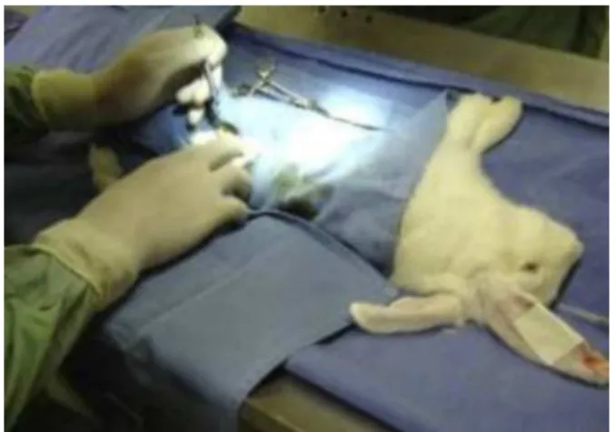
Fig.1–Surgicalpreparation.
Anesthetizedrabbitswereplacedinrightlateraldecubitus position;theposterolateralretroperitonealaccessroute wasused.
needleatadepthof5mm,whichremainedinsidetheIVDfor 5s(Fig.2).
Theprocess ofisolation and collection ofmononuclear
stemcells(SC)wasdonebypuncture-aspirationofbone mar-rowfromtheiliaccrest,andispartoftheprotocolthathas alreadybeendevelopedandconsolidatedinthiseducational institution.16
After cells were collected and isolated, they were
introducedintotheanimalIVDwiththesamesurgical
tech-niqueas described above. Thematerial was placedin the
upperlimitofthepre-drilledIVD,withasmaller-caliberneedle (13×4.5; 26G
½
). Exactly analogous procedures were per-formedinanimalsthatreceivedonlyisotonicsalinesolution (SS);theamountinjectedwasequaltothecellvolume,follow-ingtheabovementionedconditions.
Thestudyincluded32animals,dividedintoeightgroups:
four groups that received SC and four controlgroups that
receivedSS.
ThegroupsweredividedaccordingtothemomentofSC
injection:
- SC2M(fouranimals):transplantationofautologous
mono-nuclear SCs after two months of disk degeneration
inductionsurgery;
- SC2W(fouranimals):transplantationofautologous
mono-nuclearSCsaftertwoweeksofdiskdegenerationinduction surgery;
Fig.2–Needlepreparationtoperformthepuncture. Thefinal5mmofa40mm×12mm(18G1
½
)needlewas delimitedfromitsbevelandfoldedinanS-shapeinorder tostandardizethedepthofthepunctures.- SCCP(fouranimals):transplantationofautologous
mono-nuclearstemcellsimmediatelyafterthediskdegeneration inductionsurgery.
This differentiation allowed for a periodic comparative
critical analysis regarding the most appropriate period of
implementation ofcell therapyinlightofthe degenerative process.
- SCSP(fouranimals),thegroupthatreceivedmononuclear
stemcells,butdidnotundergodiskdegenerationinduction surgery.
Similarly,theanimalsthatwerepartofthecontrolgroup weresubdividedaccordingtothepairingwithrespecttothe experimentalgroupandreceivedisotonicSSinjection,as fol-lows:SS2M(fouranimals),correspondingtoSC2M;SS2W(four animals),correspondingtoSC2W;SSCP(fouranimals), corre-spondingtoSCCP;andSSSP(fouranimals),correspondingto SCSP.
Ofthe32rabbits,sixdiedduringsurgeryorpostoperatively. Thus,26rabbitswerehistologicallyexamined,divided
accord-ingtoTables1and2.
Eight weeks after the injection (CS/SS) in the IVD, the
rabbitswereeuthanizedbyinjectionofanoverdoseof
pen-tobarbital (90mg/kg) and their spines were harvested for
histologicalanalysis.
Afterthesampleswereprocessed,dehydrated,and embed-dedinparaffin, sectionsof6micronthicknessweretaken. Thesectionswerestainedwithsiriusredforcollagen analy-sis.Throughamicroscopewithpolarizedlight,aspectsrelated
tothecollagenarrangementintheIVDswereobservedwith
20-foldmagnification.
FromeachslidepreparedwithanIVD,sixfieldsofvision werephotographedbythesoftwareDinoCapture®2.0v.1.2.7 (AnMoElectronicsCorporation).Thesixfieldshadbeen pre-definedforalldiscs,startingfromthemostperipherallayerof theAFlamellae(Fig.3).Thisconventionallowsforapanoramic
andcomprehensivesampleoftheAFstructure.
TheimageswereprocessedbythesoftwareImageProPlus® v.4.50(MediaCybernetics,SilverSpring,MD),whichquantified thegreen(collagentypeIII)andreddots(collagentypeIII)of theslide,andstratifiedthemwiththeirrespectivepercentages (Fig.4).
Results
Inordertocomparegroupsandfindpossiblesignificant differ-encesbetweenthem,thefollowingvariableswereindividually assessed:theinnerlayeroftheAF(ILAF);theexternallayerof theAF(ELAF);andtheentireAF.
Foreachvariableandeachtypeofcollagen,ineach appli-cationtime,thenullhypothesisthatthecollagenresultsinthe SCgroupwouldbeequaltothoseoftheSSgroupwastested.
Tables3–5showsthedescriptivestatisticsandp-valuesofthe
statisticaltests.
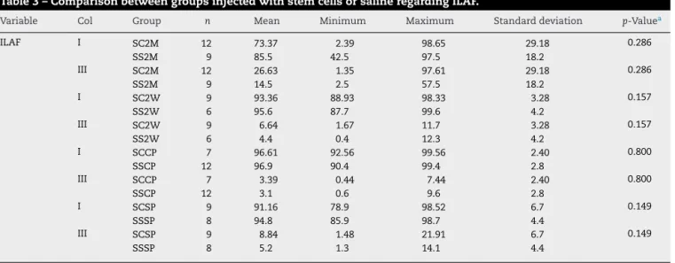
WhencomparingtheILAFofthegroupinjectedwithSC
Table1–Distributionofrabbitsingroupsinjectedwithstemcells.
Group Stemcells
SC2M SC2W SCCP SCSP
Rabbits (n=14) 4 3 4 3
Discs–intervention(n=37) 12 9 7 9
Discs–control (n=60) 16 14 16 14
SC2M,SCinjection2monthsafterpuncture;SC2W,SCinjection2weeksafterpuncture;SCCP,SCinjectionatthesametimeofpuncture;SCSP, SCinjectionwithoutpuncture.
Table2–Distributionofrabbitsingroupsinjectedwithsalinesolution.
Group Saline
SS2M SS2W SSCP SSSP
Rabbits (n=12) 3 2 4 3
Discs–intervention(n=35) 9 6 12 8
Discs–control (n=55) 15 10 17 13
SS2M,salineinjection2monthsafterpuncture;SS2W,salineinjection2weeksafterpuncture;SSCP,salineinjectionatthesametimeof puncture;SSSP,salineinjectionwithoutpuncture.
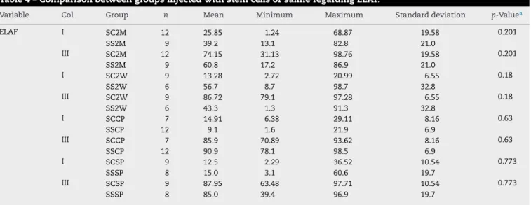
WhencomparingtheELAFofthegroupinjectedwithSC withthatofthegroupsinjectedwithSS,astatistically signif-icantdifferencewasobservedbetweentheSC2WandSS2W groups(p=0.018).Thisdifferencewasduetoanincreasein typeI collageninSS2Wgroup(56.7%)whencomparedwith SC2S(13.28%).CollagentypeI andIII valuesandgroup com-parisonsareshowninTable4andFig.5.
WhencomparingtheAFofthegroupinjectedwithSCwith thatofthegroupsinjectedwithSS,astatisticallysignificant
differencewasobservedbetweentheSC2WandSS2Wgroups
(p=0.025).Onceagain,thisdifferencewasduetoanincrease intypeIcollageninSS2Wgroup(76.1%)whencomparedwith theSC2W(53.32%).CollagentypeIandIIIvaluesandgroup comparisonsareshowninTable5.
Control
Fig.3–DinoCaptureTool.
ELAF
ILAF
Fig.4–ImageProPlusTool.
Exampleofthesoftwareusedtomakethepixelcountonaphotographoftheinnerlayeroftheannulusfibrosus(ILAF)and theexternallayeroftheannulusfibrosus(ELAF).Threepiecesofinformationaregivenforeachimage:1,numberofobjects; 2,percentageofobjectsintheimage;and3,percentageofthetotalimagearea.
Discussion
Disk degeneration induction hasbeen studied indepth in
another branch of this research through slides stained in
hematoxylin-eosin,fastgreen,andsiriusred.10Thepresent
studyusedslidesobtainedfromthesamespeciesand
popu-lationofrabbits,andtheinductionofdiskdegenerationwas
performedinthesamemanner.Thestandardizationofdisk
degenerationinductioninrabbitsinthisexperiment,through
the needle puncturemethod, waseffective and was
repro-ducedinasimilarmannertotheresultsofotherstudiesthat usedthesametechnique.10,17–20
Manycontemporarystudiesusedimmunohistochemistry
toassessdifferenttypesofcollagen.1,21–23Theforerunnersin
Table3–ComparisonbetweengroupsinjectedwithstemcellsorsalineregardingILAF.
Variable Col Group n Mean Minimum Maximum Standarddeviation p-Valuea
ILAF I SC2M 12 73.37 2.39 98.65 29.18 0.286
SS2M 9 85.5 42.5 97.5 18.2
III SC2M 12 26.63 1.35 97.61 29.18 0.286
SS2M 9 14.5 2.5 57.5 18.2
I SC2W 9 93.36 88.93 98.33 3.28 0.157
SS2W 6 95.6 87.7 99.6 4.2
III SC2W 9 6.64 1.67 11.7 3.28 0.157
SS2W 6 4.4 0.4 12.3 4.2
I SCCP 7 96.61 92.56 99.56 2.40 0.800
SSCP 12 96.9 90.4 99.4 2.8
III SCCP 7 3.39 0.44 7.44 2.40 0.800
SSCP 12 3.1 0.6 9.6 2.8
I SCSP 9 91.16 78.9 98.52 6.7 0.149
SSSP 8 94.8 85.9 98.7 4.4
III SCSP 9 8.84 1.48 21.91 6.7 0.149
SSSP 8 5.2 1.3 14.1 4.4
Therewasnostatisticallysignificantdifferencebetweengroups.
Col,collagen;SC2M,SCinjection2monthsafterpuncture;SC2W,SCinjection2weeksafterpuncture;SCCP,SCinjectionatthesametimeof puncture;SCSP,SCinjectionwithoutpuncture;SS2M,SSinjection2monthsafterpuncture;SS2W,SSinjection2weeksafterpuncture;SSCP, SSinjectionatthesametimeofpuncture;SSSP,SSinjectionwithoutpuncture.
Table4–ComparisonbetweengroupsinjectedwithstemcellsorsalineregardingELAF.
Variable Col Group n Mean Minimum Maximum Standarddeviation p-Valuea
ELAF I SC2M 12 25.85 1.24 68.87 19.58 0.201
SS2M 9 39.2 13.1 82.8 21.0
III SC2M 12 74.15 31.13 98.76 19.58 0.201
SS2M 9 60.8 17.2 86.9 21.0
I SC2W 9 13.28 2.72 20.99 6.55 0.18
SS2W 6 56.7 8.7 98.7 32.8
III SC2W 9 86.72 79.1 97.28 6.55 0.18
SS2W 6 43.3 1.3 91.3 32.8
I SCCP 7 14.91 6.38 29.11 8.16 0.63
SSCP 12 9.1 1.6 21.9 6.9
III SCCP 7 85.9 70.89 93.62 8.16 0.63
SSCP 12 90.9 78.1 98.5 6.9
I SCSP 9 12.5 2.29 36.52 10.54 0.773
SSSP 8 15.0 3.1 60.6 19.7
III SCSP 9 87.95 63.48 97.71 10.54 0.773
SSSP 8 85.0 39.4 96.9 19.7
AstatisticallysignificantdifferencewasobservedbetweentheSC2WandSS2Wgroups(p=0.018).
Col,collagen;SC2M,SCinjection2monthsafterpuncture;SC2W,SCinjection2weeksafterpuncture;SCCP,SCinjectionatthesametimeof puncture;SCSP,SCinjectionwithoutpuncture;SS2M,SSinjection2monthsafterpuncture;SS2W,SSinjection2weeksafterpuncture;SSCP, SSinjectionatthesametimeofpuncture;SSSP,SSinjectionwithoutpuncture.
a NonparametricKruskal–Wallistest;p<0.05.
thisareadatebacktothelate1990s.1,21Thisassessmenthasas amainadvantageitsspecificity;however,itisstillan expen-sivemethodinBrazil.Inthepresentstudy,siriusredproved tobeasimpleandinexpensivemethodtoassesstypeIandIII collagen.
Regardingthenumberofcellsinjectedintothetreatment, currentstudiesarenotstandardized.Somesuggestthatcell cultureisessentialtothesuccessoftreatment;however,most studiesdonotdiscussconsiderationsforoptimalcellnumber. Seriganoetal.22indicatedthattheoptimumdoseof autolo-gousmesenchymalstemcells(MSCs)indogsis1×106cells.
Inturn,Ghoshetal.23suggestedthatalowerdoseof0.1
×106
maybemoreeffective.Accordingtotheauthors,an
exagger-atednumberofcellsinthisenvironmentwithlownutrient
supplymaycausecellstocompeteforthesupplement,which maybedestructivetotheNPduetotheaccumulationofdead cellsandcellulardegradationproducts.23
Furthermore,thediscogenicdifferentiationofMSCsmay
alsobestimulatedbyco-culture.MSCscanbedirectly culti-vatedincontactwithIVDcells.Duringtheco-cultureofbone
marrow-derivedMSCandNPcells,ithasbeenobservedthat
theycommunicateinabidirectionalmanner,whichresults
Table5–ComparisonbetweengroupsinjectedwithstemcellsorsalineregardingAF.
Variable Col Group n Mean Minimum Maximum Standarddeviation p-Valuea
Entire AF
I SC2M 12 49.61 2.16 78.59 22.21 0.155
SS2M 9 62.3 27.8 88.9 16.1
III SC2M 12 50.39 21.41 97.84 22.21 0.155
SS2M 9 37.7 11.1 72.2 16.1
I SC2W 9 53.32 47.90 59.15 3.62 0.25
SS2W 6 76.1 48.2 97.4 17.5
III SC2W 9 46,68 40.85 52.10 3.62 0.25
SS2W 6 23.9 2.6 51.8 17.5
I SCCP 7 55.76 51.10 64.33 4.57 0.151
SSCP 12 53.0 48.6 60.7 3.5
III SCCP 7 44.24 35.67 48.90 4.57 0.151
SSCP 12 47.0 39.3 51.4 3.5
I SCSP 9 51.61 43.83 64.43 6.59 0.534
SSSP 8 54.9 45.5 79.5 11.1
III SCSP 9 48.39 35.57 56.17 6.59 0.564
SSSP 8 45.1 20.5 54.5 11.1
AstatisticallysignificantdifferencewasobservedbetweentheSC2WandSS2Wgroups(p=0.025).
Col,collagen;SC2M,SCinjection2monthsafterpuncture;SC2W,SCinjection2weeksafterpuncture;SCCP,SCinjectionatthesametimeof puncture;SCSP,SCinjectionwithoutpuncture;SS2M,SSinjection2monthsafterpuncture;SS2W,SSinjection2weeksafterpuncture;SSCP, SSinjectionatthesametimeofpuncture;SSSP,SSinjectionwithoutpuncture.
100%
90%
80%
70%
60%
50%
40%
30%
20%
Collagen distribution, %
10%
0%
SC2M
P=.201 P=.018 P=.063
Groups
P=.773 SCSP SCCP
SC2W SS2W SSCP SSSP
SS2M
Col 3
Col 1
Fig.5–Comparisonofpercentageofcollagendistributionintheexternallayerofannulusfibrosus(ELAF)betweendifferent groupsinjectedwithstemcellsandsaline.
AstatisticallysignificantdifferencewasobservedbetweentheSC2WandSS2Wgroups(p=0.018).Col1,type1collagen;Col
3,type3collagen;SC2M,SCinjection2monthsafterpuncture;SC2W,SCinjection2weeksafterpuncture;SCCP,SC injectionatthesametimeofpuncture;SCSP,SCinjectionwithoutpuncture.SS2M,salineinjection2monthsafterpuncture; SS2W,salineinjection2weeksafterpuncture;SSCP,salineinjectionatthesametimeofpuncture;SSSP,salineinjection withoutpuncture.
inanimprovementinthephenotypeoftheNPcellsandthe
differentiationofMSCcells.24Thissuggeststhatthe implanta-tionofMSCscanexertparacrineeffectsindegenerateNPcells thatresideonthedisk,andhelprestorenormalcellfunction andthediskrepairprocess.
Inthepresentstudy,mononuclearstemcellswereused.
Thefact that theyare obtainedthrough a simplerprocess
thantheMSCwastakenintoaccount,astheyonlyneedto
undergotwoprocessesofcentrifugationandFicoll-Hypaque
densitygradient,whiletheMSCneedtoundergothesesame
processes,inadditiontocultureandcellgrowthfor approx-imately14–16days,thuspresentinghighercostsandgreater riskofcontamination.16
InjectionofMSCs and mononuclearmesenchymal stem
cells istypicallysafe, althoughthereis apotentialfor for-mationofperipheralosteophytes,suggestingtheimportance ofaproperand safe carriertoinjectcells in thisregion.25 Inthisregard,thecarriermayallowthecelltoreceiveaxial
loads, which are important to stimulate the synthesis of
extracellularmatrixandinduceMSCdifferentiationwithout furtherexogenous stimulus.26 Different carriershave been usedintheliterature,suchassomehydrogels26,27 andfibrin glue.28Althoughthesestudiesadvocate theuse ofamobile carrierintheapplicationofstemcells,theliteratureis con-troversialinthisregard.OtherstudieshaveusedonlySS29or evennocarrier.22Inthepresentstudy,DMEMculturemedium with20%fetalbovineserumwasused,amethodthathasalso beenusedsuccessfullyintheliterature.30
Intheanalysisofthepresentresults,whenassessingthe entireAF,adifferencebetweenthegroupsthatwereinjected
two months after the puncture was observed. The SC2M
showed a slight increase in type III collagen, divided into
49.61%typeIand50.39%typeIII.TheSS2Mshowed62.35%
typeIand37.65%typeIIIcollagen.Thegroupthatreceived SChadadivisionclosertothatofthegroupsinwhichdisk
degenerationwasnotinduced,whiletheSSgroupshoweda
significantincreaseintheproportionoftypeIcollagen.This disproportionality occurred inELAF,indicating a disorgani-zationoftheAFstructure.However,thedifferencewasnot statisticallysignificant.
ThesameincreaseincollagentypeIinELAFandAFcould alsobeobservedbetweenthegroupsinjectedtwoweeksafter thepuncture.TheSC2Wshowed53.32%typeIand46.68%type IIIcollagen.TheSS2Wshowed76.15%typeIand23.85%type IIIcollagen.Onceagain,itwasobservedthatthegroupthat receivedSChadadivisionclosertothatofthegroupsinwhich diskdegenerationwasnotinduced,whiletheSSgrouphada significantincreaseintheproportionoftypeIcollagen,again attheexpenseofELAF.Inthestatisticalanalysis,astatistically significantdifferencewasobservedbothinELAF(p=0.018)as wellasinAF(p=0.025).
ThepresentdatasuggestthatmononuclearSCtherapywas
able toreshapethe changescausedbythe injuryfrom the
biopsyneedlewhentheSCareinjectedaftertwoweeks,but
thesamesuccesswasnotachievedwhenSCswereinjected
twomonthsafterinjury.Despitethefactthatthedatafrom
SC2M wasclosertothegroups inwhichdisk degeneration
wasnotinducedthan werethevaluesfromtheSC2W,this
differencewasnotstatisticallysignificant.
However,whencomparingthegroupsthatwereinjected
atthemomentofpuncture,SCCPshowed55.76%typeIand
44.24% type III collagen. In turn, SSCP showed 53% type I
and 47% typeIIIcollagen. Thisisthe highestmismatchin
degeneration,withachangeintheproportionoftypeIand typeIIIcollagenafterpuncture.Thissuggeststhatthedamage causedinthediscsofSSCPwerehealed.
Whencomparing thegroups thatwere injectedwithout
puncture,theSCSPshowed51.61%typeIand48.39%typeIII collagen.Inturn,theSSSPshowed54.9%typeIand45.10%type IIIcollagen.Asexpected,nolargedifferenceswereobserved
between these groups, as degeneration through puncture
wasnotinduced.Nostatisticallysignificant differencewas
observedbetweengroups.ThesedataindicatethatMSCsdid
notalterthestructureofcollagenindiscsthatwereinjected withoutdegeneration.
Conclusion
Treatmentwithmononuclearmesenchymalstemcellsisable
toreducechangesinthedistributionoftypeIandIIIcollagen intheAFofdegeneratedrabbitsdiscsuntiltwoweeksafter degenerationinduction.
Conflicts
of
interest
Theauthorsdeclarenoconflictsofinterest.
r
e
f
e
r
e
n
c
e
s
1. NerlichAG,SchleicherED,BoosN.1997VolvoAwardwinner
inbasicsciencestudies.Immunohistologicmarkersfor
age-relatedchangesofhumanlumbarintervertebraldiscs.
Spine(PhilaPa1976).1997;22(24):2781–95.
2. BrisbyH.Pathologyandpossiblemechanismsofnervous
systemresponsetodiscdegeneration.JBoneJointSurgAm.
2006;88Suppl.2:68–71.
3. HughesSP,FreemontAJ,HukinsDW,McGregorAH,RobertsS.
Thepathogenesisofdegenerationoftheintervertebraldisc
andemergingtherapiesinthemanagementofbackpain.J
BoneJointSurgBr.2012;94(10):1298–304.
4. Cavalli-SforzaL,FeldmanM,DornbuschS,ChenKH.Cultural
evolution.Anthropologyandculturaltransmission.Nature.
1983;304(5922):124.
5. CollocaCJ,KellerTS,MooreRJ,HarrisonDE,GunzburgR.
Validationofanoninvasivedynamicspinalstiffness
assessmentmethodologyinananimalmodelof
intervertebraldiscdegeneration.Spine(PhilaPa1976).
2009;34(18):1900–5.
6. KatzJN.Lumbardiscdisordersandlow-backpain:
socioeconomicfactorsandconsequences.JBoneJointSurg
Am.2006;88Suppl.2:21–4.
7. MurrayCJ,AtkinsonC,BhallaK,BirbeckG,BursteinR,Chou
D,etal.ThestateofUShealth,1990–2010:burdenofdiseases,
injuries,andriskfactors.JAMA.2013;310(6):591–608.
8. AnHS,MasudaK.Relevanceofinvitroandinvivomodelsfor
intervertebraldiscdegeneration.JBoneJointSurgAm.
2006;88Suppl.2:88–94.
9. EvansC.Potentialbiologictherapiesfortheintervertebral
disc.JBoneJointSurgAm.2006;88Suppl.2:95–8.
10.VialleEN,VialleLR,ArrudaAO.Histomorphometricanalysis
ofexperimentaldiscdegeneration.GlobalSpineJ.
2012;2(3):129–36.
11.VialleEN,VialleLR,Bleggi-TorresLF,SakamotoKS.
Histologicalevaluationoftheeffectsofmethylprednisolone
onexperimentalmedullarylesioninrats.RevBrasOrtop.
2007;42(4):101–13.
12.CarvalhoKA,CunhaRC,VialleEN,OsieckiR,MoreiraGH,
SimeoniRB,etal.Functionaloutcomeofbonemarrowstem
cells(CD45(+)/CD34(−))aftercelltherapyinacutespinalcord
injury:inexercisetrainingandinsedentaryrats.Transplant
Proc.2008;40(3):847–9.
13.LipsonSJ,MuirH.1980Volvoawardinbasicscience.
Proteoglycansinexperimentalintervertebraldisc
degeneration.Spine(PhilaPa1976).1981;6(3):194–210.
14.MasudaK,AotaY,MuehlemanC,ImaiY,OkumaM,Thonar
EJ,etal.Anovelrabbitmodelofmild,reproducibledisc
degenerationbyananulusneedlepuncture:correlation
betweenthedegreeofdiscinjuryandradiologicaland
histologicalappearancesofdiscdegeneration.Spine(PhilaPa
1976).2005;30(1):5–14.
15.RousseauMA,UlrichJA,BassEC,RodriguezAG,LiuJJ,LotzJC.
Stabincisionforinducingintervertebraldiscdegenerationin
therat.Spine(PhilaPa1976).2007;32(1):17–24.
16.Guarita-SouzaLC,AtahydeK,CarvalhoT,RebelattoC,Hansen
P,FurutaM,etal.Acomparac¸ãoentreotransplantede
célulastroncomononuclearesemesenquimaisnoinfartodo
miocárdio.RevBrasCir.2005;20(3):270–8.
17.KimDW,ChunHJ,LeeSK.Percutaneousneedlepuncture
techniquetocreatearabbitmodelwithtraumatic
degenerativediskdisease.WorldNeurosurg.
2015;84(2):438–45.
18.VialleE,VialleLR,ArrudaADO,RietRN,KriegerABD.Análise
radiológicadadegenerac¸ãodiscalexperimentalemcoelhos.
RevBrasOrtop.2009;44(4):313–9.
19.SobajimaS,KompelJF,KimJS,WallachCJ,RobertsonDD,Vogt
MT,etal.Aslowlyprogressiveandreproducibleanimalmodel
ofintervertebraldiscdegenerationcharacterizedbyMRI,
X-ray,andhistology.Spine(PhilaPa1976).2005;30(1):
15–24.
20.KimKS,YoonST,LiJ,ParkJS,HuttonWC.Discdegeneration
intherabbit:abiochemicalandradiologicalcomparison
betweenfourdiscinjurymodels.Spine(PhilaPa1976).
2005;30(1):33–7.
21.NerlichAG,BoosN,WiestI,AebiM.Immunolocalizationof
majorinterstitialcollagentypesinhumanlumbar
intervertebraldiscsofvariousages.VirchowsArch.
1998;432(1):67–76.
22.SeriganoK,SakaiD,HiyamaA,TamuraF,TanakaM,Mochida
J.Effectofcellnumberonmesenchymalstemcell
transplantationinacaninediscdegenerationmodel.JOrthop
Res.2010;28(10):1267–75.
23.GhoshP,MooreR,Vernon-RobertsB,GoldschlagerT,Pascoe
D,ZannettinoA,etal.ImmunoselectedSTRO-3+
mesenchymalprecursorcellsandrestorationofthe
extracellularmatrixofdegenerateintervertebraldiscs.J
NeurosurgSpine.2012;16(5):479–88.
24.StrassburgS,RichardsonSM,FreemontAJ,HoylandJA.
Co-cultureinducesmesenchymalstemcelldifferentiation
andmodulationofthedegeneratehumannucleuspulposus
cellphenotype.RegenMed.2010;5(5):701–11.
25.VadalàG,SowaG,HubertM,GilbertsonLG,DenaroV,KangJD.
Mesenchymalstemcellsinjectionindegenerated
intervertebraldisc:cellleakagemayinduceosteophyte
formation.JTissueEngRegenMed.2012;6(5):348–55.
26.RichardsonSM,HughesN,HuntJA,FreemontAJ,HoylandJA.
HumanmesenchymalstemcelldifferentiationtoNP-like
cellsinchitosan-glycerophosphatehydrogels.Biomaterials.
2008;29(1):85–93.
27.FrithJE,CameronAR,MenziesDJ,GhoshP,WhiteheadDL,
GronthosS,etal.Aninjectablehydrogelincorporating
intervertebraldiscregeneration.Biomaterials. 2013;34(37):9430–40.
28.AllonAA,AurouerN,YooBB,LiebenbergEC,BuserZ,LotzJC.
Structuredcocultureofstemcellsanddisccellsprevent
discdegenerationinaratmodel.SpineJ.2010;10(12):
1089–97.
29.SobajimaS,VadalaG,ShimerA,KimJS,GilbertsonLG,Kang
JD.Feasibilityofastemcelltherapyforintervertebraldisc
degeneration.SpineJ.2008;8(6):888–96.
30.HoG,LeungVY,CheungKM,ChanD.Effectofseverityof
intervertebraldiscinjuryonmesenchymalstemcell-based