Mailing Address: Marco Antonio Rodrigues Torres • Rua Faria Santos, 394/601 - 90670-150 – Porto Alegre, RS - Brazil
E-mail: [email protected] Received on 08/24/05 • Accepted on 03/17/06
Assessment of Left Ventricular Diastolic Function in
Hypertensive Patients Under Different Hemodynamic
Conditions: Comparative Study Between Transmitral and
Tissue Doppler Echocardiography
Luiz Cláudio Danzmann, Valéria Centeno de Freitas, Luiz Felipe Araújo, Marco Antonio Rodrigues Torres Hospital de Clínicas de Porto Alegre e Universidade Federal do Rio Grande do Sul - Porto Alegre, RS - Brazil
O
BJECTIVETo evaluate the behavior of LV diastolic displacement velocities of basal posterior septum near mitral ring segment obtained by tissue Doppler in relation to mitral fl ow velocities of hypertensive subjects, submitted to preload increase by left inferior limbs elevation for 5 minutes, and during 1 minute of handgrip (conditions 1 and 2, respectively).
M
ETHODSTwenty fi ve outpatients (15 men, 52±11 years), with arterial hypertension > 3 years, by the transmitral fl ow Doppler variables: E, A and E/A, and of tissue Doppler: E’, A’ and E’/A’, in basal situation and in conditions 1 and 2. For comparison of results in basal situation with conditions 1 and 2 a paired t test was applied.
R
ESULTSE(cm/s) varied from 68,88 ± 11,94 to 75,82 ± 15,71* and E’(cm/s) varied from 8,22 ± 2,30 to 8,31 ± 2,24 in condition 1 (p<0.05). In addition, a comparison of variation percent (%) of Doppler transmitral indices with corresponding tissue Doppler indices variation % showed a signifi cant difference between E and E’, with preload increase (p=0.01).
C
ONCLUSIONPatients with left ventricular adaptation to systemic arterial hypertension showed less modifi cation of the index E´ than E, after preload increase maneuver, an evidence that may suggest its more ample utilization in the context of echocardiographic left ventricular diastolic function evaluation.
K
EY WORDSLeft ventricular (LV) diastolic dysfunction has been recognized as one of the leading causes of congestive heart failure (CHF)1. Several studies have indicated that 30% to
50% of CHF patients have normal or slightly impaired left ventricular systolic function. Some researchers propose that these patients should be designated as a “diastolic” heart failure group2.
Since then, transmitral fl ow velocity profi le at the LV entry pathway has been routinely assessed by spectral Doppler in the search for patterns that may characterize such a diagnosis and reveal something about disease prognosis3. Although this method had initially determined
what would become known as diastolic measurement pattern, soon it was realized that evaluation results varied and reproducibility was limited4.
A few studies showed the lack of a pattern of normality even with a clear clinical condition of diastolic dysfunction and heart failure. This lack of a pattern began to be designated as “pseudonormal pattern”5. Such a pattern
results from increased left atrial pressure that becomes higher than the effects of LV relaxation. This dependence on velocity and the isovolumetric relaxation time in relation to LV relaxation, as well as to left atrial pressure, was noticed long ago, and these variables set a framework that conveys the limitations of the methods based on transmitral fl ow assessment5.
This pattern may take place due to the fact that fl ow variables at the LV entry pathway are data determined by pressure differences between the left atrium and the LV during diastole. Evaluation of volume changes has the theoretical advantage of being less preload-dependent than TMD variables6.
Tissue Doppler (TD) assesses high-amplitude and low-velocity signals from the myocardium7. This resource
has been useful as a quantitative index of LV segmental function for analysis of both diastole8 and systole9. Diastolic
dysfunction studies have shown that this is relatively independent of preload6 and it has been documented
that probability of the same “pseudonormalizaton” seen with TMD appearing is low
TD assessment of the diastolic function may even be used to yield a highly accurate diastolic segmental analysis, as shown in a recent study10, in which
interrogation of multiple sites with this technique was possible using color tissue Doppler data and processing signals from all LV segments.
This clinical study was designed to assess the behavior of LV diastolic indices obtained by TDI as compared to those obtained by TMD under hemodynamic overload conditions – induced by changes in preload (leg-lifting) – and postload – by isometric exercise, (handgrip manouvre) – during analysis of the diastolic function of this chamber. The study population consisted of patients with systemic arterial hypertension (SAH) for at least three years, whose left ventricles had adapted to this condition.
M
ETHODSPatient selection - Twenty-fi ve outpatients with SAH for at least three years were consecutively evaluated. Exclusion criteria were: clinical, electrocardiographic or echocardiographic signs of myocardial ischemia, valvar diseases, cardiac arrhythmias, coronary heart diseases and peripherical diseases. Moreover, chronic renal failure and diabetic patients were also excluded.
Investigators did not interfere with cardiac medications for SAH treatment. All individuals who underwent the test signed the written consent form expressing their agreement with the protocol approved by the institution Ethics Committee under the name “Assessment of the left ventricle diastolic function by pulsed-wave tissue Doppler in hypertensive patients”, at the meeting held on January 19, 2001 (number 99376).
Echocardiography - The echocardiograms were performed with the HDI 5000, Philips-ATL, Seattle, WA equipment with patients at rest, lying down in the left lateral decubitus position. The images were acquired in standard tomography slices of the LV (longitudinal and transversal parasternal and, apical 4-, 5-, and 2- chamber views) with a 3.5 MHz transducer in harmonic mode. The images were archived on VHS tapes, and all analysis of spectral mitral and tissue Doppler, as well as the measurements of cavities and analysis of LV kinesis were performed on-line and off-line.
Parietal thicknesses of the interventricular septum (Psiv) and of the posterior wall (Pw), as well as LV diastolic (Dd) and systolic (Sd) diameters were obtained using the M mode; LV hypertrophy (LVH) was defi ned as a mass index >110g/sq m for women and 125g/sq m for men; AE dimension was also measured during late systole. Ventricular mass was automatically calculated by the equipment (mass formula: LV = 1.04 x [(Dd + Pw + Psiv)3 – Dd3]), and LV systolic and diastolic
volumes, as well as ejection fraction, were obtained by the Teichholz method.
Transmitral fl ow velocities - LV apical four-chamber transthoracic images were recorded with standard 2.5 MHz transducer equipment. The volume sample was placed on the free-edge of mitral valve leafl ets, at the apical four-chamber view. Flow velocity was recorded with the pulsed-wave Doppler method using the mean of three representative heart beats. Peak early diastolic velocity (E), peak atrial systolic velocity (A) and E/A ratio were determined.
atrial systole or late diastolic fi lling (A’ cm/s) and the E’/A’ ratio were recorded11.
Changes induced to hemodynamic conditions - Flow velocity profi les were obtained under baseline conditions, after manoeuvres to increase LV preload (condition 1) –by uniform and supported lifting of both legs to a 45-degree angle in relation to the bed during a continuous fi ve-minute period – and after manoeuvres to increase LV postload (condition 2) – during a continuous one-minute period of sustained handgrip with a manual dynamometer at 30% of its maximum capacity.
Reproducibility - Interobserver variability – calculated as the difference between two measurements taken on the same patient by two different observers who had undergone the same training, divided by the mean value of the difference – varied from 1.5 to 2.6% for transmitral fl ow indices, and 0.8 to 1.7% for pulsed-wave tissue Doppler.
Statistical analysis - All values were presented as mean ± standard deviation (SD). The variables obtained at rest (baseline), through mitral Doppler and tissue Doppler were compared to the conditions associated with lower limb lifting (condition 1) and to the handgrip manouevre (condition 2). The mean TMD and TD differences between the baseline state and conditions 1 and 2 were compared by paired Student’s t-test.
The percentage of variation of TMD indices under conditions 1 and 2, in relation to the baseline and compared to corresponding percentage values (%) of the variation of TD indices under conditions 1 and 2, also in relation to the baseline, were calculated in a second assessment. It was then verifi ed that the mean values of the difference in percentages of variation (∆ %) were
smaller than the standard deviation. The conclusion that data did not have a normal distribution led to the use of the Wilcoxon t-test to compare these variables. A value of P < 0.05was considered statistically signifi cant.
R
ESULTSSample characteristics - The sample consisted of twenty-fi ve patients, aged 32 to 75 years (52 ± 10.9), withborderlineanthropometric indices (body mass index = 27.9) who did not present extreme variations in mean arterial blood pressure or heart rate during the exam (MAP=108±20.7mmHg and HR=69.7±13.1bpm). Mean indices of systolic functions were normal, with an ejection fraction of 66.9±9.6% and a systolic shortening fraction of 37.5±7.1%, and adequate left ventricular mass (182.6±65.3g.) (table 1).
Baseline values-Under baseline conditions, values for velocities calculated by TMD were: E = 68.88 ± 1.94cm/ s. A = 61.88 ± 12.99cm/s. E/A = 1.16 ± 0.31. Under baseline conditions, values for velocities calculated by TD were: E’ = 8.31 ± 2.31cm/s. A’ = 9.12 ± 1.93cm/s. E’/A’ = 0.92 ± 028 (Table 2).
Comparison between variations recorded by mitral Doppler and by tissue Doppler -The comparison between the percentages of variation in mitral fl ow velocities after changes in hemodynamic conditions and the percentages of variation obtained by tissue Doppler analysis revealed a signifi cant difference between the ∆% recorded for
the E wave ∆% vs. that of the E’ wave (6.1% vs. 0%,
P=0.01), which corroborates the nonvariability of E’ following increase in preload (Table 2).
By comparing the magnitude of changes observed in the variables evaluated by both methods under baseline hemodynamic conditions, lower limb lifting and handgrip, and by applying the Wilcoxon t-test to evaluate differences between the measurement medians, it becomes clear that there is a signifi cant difference between variations in the E waves measured by mitral Doppler and the E’ waves measured by tissue Doppler.
D
ISCUSSIONDiastole and preload/postload inter ference -
Transmitral fl ow pulsed-wave Doppler echocardiography is the most conventional method to evaluate diastole. The test, however, shows an important variation in E and A wave indices, and in the ratio between both waves (E/A) in view of changes induced by LV preload.
Tissue Doppler is the method that assesses segmental myocardial velocity at both diastole and systole. In a study comparing the interventricular septum on TD with that of transmitral fl ow in twenty patients with standard relaxation defi cit (E/A wave ratio <1), who received an infusion of 700 ml of physiological saline, a signifi cant change was observed in the E/A velocity ratio measured by transmitral fl ow (E/A=0.7 ± 0.1 vs. 0.9 ± 0.1 p < 0.01), which was not observed in the TD measurements (E’/A’=0.5 ± 0.1 vs. 0.5 ± 0.1. p=NS) 6.
These results point to the fact that assessment of segmental ventricular wall motion by TD is relatively independent from ventricular preload, as has already been suggested12.
Table 1 - Sample clinical, echocardiographic and anthropometric data
Mean SD(±)
Age (years) 52.0 10.9
EF (%) 66.9 9.6
SF (%) 37.5 7.1
LV MASS (g.) 182.6 65.3
MAP (mmHg) 108.7 20.7
HR (bpm) 69.7 13.1
BMI 27.9 4.8
Concerning postload, both transmitral fl ow study and TD show variations. Oki et al13, conducted a detailed
study of pulsed-wave tissue Doppler. The authors13
evaluated systolic and diastolic functions in twenty healthy individuals by echocardiogram before and after acute postload elevation, secondary to the infusion of angiotensin II (until they obtained a 30 mmHg elevation in mean arterial blood pressure). By doing so, the authors observed that all diastolic parameters (TMD and TD) underwent signifi cant variations.
These findings suggested that neither method is
Table 2 - Transmitral fl ow Doppler and Tissue Doppler under baseline conditions and changes to pre- and postload
Transmitral fl ow Doppler Tissue Doppler
E A E/A E` A` E`/A`
Mean ± SD Mean ± SD Mean ± SD Mean ± SD Mean ± SD Mean ± SD
Baseline 68.88 ± 11.94 61.88 ± 12.99 1.16 ± 0.31 8.22 ± 2.30 9.12 ± 1.93 0.92 ± 0.28
Condition 1 75.82 ± 15.71* 63.34 ± 16.61 1.30 ± 0.52 8.31 ± 2.24 10.05 ± 2.36* 0.86 ± 0.25*
Condition 2 70.30 ± 17.49 66.44 ± 17.55 1.14 ± 0.47 7.54 ± 1.86* 9.60 ± 2.20 0.81 ± 0.27*
* statistically signifi cant difference compared to baseline with p<0.05. E- early transmitral diastolic fl ow velocity; A- late transmitral diastolic fl ow velocity; E’- early diastolic myocardial wall motion velocity; A’- late diastolic myocardial wall motion velocity.
Table 2 - Differences between percentages of variation (∆%) of TMD and TDI indices under conditions of
pre- and postload elevation (C.1 and C.2, respectively) in relation to the baseline state
TMD TD P
∆% E-E’ C.1/baseline 6.1 0.0 0.010
∆% E-E’ C.2/baseline 1.1 -7.1 0.045
∆% A-A’ C.1/baseline -1.2 10.0 NS
∆% A-A’ C.2/baseline 10.7 8.2 NS
∆% E/A-E’/A’ C.1/baseline 8.0 -6.3 0.001
∆% E/A-E’/A’ C.2/baseline -8.0 -14.3 NS
∆% E-E’ C.1/baseline: difference in percentages of variation between E and E’ waves after preload elevation; ∆% E-E’ C.2/baseline: difference in percentages of variation between E and E’ waves after postload elevation; ∆% A-A’ C.1/baseline: difference in percentages of variation between A and A’ waves after preload elevation; ∆% A-A’ C.2/baseline: difference in percentages of variation between A and A’ waves after postload elevation;
∆% E/A-E’/A’ C.1/baseline: differences in percentages of variation between E/A and E’/A’ ratios after preload elevation; ∆% E/A-E’/A’ C.2/baseline: differences in percentages of variation between E/A and E’/A’ ratios after postload elevation.
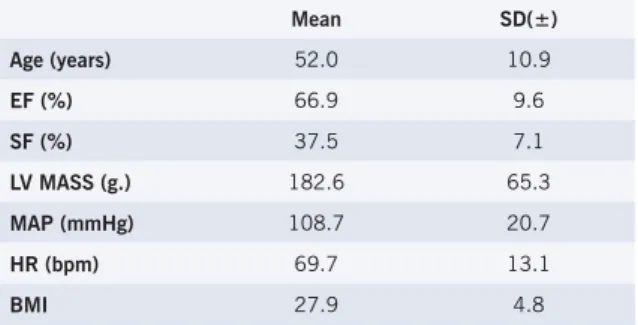
Fig. 1 - Change in Etransmitral fl ow velocity at baseline state and after LV preload elevation by tilting lower limbs to 45°.
VELOCIDADE E
40
60
80
100
P = 0,002
cm/s
Baseline ↑ Lower limbs
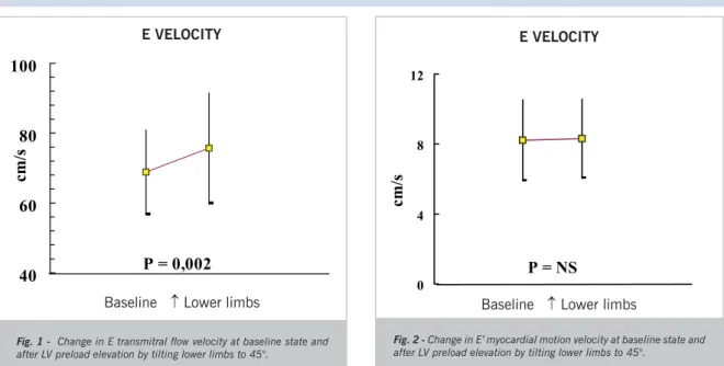
Fig. 2 - Change in E’ myocardial motion velocity at baseline state and after LV preload elevation by tilting lower limbs to 45°.
Velocidade E'
0 4 8 12
P = NS
cm/s
Baseline ↑ Lower limbs
entirely independent of postload-variations. This fact, however, enables their utilization to detect and follow-up the effect of LV postload variations, which could be useful, for instance, to keep track of clinical progression or, perhaps, to quantify response to drug therapies.
Motion of mitral annulus and adjacent segments of the ventricular myocardium - In view of the relatively fi xed position of the ventricular apex during the cardiac cycle14 – with LV relatively preserved as to its geometry
– changes on the longitudinal axis would refl ect volumetric changes, as indicated by Sohn et al16. In this study,
the analysis of the wall motion in the basal portion of the interventricular was deliberately chosen, since this myocardial portion adjacent to the mitral annulus moves in a more parallel fashion than the others (such as the anterior, lateral and inferior face), as already highlighted by Sohn et al16.
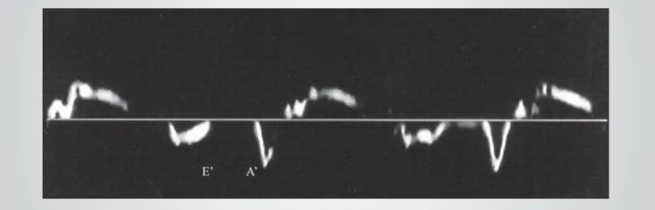
As seen on Figure 3, the tissue Doppler exam captured, on the segment analyzed, two different movements towards the atrial chamber: one known as (E’), which starts simultaneously with the beginning of blood fl ow towards the LV entry pathway, where its peak velocity always occurs earlier than the peak fl ow velocity measured by mitral Doppler and is ended well before the blood infl ow through the mitral orifi ce ends. The E’ peak velocity is higher than the peak of another wave (A’), which is similar to the normal pattern of transmitral fl ow. As emphasized by Sohn et al6, on their classical study: “Mitral infl ow after
the end of left ventricular lengthening in the long-axis dimension. whether it is driven by pressure gradient or inertia. would lead to an increase in left ventricular volume in the short-axis dimension. which would mean higher compliance of the ventricular chamber in the short-axis dimension. Therefore, it may be assumed that a relaxation abnormality refl ected in the long-axis dimension could potentially be evident earlier than clinical manifestation of global left ventricular relaxation abnormality”.
This particular aspect in the methodology suggests that mitral Doppler analysis of the variables yields data concerning velocities, whereas with tissue Doppler measurements, data would refl ect changes in volume.
Tissue Doppler: independence of preload changes
- Data analysis in this study showed independence of E’ values to the leg-lifting manouevre (condition 1, corresponding to change in preload). Other researchers – such as Garcia et al12 – had already observed that E’
velocity had only a slight relationship to E peak velocity, which indicated the relative independence of E’ peak velocity obtained by tissue Doppler.
With this study, it is shown for the fi rst time that, even with simpler and noninvasive bedside manoeuvres, no signifi cant changes were observed in the peak early
E’ velocity of the mitral annulus on tissue Doppler (in contrast to E velocity on mitral Doppler), after a change in preload induced by elevation of the lower limbs.
Lower limb elevation and handgrip manoeuvres as a means to study adaptation of the heart to new hemodynamic conditions - Both manoeuvres used in this study, aiming at provoking new hemodynamic levels as a means to evaluate adaptive responses of circulation and the myocardium, have been employed for a long time. The preload increase mechanism with corresponding elevation of systemic arterial blood pressure was recently correlated to the release of powerful endothelial-action vasoconstrictors15.
The mechanism of hemodynamic change induced by increase in preload (lower limb lifting), using the same protocol applied to the subjects in this study, has also already been established16. Comments made concerning
magnitude of the impact of the manoeuvres on these hemodynamic variables warrant analysis. As seen in Figures 1 and 2 (with > preload), the magnitude of the changes was enough to interfere with behavior of the E variable, obtained by mitral Doppler in this study, but did not have any impact on the E’ variable measured by tissue Doppler.
Tak et al obser ved a significant effect of the handgrip manoeuvre on preload and, consequently, on diastolic indices17. Other publications ratifi ed the use
of the manoeuvre to produce isometric exercise and an effective postload elevation, by applying 30% to 50% of maximal force over a period of 1 to 5 minutes18,19.
We observed that a few patients had diffi culties to maintain the same handgrip strength during the set time (fi ve minutes), and a few individuals were only able to sustain a smaller load over a shorter time, even though this has been a commonly reported problem in studies previously published regarding utilization of similar-type isometric exercises.
Diagnosis of diastolic dysfunction of the basal segment of the interventricular septum as measured by tissue Doppler - The fi ndings in this study point to a preload-independence, considering the E’ wave
velocity. Statistical data and results obtained from the analysis of all variables are consistent with fi ndings in literature6,12,13,16.
Application of the fi ndings in this study to clinical practice - In patients with the ‘pseudonormalization” pattern, whose parameters may be obtained both by mitral Doppler and tissue Doppler, the variation of peak E and E’ velocities, as well as the E/A ratio in relation to the E’/A’ ratio, may be employed to differentiate this ‘pseudonormal’ pattern from a normal fi lling pattern.
Considering that the most common practice in echocardiographic procedures has been to try to differentiate this ‘pseudonormal’ pattern from normal diastolic function pattern by analyzing the pulmonary vein fl ow pattern – which is not always useful for this purpose in this subgroup of patients – the adoption of the simple manoeuvres proposed in this study may be useful for these patients. Moreover, variation in TD indices after the postload changes reported in this study suggest that these indices may be used to monitor the diastolic function of hypertensive patients being treated with medication to reduce postload.
Study limitations - This study, similarly to others previously published, has several practical limitations. Among them: 1) considering that patients with left ventricular asynergy were left out, it is possible that patients with kinesis abnormalities (for instance, those affecting the basal segment of the interventricular septum) may present abnormalities in the profi les of the variables analyzed, which does not express the overall behavior of the ventricle, which may be normal for the behavior of its diastolic function parameter16;2) we have
opted to measure myocardial velocity at the region more
adjacent to the annulus on the interventricular septum, which corresponds more exactly to the basal segment of the posterior interventricular septum. However, due to the fact that it is subject in a lower degree to the translational movement of the myocardium, this approach has the disadvantage of being affected whenever the right ventricular function is altered. For instance, secondarily to pulmonary hypertension, especially if this chamber also presents signs of associated dysfunction16; 3) since one
of the exclusion criteria was that patients should not be in sinus rhythm, it is possible that these fi ndings may not be applicable to those with atrial fi brillation, for instance; 4) although less affected than mitral Doppler analysis, the constraining aspect of an inadequate acoustic window has to be taken into consideration. The Doppler diastolic evaluation is usually also affected by tachycardia, valvar diseases and cardiac rotational movement20.
Conclusions - In patients with the left ventricle adapted to arterial hypertension, the diastolic motion of the myocardial segment adjacent to the mitral annulus measured by tissue Doppler was less dependent on preload than when assessed by the transmitral fl ow Doppler. This fact suggests the validity of the method as a coadjuvant index for assessment of LV diastolic function in this group of patients. We may also conclude that TD indices were signifi cantly affected by postload variation, and that they could be used to monitor and follow-up the diastole after antihypertensive treatment with postload-reducing drugs.
Potencial Confl ict of Interest
No potential confl ict of interest relevant to this article was reported.
1. Vasan R S, Benjamin E J. Diastolic heart failure – no time to relax. Editorials. N Engl J Med. 2001; 344:56-59.
2. Grossman W. Diastolic dysfunction and congestive heart failure. Circulation. 1990;81 (Suppl III):III-1-7.
3. Appleton CP, Firstenberg MS, Garcia MJ, Thomas JD. The echo-Doppler evaluation of left ventricular diastolic function. A current perspective. Cardiol Clin. 2000;18:513-46.
4. Nishimura RA, Abel MD, Hatle LK, Tajik AJ. Assessment of the diastolic function of the heart: background and current application of Doppler echocardiography. Part II. Clinical studies. Mayo Clin Proc. 1989;64:977-90.
5. Nagueh SF, Middleton KJ, Kopelen HA, et al. Doppler tissue imaging: a noninvasive technique for evaluating of left ventricular relaxation and estimation of fi lling pressures. J Am Coll Cardiol. 1997;30:1527-33. 6. Sohn D, Chai I, Lee D, et al. Assessment of mitral annulus velocity by Doppler tissue imaging in the evaluation of left ventricular diastolic function. J Am Coll Cardiol. 1997;30:474-80.
7. Hatle L, Sutherland GR. Regional myocardial function--a new approach. Eur Heart J. 2000;21:1337-57.
8. Wilkenshoff U, Sovany A, Wigström L, et al. Regional mean systolic myocardial velocity estimation by real time color Doppler myocardial
R
EFERENCESimaging: a new technique for quantifying regional systolic function. J Am Soc of Echocardiogr. 1998;11:683-692.
9. Pasquet A, Armstrong G, Beachler L, Lauer M, Marwick T. Use of segmental tissue Doppler velocity to quantitate exercise echocardiography. J Am Soc Echocardiog. 1999;12:901-12. 10. von Bibra H, Tuchnitz A, Klein A, et al. Regional diastolic function by
pulsed Doppler myocardial mapping for the detection of left ventricular ischemia during pharmacologic stress testing. J Am Coll Cardiol. 2000;36:444-452.
11. Pai R, Gill K. Amplitudes, durations, and timings of apically directed left ventricular myocardial velocities: their normal pattern and coupling to ventricular fi lling and ejection. J Am Soc Echocardiogr. 1998;11:105-11.
12. Garcia MJ, Thomas JD, Klein AL. New echocardiographic applications for the study of diastolic function. J Am Coll Cardiol. 1998;32:865-75.
13. Oki T, Fukuda K, Tabata T, et al. Effect of an acute increase in afterload on left ventricular regional wall motion velocity in healthy subjects. J Am Soc Echocardiogr. 1999;12:476-83.
of wall motion. J Am Soc Echocardiogr. 1988;1:393¯405.
15. Mangieri E, Tanzilli G, Barilla F, Ciavolella M, Puddu PE, De Angelis C, et al. Handgrip increases endothelin-1 secretion in normotensive young male offspring of hypertensive parents. J Am Coll Cardiol. 1998;31:1362-6.
16. Shimizu Y, Masaaki U, Shimizu H, et al. Peak negative myocardial velocity gradient in early diastole as a noninvasive indicator of left ventricular diastolic function. J Am Coll Cardiol. 1998;32:1418-25. 17. Tak T, Choudhary RS, Chatterjee S, et al. Effect of loading conditions on Doppler-derived transmitral flow indices in normal subjects and patients with coronary artery disease. Echocardiography.
1992;9:467-474.
18. Campaniello M, Antonelli G, Di Venere N, et al.The echocardiographic assessment of the functional variations in the left ventricle induced by isometric stress in subjects with primary cardiomyopathy in the pre-dilated phase. Cardiologia. 1994;39:543-9.
19. Heng MK, Bai JX, Marin J. Changes in left ventricular wall stress during isometric and isotonic exercise in men. Am J Cardiol. 1998;62(10 pt 1):794-8.