*e-mail: [email protected]
Received: 21 July 2013 / Accepted: 27 January 2014
Dissolved ozone in biological luid monitored by optical device
operating in the red-infrared region
Henrique Cunha Carvalho*, Milene da Silva Melo, Carlos José de Lima, Renato Amaro Zângaro
Abstract Introduction: When a gas is used for therapy, often the kinetic behavior and their distribution in biological systems is not known, leading to unsatisfactory results for clinical application. The use of ozone in living organisms has been scientiically released worldwide under the name of ozone therapy. The eficacy of this technique is determined primarily by the diffusion of gas within the tissues or luids and which determines their action in the entire target region. We propose the development of technique to monitoring the O3 dissolved in the biological luid using an optical device operating in the red-infrared region. Methods: The recombination of O3 in O2 enables the monitoring of the latter by the measurement of SpO2, and, based on this phenomenon, we propose to use an optical device operating in the red-infrared region to monitoring indirectly the diffusion of O3 in luids. The system was based on optomechanical arrangement using a capsule containing luid that was ozonated or oxygenated during the process. A pulse oximeter is a noninvasive device used for continuously measure of SpO2 resulting from the recombination of ozone. Results: The measurements of SpO2 when subjected to ozone and oxygen, showed an increased rate of SpO2 function of time for both cases reaching its peak in 80s and 160s, respectively. The experimental data concerning the SpO2 saturation as a function of time can be itted by the theoretical model, showing a good correlation between them. Conclusion: A technique was developed using an optical device operating in the red-infrared region to monitoring ozone dissolved in biological luid, showing a simple and effective way to indirectly monitoring the presence of ozone in luids. Keywords Ozone, Biological luid, Diffusion, Recombination, Optical device, Red-infrared region.
Introduction
The use of ozone in living organisms has been widely publicized primarily on its action on bacteria, fungi, protozoa and viruses (Bocci et al., 2011a; Loeb, 2011). Numerous other biological and medical applications
has been scientiically tried and reported around the
world under the name of ozone therapy.
Ozone involved in redox balance, being a powerful oxidant that reacts immediately when in contact
with biological luids, inducing molecules directly
on the balance of reactive oxygen species (ROS),
which inluence many biochemical events in the cell metabolism, providing antimicrobial effects beneits in
addition to repair and balance of the target organism (Bocci et al., 2011a).
Thus, satisfactory results have been obtained in the treatment of diabetic foot (Wainstein et al., 2011), hepatitis (Zaky et al., 2011), cancer (Schulz et al., 2008), coronary heart disease (Martínez-Sánchez et al.,
2012), atherosclerosis (Delgado-Roche et al.,
2013), as a disinfecting agent in microorganisms
causing nosocomial infections (Doan et al., 2012;
Zoutman et al., 2011), and others.
When a gas is used for therapeutic purposes, often their kinetic behavior and their distribution in biological systems is not known, leading to unsatisfactory results when clinical application. The application of
ozone therapy eficacy is determined primarily by the diffusion of gas within the tissues or luids and which
determines their action in the entire target region. The techniques commonly used to monitoring the
diffusion of ozone luids are invasive (Buchan et al.,
2005; Loeb, 2011), which prevents monitoring systems such as sterile blood. Noninvasive optical techniques may be used in the UV region at 254 nm (Gao et al., 2012; Kalnajs and Avallone, 2010), but this spectral
region cannot be applied in biological luids due to
high radiation absorption UV blood.
The recombination of O3 in O2, allows the monitoring of the latter by the measurement of
SpO2 and based on this phenomenon, we propose
a new approach to monitoring the O3 dissolved in
the biological luid using a simple and ingenious
Methods
The present study aims to determine the diffusion of
ozone gas in luids by measuring SpO2. The model
was proposed in order to be able to perform real-time monitoring, noninvasively and nondestructively, using such a pulse oximeter operating in the region red - infrared. A system was developed based on the
use of an optical oximeter (MD-300C - Moriya JG),
which operates at two emission wavelengths, λ1 and
λ2, the irst of which presents high absorption by
oxyhemoglobin and second low absorption. The SpO2 absorption corresponds to the difference between
these two variables. As a biological luid, was used
0.8 mL of whole bovine milk, type C, with 10 samples,
ive submitted to the low of oxygen and ive others submitted to the low of ozone. This luid was used due
to optical scattering characteristics, which facilitates the detection of the SpO2, and the model can be easily implemented in blood.
Pulse oximetry is based on light transmittance
of hemoglobin. The light transmission coeficient
of a substance is determined by the Beer-Lambert Law, which states that the concentration of an unknown solute in a solvent may be determined by absorption/scattering of light. In this case, solutes are oxyhemoglobin and reduced hemoglobin, and blood as the solvent.
During systole, the blood system is illed, increasing blood volume and absorption/scattering of light. During
diastole, blood volume and light absorption/scattering reach their lowest point. The pulse oximeter operates synchronously with the cardiac pulse and SpO2 is obtained from the difference between the maximum and minimum absorption/scattering intensities during systole and diastole. In this case, two wavelengths are used, λ1 (660 nm) that is highly absorbed by oxyhemoglobin and λ2 (910 nm) which is absorbed by all the others components present in the tissue (Figure 1). The difference of intensity between the two signals expressed SpO2. In this work, the solvent is milk and solutes are ozone and oxygen.
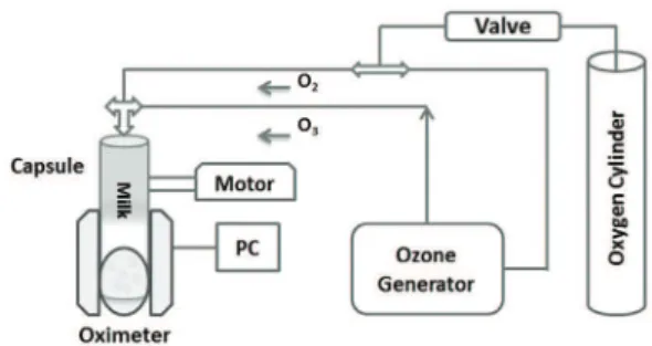
The system of SpO2 was developed according to the diagram shown in Figure 2, using an optical oximeter having in its interior a capsule containing
milk that received the gas low. In this case, the inger
is a capsule and the biological tissue is the milk inside of the capsule.
To observe the ability of diffusion in the milk a preliminary experiment used pure oxygen, and the results were used as reference of oxygen saturation. A second experiment was then conducted in order to determine the recombination of O3 in O2, and in this case, ozone was infused into the milk and SpO2 were monitored by the oximeter.
For their timing, the oximeter requires mimicking the human physiological system, which is based on the systole/diastole. In this case, this condition was obtained by moving the milk contained in the capsule. The movement is developed with the aid of an eccentric device, powered by a stepper motor operating at 60 rpm. The diffusion in the milk was provided by two gas lines, O2 and O3, both operating
at a low rate of 1/
32 L/min, at different times, and the
ozone generator used (OzonLife - Medical Systems) was able to provide 40 mg/L.
Several models have been proposed to describe the transfer of ozone from the gas phase to the liquid phase (Bin, 2006). Generally, these models postulate that the concentration in both phases is homogeneous, except in the gas-liquid interface area. To determine the
mass transfer coeficient (KLa) is necessary to calculate
the mass balance of the limiting phase (liquid), which is given by Equation 1 (Kunz et al., 1999):
dCL/dt = KLa × (CLsat – C
L) – Kd × CL (1)
where, KLa: volumetric mass transfer coeficient [min–1]; C
L
sat: saturation concentration of gas in solution
[mg.L–1]; C
L: concentration of ozone in solution
Figure 1. Operation principle of the pulse oximeter.
[mg.L–1]; K
d: kinetic constant of ozone decomposition
[min–1]; C
L: t: time of ozonation [min].
Results
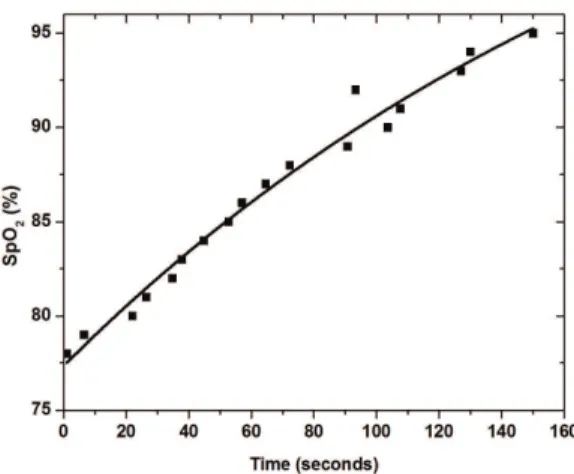
The experiment was conducted to determine the SpO2 sample of 0.8 mL of milk, when this was ozonated or oxygenated. The results showed that in both cases, saturation occurs at relatively short times, of the order of a few minutes, which can be seen in Figures 3 and 4.
Because the speciics of the experiment, the
oximeter response starts from 75%, and therefore the Figures 3 and 4, has its origins located in this region. The measurements of SpO2 when subjected to ozone and oxygen, showed an increased rate of SpO2 function of time for both cases reaching its peak in 80 s and 160 s, respectively. The experimental data concerning the SpO2 saturation as a function of time
can be itted by the theoretical curves obtained from
Equation 2, showing a good correlation between them.
CO3 = a – b × e (-t/c) (2)
where, CO3: ozone concentration [mg.L–1]; a, b and
c: parameters; t: time [min].
Discussion
The ozone decomposes in water spontaneously through complex mechanisms involving free radical
generation. Due to the high water concentration in
the milk, can be considered in this case that the mechanism of decomposition of ozone in milk is similar to that of water.
During the recombination of O3 in O2, two reaction
mechanisms are understood in the liquid, the direct oxidation of compounds by the ozone and indirect oxidation of compounds by hydroxyl free radicals produced during the decomposition of O3. In this case, the free radicals present, determine the rate of reaction, and also the regeneration of the superoxide radical O2-ion or proton form HO2, derived from the hydroxyl radical (OH), which means the consumption of 1 mol of ozone (Guerrero, 2005). As a result, all species capable of consuming hydroxyl radicals without regenerating the superoxide ion will produce a stabilizing effect on the ozone molecule in the sample. This effect associated with those produced by-products, provides greater quantity of molecular oxygen, resulting in a smaller time interval for increased saturation SpO2.
The recombination of O3 in O2 depends mainly on
the type of low, gas solubility and temperature. The type of low is deined by the Reynolds number (Re)
according to Equation 3, and values of “Re” greater
than 2400, as set turbulent low. This experiment
was carried out under atmospheric pressure and the calculated Reynolds number was equal to 16405,
characterizing it as turbulent low. In order to reduce the inluence of temperature on the process, the
experience was performed at a constant temperature equal to 21 °C.
Re = D × v × ρ / µ (3)
Where, Re: Reynolds number [dimensionless];
D: diameter of capsule = 0.08 [m]; v: velocity of low = 6.217 [m.s–1]; ρ: density of milk at 21 °C = 1029
[Kg/m3] and µ: viscosity of milk at 21 °C = 2.1 [cP].
The concentration of ozone versus time expressed by Equation 2 is dependent on the solubility of the gas, which in turn directly affects the mass transfer. The data concerning the solubility of gases in liquids
Figure 3. SpO2 of milk sample under oxygen action. Dots represent average experimental values; continuous line represents the trend line of the data obtained from Equation 1.
have great theoretical and practical interest, where the processes involved in solubility/mass transfer between gas and liquid are crucial to monitoring the diffusion of gas in the interface processes.
As shown in Figures 3 and 4, to reach the oxygen saturation (SpO2) at 95% produced by ozonized luid
requires half time if compared when the luid is
oxygenated, which is supported by the calculations obtained from Equation 2. This analysis sparked numerous questions about which mechanisms are involved in this phenomenon. Analyzing the variables involved in the process, different studies have reported
a signiicant difference in solubility of ozone and
oxygen (Bin, 2006; Bocci et al., 2011a) when dissolved in water at normal temperature and pressure, and this difference according to our analysis the most plausible answer to explain this result. Bin (2006) also presented several relationships between different expressions of the solubility of ozone in liquids. Several authors have also published data on solubility of ozone in water (Rischbieter et al., 2000; Kuosa et al., 2004; Levanov et al., 2008), indicating that it is about ten times more soluble than molecular oxygen (Bocci et al., 2011b), supporting this hypothesis. Another aspect to be considered when the milk is ozonized, that is, the ozone decomposition constant (Kd) increases exponentially as a function of the presence in milk of different molecules, increasing the rate of recombination O3, with consequent reduction in the time to reach saturation. The relative high pH of the sample, in this case close to 7.0, also contributes to this phenomenon. In this situation the value of Kd can be as high enough to compete with the value of KLa, making the recombination-O3 O2 occurs immediately after diffusion of ozone in liquid medium, explaining why the SpO2 induced by the action of ozone is faster than that induced by oxygen.
The results allow us to conclude that the technique, developed using an optical device operating in the red-infrared region to monitoring ozone dissolved in
biological luid, provides a simple and effective way to indirectly monitor the presence of ozone in luids.
Acknowledgments
CAPES (Coordination for the Improvement of Higher Education Personnel) and FAPESP (São Paulo Research
Foundation) for the inancial support.
References
Bin AK. Ozone solubility in liquids. Ozone: Science & Engineering. 2006; 28(2):67-75. http://dx.doi. org/10.1080/01919510600558635
Bocci V, Zanardi I, Travagli V. Oxygen/ozone as a medical gas mixture. A critical evaluation of the variuos methods clariies positive and negative aspects. Medical Gas Research. 2011a; 1(1):1-6. PMid:22146387 PMCid:PMC3231820. http://dx.doi.org/10.1186/2045-9912-1-6
Bocci V, Zanardi I, Travagli V. Ozone acting on human blood yields a hormetic dose-response relationship. Journal of Translational Medicine. 2011b; 9(66):1-11.
Buchan KAH, Martin-Robichaud DJ, Benfey TJ. Measurement of dissolved ozone in sea water: a comparison of methods. Aquacultural Engineering. 2005; 33(3):225-31. http://dx.doi.org/10.1016/j.aquaeng.2005.02.002 Delgado-Roche L, Martínez-Sánchez G, Re L. Ozone oxidative preconditioning prevents atherosclerosis development in New Zealand white rabbits. Journal of Cardiovascular Pharmacology. 2013; 61(2):160-5. PMid:23222311. http:// dx.doi.org/10.1097/FJC.0b013e31827a820d
Doan L, Forrest H, Fakis A, Craig J, Claxton L, Khare M. Clinical and cost effectiveness of eight disinfection methods for terminal disinfection of hospital isolation rooms contaminated with Clostridium dificile 027. Journal of Hospital Infection. 2012; 82(2):114-21. PMid:22902081. http://dx.doi.org/10.1016/j.jhin.2012.06.014
Gao RS, Ballard J, Watts LA, Thornberry TD, Ciciora SJ, McLaughlin RJ, Fahey DW. A compact, fast UV photometer for measurement of ozone from research aircraft. Atmospheric Measurement Technology. 2012; 5:2201-10. http://dx.doi. org/10.5194/amt-5-2201-2012
Guerrero PJ. Air quality modeling in very complex terrains: ozone dynamics in the northeastern Iberian peninsula [thesis]. Barcelona: Universidade Politécnica da Catalunha, 2005.
Kalnajs LE, Avallone LM. A novel lightweight low-power dual-beam ozone photometer utilizing solid-state optoelectronics. Journal of Atmospheric Oceanic Technology. 2010; 5(5):869-80. http://dx.doi. org/10.1175/2009JTECHA1362.1
Kunz A, Freire RS, Rohwedder JJR, Duran N, Mansilla H, Rodriguez J. Construção e otimização de um sistema para produção e aplicação de ozônio em escala de laboratório. Química Nova. 1999; 22(3):425-28. http://dx.doi.org/10.1590/ S0100-40421999000300022
Kuosa M, Laari MA, Kallas J. Determination of the Henry’s coeficient and mass transfer for ozone in a bubble column at different pH values of water. Ozone: Science & Engineering. 2004; 26(3):277-86. http://dx.doi. org/10.1080/01919510490455746
Levanov AV, Kuskov IV, Antipenko EE, Lunin VV. The solubility of ozone in aqueous solutions of sulfuric, phosphoric, and perchloric acids. Russian Journal of Physics Chemical A. 2008; 82(7):1126-31. http://dx.doi. org/10.1134/S0036024408070133
haemostatic and oxidative stress index in coronary artery disease. European Journal of Pharmacology. 2012; 691(1-3):156-62. PMid:22796450. http://dx.doi.org/10.1016/j. ejphar.2012.07.010
Rischbieter E, Stein H, Schumpe A. Ozone solubilities in water and aqueous salt solutions. Journal of Chemical Engineering Data. 2000; 45(2):338-40. http://dx.doi. org/10.1021/je990263c
Schulz S, Häussler U, Mandic R, Heverhagen JT, Neubauer A, Dünne AA, Werner JA, Weihe E, Bette M. Treatment with ozone/oxygen-pneumoperitoneum results in complete remission of rabbit squamous cell carcinomas. International Journal of Cancer. 2008; 22(10):2360-7. PMid:18224691. http://dx.doi.org/10.1002/ijc.23382
Wainstein J, Feldbrin Z, Boaz N, Harman-Boehm I. Eficacy of ozone-oxygen therapy for the treatment of diabetic foot ulcers. Diabetes Technology Therapy. 2011; 13(12):1255-60. PMid:21751891. http://dx.doi.org/10.1089/dia.2011.0018
Zaky S, Kamel SE, Hassan MS, Sallam NA, Shahata MA, Helal SR, Mahmoud H. Preliminary results of ozonetherapy as a possible treatment for patients with chronic hepatitis C. Journal of Alternative and Complement Medicine. 2011; 17(3):256-63. PMid:21417811. http:// dx.doi.org/10.1089/acm.2010.0016
Zoutman D, Shannon M, Mandel A. Effectiveness of a novel ozone-based system for the rapid high-level disinfection of health care spaces and surfaces. American Journal of Infection Control. 2011; 39(10):873-9. PMid:21546123. http://dx.doi.org/10.1016/j.ajic.2011.01.012
Authors
Henrique Cunha Carvalho*, Milene da Silva Melo, Carlos José de Lima, Renato Amaro Zângaro
Instituto de Engenharia Biomédica, Universidade Camilo Castelo Branco – UNICASTELO, Parque Tecnológico de São José dos Campos, Estrada Doutor Altino Bondesan, 500, Eugênio de Melo, CEP 12247-016, São José dos Campos, SP, Brasil
Carlos José de Lima, Renato Amaro Zângaro