Eduardo Rollo Duarte1, Maria Luiza M. A. Frigério2, Orivaldo Tavano3,
Paulo César Razuk4, Maria Rita de Cássia M. Costa5, Carlos Henrique Ferreira Martins6,
Maurício Serejo Ribeiro7, Éderson A. G. Betiol8
Treatment of snoring and sleep apnea syndrome with a removable
mandibular advancement device in patients without TMD
Introduction: Among the sleep disorders reported by the American Academy of Sleep, the most common is obstructive sleep apnea-hypopnea syndrome (OSAHS), which is caused by difficulties in air passage and complete interruption of air flow in the airway. This syndrome is associated with increased morbidity and mortality in apneic individuals.
Objective: It was the objective of this paper to evaluate a removable mandibular advancement device as it provides a noninvasive, straightforward treatment readily accepted by patients.
Methods: In this study, 15 patients without temporomandibular disorders (TMD) and with excessive daytime sleepiness or snoring were evaluated. Data were collected by means of: Polysomnography before and after place-ment of an intraoral appliance, analysis of TMD signs and symptoms using a patient history questionnaire, muscle and TMJ palpation.
Results: After treatment, the statistical analysis (t-test, and the “before and after” test) showed a mean reduction of 77.6% (p=0.001) in the apnea-hypopnea index, an increase in lowest oxyhemoglobin saturation (p=0.05), decrease in desaturation (p=0.05), decrease in micro-awakenings or EEG arousals (p=0.05) and highly significant improvement in daytime sleepiness (p=0.005), measured by the Epworth Sleepiness Scale. No TMD appeared during the monitor-ing period.
Conclusion: The oral device developed in this study was considered effective for mild to moderate OSAHS.
Keywords: Snoring. Obstructive sleep apnea. Mandibular advancement. Occlusal splints.
How to cite this article: Duarte ER, Frigério MLMA, Tavano O, Razuk PC, Costa MRCM, Martins CHF, Ribeiro MS, Betiol EAG. Treatment of snoring and sleep ap-nea syndrome with a removable mandibular advancement device in patients without TMD. Dental Press J Orthod. 2012 Mar-Apr;17(2):90-6.
Submitted: September 12, 2008 - Revised and accepted: April 26, 2009 » The authors report no commercial, proprietary, or financial interest in the products or companies described in this article.
Contact address: Eduardo Rollo Duarte – Rua Felício Soubihe 3-74, Jd. Planalto – CEP: 17.012-623 – Bauru/SP – Brazil – E-mail: [email protected]
1 Head of the department of Orofacial Pain (TMJ) and Sleep Dentistry, Clinic of
Sleep Disorders, Bauru, Brazil.
2 Assistant Professor, Department of Dental Prosthesis, FOUSP, Brazil.
3 Head Professor of Department of Surgery and Radiology, Bauru Dental School,
FOB-USP, Brazil.
4 Head Professor, Department of Engineering, UNESP, Bauru, Brazil.
5 Neurologist and Sleep physician. Head of the Clinic of Sleep Disorders, Bauru,
Brazil.
6 Otolaryngologist, Hospital for Rehabilitation of Craniofacial Anomalies, USP. Head
of the Respiratory Department, Clinic of Sleep Disorders, Bauru, Brazil. 7 Head of Dental Prosthesis, FOC UNIFLU, Rio de Janeiro, Brazil.
8 Professor of Department of Dental Prosthesis of Paraná Federal University,
INTRODUCTION
One often hears that when a person is asleep and snoring, it means that they are fast and sound asleep. It couldn’t be farther from the truth. A partial obstruc-tion in the upper airway causes snoring while a com-plete obstruction leads to sleep apnea, which is the total cessation of airflow. These events comprise what is known as Sleep Apnea and Hypopnea Syndrome (OSHAS) and lead to serious consequences such as systemic and pulmonary hypertension, excessive daytime sleepiness, impaired memory, irritability, de-pression, decreased libido, impotence, burnout, head-ache and a proneness to workplace and car accidents. Apnea is characterized by an interruption in airflow (breathing) for at least ten seconds. Hypopnea is a 50% reduction in airflow accompanied by a “micro-awakening” or EEG arousal and/or a drop in blood
oxyhemoglobin saturation (SaO2) of about 2% to 4%.4
It is diagnosed by examining sleep with polysom-nography (PSG), which consists of simultaneously recording activities occurring in the organism during a given period and tracking not only the number of ap-neas and hypopap-neas during the patient’s sleep but also defining OSHAS severity using the apnea-hypopnea index (AHI), which is the sum of apnea and hypopnea
events per hour of sleep.6
Several authors have confirmed the validity and effectiveness of oral appliances for OSHAS
treat-ment.1,2,5,8,9,10,16 Appliances that reposition the
man-dible anteriorly are indicated for primary snoring and
mild to moderate OSAHS with an AHI of up to 30 4,9.
In OSAHS patients this therapy can be effective even in the presence of other diseases such as
conges-tive heart failure and arterial hypertension,8,19,30
there-by contributing to clinical control.
In treating OSHAS a correct diagnosis is of para-mount importance, as well as a multidisciplinary team with training and knowledge of sleep disorders, and a reliable treatment indication for each case. Dentists should be part of this team as they play a key role both in the initial diagnosis, by referring the pa-tient to polysomnography, and in the treatment itself after the examination.
The purpose of this study was to develop an alter-native intraoral appliance model for treating OSHAS, evaluate its effectiveness and magnitude of changes in AHI, and observe the side effects and development
of potential TMD (temporomandibular dysfunction) signs and symptoms in the follow-up phase.
MATERIAL AND METHODS
The study evaluated 15 patients among those who presented voluntarily at the Clinic of Sleep Disorders located in Bauru, Brazil, reporting exces-sive daytime sleepiness and/or snoring. Patients had a mean age of 49.9 years.
Initially, patients answered a questionnaire, the Epworth Sleepiness Scale (ESS), designed to
mea-sure their daytime sleepiness in specific situations,15
including patients with ESS scores greater than five. Examination to check for the presence of Temporo-mandibular Disorders (TMD) began with the pa-tient’s clinical history using the Anamnesis Index
(AI) questionnaire.7,11,18 The results were compared
with the AI table, which classifies patients’ TMD, thus: 0-15, no TMD, 20-40, mild TMD, 45-65, moder-ate TMD, 70-100, severe TMD. Thereafter, a physi-cal and cliniphysi-cal examination was performed by digi-tal palpation of the right and left TMJs and muscles (superficial and deep masseter, medial pterygoid and temporal). Patient response to TMJ palpation was
graded as suggested by Pertes and Gross21 and as
per-formed by Miranda.18 Therefore, assessment of the
development of TMD signs and symptoms was per-formed by comparing AI before and after treatment, and also by physical examination, i.e., palpation of masticatory muscles and TMJ.
Exclusion criteria were as follows: AI higher than 15 (since subjects with AI between zero and 15 are considered without TMD, and above 15, with TMD) or presence of TMD signs and symptoms, moderate or severe periodontal disease, lower than 6 mm maximum mandibular protrusion and less than 35 mm mouth opening.
All patients underwent polysomnography (PSG) in the Clinic of Sleep Disorders before and after wear-ing the appliance. The average time interval between PSGs was five months.
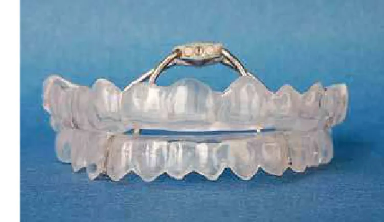
employed to join the maxillary to the mandibular portion of the splint (Fig 1).
At each visit after splint insertion it was advanced by 0.25 mm until the mandibular advancements reached 1 mm within a two-week period. Patients were monitored on a weekly basis and, after reporting an improvement in snoring and sleepiness, were referred for a second PSG. After approximately six months wearing the splint, the patients were examined again to assess TMD development.
The tests used for statistical analysis of the re-sults included Student’s t-distribution test for small samples, with degree of freedom (df ) lower than 120 and the “before and after” test which, in simple com-parative experiments with samples that have the same origin, ensures greater accuracy than t-test. Both tests were applied to ensure outcome reliability.
RESULTS
Anthropometric data of the subjects who partici-pated in the research are presented in Table 1.
The data were statistically evaluated by Student’s t-test and confirmed by the “before and after” test. Assessment results of daytime sleepiness before and after splint use are depicted in Table 2.
All patients exhibited decreased ESS values, not-ing that the treatment reduced excessive daytime sleepiness in a highly significant manner in 87% of the patients (p=0.005).
It is noteworthy that AI variation was not sufficient to affect the TMD classification of any patient, i.e., it did not increase over 15. On physical examination by
muscle palpation 14 patients showed no significant differences after treatment compared to muscle pal-pation values found prior to treatment.
During mandibular advancement therapy patients
6 and 15 (13.3%) developed morning muscle pain.10
Treatment included the prescription of oral medica-tion (Celecoxib 200 mg) once a day for three days. The mandibular advancement was halted for a week and
then restarted at 0.25 mm per week.2 The two patients
were left with the splint positioned 1 mm below the 75% of maximum protrusion mark. These two patients never again exhibited TMD signs and symptoms.
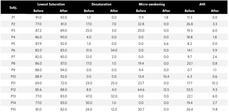
Table 4 presents the oxyhemoglobin saturation index, number of arousals and number of apneas and hypopneas occurring per hour during the first and second PSG.
On the one-tailed Student’s table for p=0.05 and df=14, tc = 1.761 with 95% confidence for the situation “with splint”, the amount of oxyhemoglobin desatura-tion events decreased significantly.
For the one-tailed test tc=1.761 (hence to>tc), the number of arousals “after” is significantly low-er than “before.”
Snoring, as evaluated by PSG, improved in all pa-tients to acceptable levels and was suppressed or not observed in patients 4 and 15 (moderate frequency level) and in patients 5 and 8 (high frequency).
AHI before treatment had a baseline mean of 18.3 (SD=11.8). After treatment, the AHI displayed a mean of 4.1 (SD=3.8, p=0.001). Thus, the outcome for “after” is significantly less than for “before”, i.e., there was a highly significant reduction in AHI.
Table 1 -Anthropometric data of research participants.
Table 2 -Results of the Epworth Sleepiness Scale (ESS) before and after splint use.
Table 3 -Anamnesis Index (AI) results before and after splint use.
Subjects Gender Age (years) Height
(m)
Weight (kg) BMI
Before After Before After Before After
P1 Male 35 37 1.72 83 80 28 27
P2 Male 39 42 1.69 102 103 36 36
P3 Male 40 43 1.72 78 76 26 26
P4 Female 50 52 1.48 51 52 23 24
P5 Female 59 60 1.61 70 78 27 30
P6 Female 63 64 1.8 97 97 30 30
P7 Male 28 28 1.87 114 116 33 33
P8 Female 54 55 1.57 69 69 28 28
P9 Female 40 42 1.66 63 63 23 23
P10 Female 55 55 1.57 94 94 38 38
P11 Male 63 65 1.68 92 93 33 33
P12 Male 54 56 1.69 79 76 28 27
P13 Male 57 58 1.58 66 66 26 26
P14 Male 56 57 1.67 83 83 30 30
P15 Male 47 47 1.68 75 75 27 27
Subjects Before After
P1 8 3
P2 6 2
P3 7 2
P4 11 3
P5 12 6
P6 8 7
P7 14 6
P8 13 10
P9 6 2
P10 6 2
P11 10 5
P12 6 3
P13 10 4
P14 13 3
P15 13 6
Subjects Before After
P1 8 4
P2 6 6
P3 15 0
P4 5 5
P5 15 10
P6 13 7
P7 0 0
P8 3 0
P9 0 0
P10 7 7
P11 6 0
P12 0 0
P13 15 4
P14 15 5
DISCUSSION
The aim of this study was to evaluate the effec-tiveness of an adjustable intraoral mandibular
ad-vancement splint in the treatment of OSAHS.5,9,19
Currently, it is recommended that to be consid-ered successful OSHAS treatment using oral splints must comply with the following polysomnographic parameters: A reduction in AHI of at least 50% or a
reduction in AHI values below ten.16
A highly significant reduction in AHI found in 15 patients, i.e., 77.6% mean reduction, demonstrates that the splints can be indicated for treating snor-ing, increased upper airway resistance syndrome and OSAHS cases of mild to moderate severity, al-though some patients with severe OSAHS can also benefit from the therapy, such as patient 12 in this research. When patient 12 was excluded so that the data analysis could be restricted to patients with AHI<30 only, a highly significant improvement in AHI was still found, i.e., a mean 75.9% (p=0.001). Nine (81.8%) out of 11 patients with AHI>10 showed a reduction in AHI to values below ten. It was ob-served that mandibular repositioning caused a re-duction in AHI. Upper airway narrowing due to ana-tomical factors such as a posterior positioning of the
Subj. Lowest Saturation Desaturation Micro-awakening AHI
Before After Before After Before After Before After
P1 91.0 92.0 1.0 0.0 11.9 1.8 11.3 0.0
P2 77.0 81.0 17.0 7.0 32.8 0.0 26.8 3.3
P3 87.2 89.0 25.0 3.0 20.0 0.0 19.3 6.0
P4 86.0 90.0 4.0 0.0 0.0 0.0 18.8 1.8
P5 87.9 92.0 1.0 0.0 0.0 6.6 8.2 0.0
P6 82.0 83.0 31.0 34.0 0.0 0.0 14.1 5.9
P7 82.0 85.0 12.0 2.0 0.0 0.0 9.7 2.6
P8 86.0 87.0 17.0 1.0 19.4 0.0 20.1 0.8
P9 88.0 94.0 0.0 0.0 9.4 0.0 0.7 1.1
P10 88.9 92.0 0.0 0.0 13.4 10.4 6.3 0.6
P11 69.0 72.0 23.0 25.0 23.7 0.0 17.7 10.2
P12 85.6 88.0 8.0 4.0 64.6 12.5 53.5 9.3
P13 77.0 83.0 47.0 32.0 0.0 0.0 22.1 6.0
P14 77.0 83.0 30.0 1.0 0.0 0.0 19.4 2.7
P15 81.0 82.0 24.0 12.0 30.7 0.0 26.4 11.8
Table 4 -Oxyhemoglobin saturation index (%) and number of arousals and obstructive events per hour (AHI).
mandible is a risk factor for sleep apnea. In advanc-ing the mandible, the splints widen and stretch the muscles, which concurrently produce an increase in electromyographic activity, widening the airway
and improving airflow.1,2,14,17,19,26
Thirteen patients (86.7%) exhibited some de-gree of tooth sensitivity, especially on awakening in the morning. With splint use, five patients (33%) complained of excessive salivation at night and one patient (6.6%) complained of dry mouth at night. In this study, specifically in regard to the development of joint or muscle pain, therapy with splints did not produce definitive or reversible TMD signs or
symp-toms. Yoshida29 found these effects in 8.5% of cases
and described them as transient. None of the partic-ipants was diagnosed with TMD prior to treatment, and after treatment completion (12-month follow-up, on average), no signs or symptoms of TMD-re-lated muscle or joint dysfunction were identified.
It is noteworthy that harmful side effects such as tooth sensitivity (86.7%), dry mouth or exces-sive salivation (40%) occurred after splint place-ment but on examination were considered minor
Authors have indicated that the advancement should be between 50% and 75% of maximum
man-dibular protrusion2 since such repositioning would
be within the measurements of a physiological
man-dibular protrusion.22 Six patients had muscle pain
and two patients (P6 and P15) presented with mod-erate pain, which prevented further advancement to 75% maximum protrusion, although the pain was controlled with non-steroidal anti-inflammatory medication. No other changes occurred in AI, indi-cating no development of TMD signs and symptoms. The splints were inserted at 60% maximum pro-trusion and advanced in increments of 0.5 mm to 1 mm within two weeks, unlike other studies, which
made weekly advancements of 0.25 mm.20
In this study, the mean mandibular advance-ment was 9.17 mm, ranging from 7 to 12 mm, there-fore within what is considered a physiological pro-trusion length. These results are similar to several studies that proved the effectiveness of splints in
mild and moderate OSAHS.5,9,30
Advancement was discontinued based on the follow-ing criteria: A position that coincided with 75% maxi-mum protrusion and improvement in subjective criteria reported by patients. Clinically, however, defining such criteria can prove elusive. Some authors have therefore suggested an association with objective parameters at the end of mandibular advancement therapy, such as
nocturnal pulse oximetry performed at home.10,28
Reports of improvement in snoring and daytime sleepiness led four patients (26.7%) to an early referral for a second PSG, but the results showed no change in AHI. This situation showed that the reports of patients and/or of their room-mates are indeed subjective and not a substitute for polysomnography as a therapeutic
assessment tool.9,16 Further advancements were made
in these patients and another PSG was performed for each one, except for patient P11, who refused to repeat it and was the only patient who showed no reduction in AHI above 50% (42.4% reduction).
Therapy with the splint caused a direct effect on sleep fragmentation as there was an increase in the lowest oxyhemoglobin saturation index (p=0.05) and a significant decrease in total desaturation and num-ber of arousals (p=0.05). Clinically, improvement in polysomnographic parameters was found to be
relat-ed to a significant decrease (p=0.05) in ESS.8,19
Improvement in snoring levels was observed in all cases and the subjective assessment of spouse confirmed this result. Spouse satisfaction is ex-tremely important since for him or her snoring can
cause another sleep disorder, i.e., insomnia.4
Long-term studies are warranted to compare the designs and materials used in the splints and to verify the occurrence of any potential desirable
or undesirable effects.3 However, even if
signifi-cant side effects do occur, a thorough evaluation - pinpointing favorable and unfavorable biological factors - should be conducted and discussed to en-able treatment decisions that benefit the patient in the best possible way while gaining control over the important and severe complications of OSAHS. Although treatment using a splint is regarded as a non-invasive therapy, side effects are likely to oc-cur during treatment. Occlusal changes may ococ-cur with long-term use, so patients should be strictly
controlled.27
When patients feel the benefits of therapy us-ing the splint and realize that they are wakus-ing up feeling rested, they start wearing it on a daily and
continuous basis.13
Although this splint was fabricated with materi-als normally used in dental practice (such as acrylic resin), in light of the limitations of this study and the success criteria adopted for the AHI reduction (above 50%), splint effectiveness was successfully evaluated and the results consistent with those found in the literature.
CONCLUSIONS
1. Aarab G, Lobbezoo F, Wicks DJ, Hamburger HL, Naeije M. Short-term effects of a mandibular advancement device on obstructive sleep apnea: an open-label pilot trial. J Oral Rehabil. 2005;32(8):564-70.
2. Almeida FR, Bittencourt LR, Almeida CI, Tsuiki S, Lowe AA, Tufik S. Effects of mandibular posture on obstructive sleep apnea severity and the temporomandibular joint in patients fitted with an oral appliance. Sleep. 2002;25(5):507-13.
3. Almeida FR, Lowe AA, Otsuka R, Fastlicht S, Farbood M, Tsuiki S. Long-term sequelae of oral appliance therapy in obstructive sleep apnea patients: Part 1. Cephalometric analysis. Am J Orthod Dentofacial Orthop. 2006;129(2):205-13. 4. American Academy of Sleep Medicine. Sleep related breathing disorders in adults:
recommendations for syndrome definitions and measurements techniques in clinical research. Sleep. 1999;22:667-89.
5. Barthlen GM, Brown LK, Wiland MR, Sadeh JS, Patwari J, Zimmerman M. Comparison of three oral appliances for treatment of severe obstructive sleep apnea syndrome. Sleep Med. 2000;1(4):299-305.
6. Bittencourt LR, Suchecki D, Tufik S, Peres C, Togeiro SM, Bagnato MC, et al. The variability of the apnoea-hypopnoea index. J Sleep Res. 2001;10(3):245-51. 7. Conti PC, Ferreira PM, Pegoraro LF, Conti JV, Salvador MC. A cross-sectional study
of prevalence and etiology of signs and symptoms of temporomandibular disorders in high school and university students. J Orofac Pain. 1996 Summer;10(3):254-62. 8. Eskafi M. Sleep apnea in patients with stable congestive heart failure. An
intervention study with a mandibular advancement device. Swed Dent J Suppl. 2004;(168):1-56.
9. Ferguson KA, Cartwright R, Rogers R, Schmidt-Nowara W. Oral appliances for snoring and obstructive sleep apnea: a review. Sleep. 2006;29(2):244-62. 10. Fleury B, Rakotonanahary D, Petelle B, Vincent G, Pelletier Fleury N, Meyer B, et
al. Mandibular advancement titration for obstructive sleep apnea: optimization of the procedure by combining clinical and oximetric parameters. Chest. 2004;125(5):1761-7.
11. Fonseca DM, Bonfante G, Valle AL, Freitas SFT. Diagnóstico pela anamnese da disfunção craniomandibular. RGO: Rev Gaúcha Odontol. 1994;42(1):23-8. 12. Gavish A, Vardimon AD, Rachima H, Bloom M, Gazit E. Cephalometric and
polysomnographic analyses of functional magnetic system therapy in patients with obstructive sleep apnea. Am J Orthod Dentofacial Orthop. 2001;120(2):169-77. 13. Jauhar S, Lyons MF, Banham SW, Cameron DA, Orchardson R. Ten-year follow-up
of mandibular advancement devices for the management of snoring and sleep apnea. J Prosthet Dent. 2008;99(4):314-21.
14. Johal A, Battagel JM. An investigation into the changes in airway dimension and the efficacy of mandibular advancement appliances in subjects with obstructive sleep apnoea. Br J Orthod. 1999;26(3):205-10.
15. Johns MW. A new method for measuring daytime sleepiness: the Epworth Sleepiness scale. Sleep. 1991;14:540-5.
REFERENCES
16. Kushida CA. Practice parameters for the treatment of snoring and obstructive sleep apnea with oral appliances: an update for 2005. An American Academy of Sleep Medicine Report. Sleep. 2006;29(2):240-3.
17. Lowe AA, Sjöholm TT, Ryan CF, Fleetham JA, Ferguson KA, Remmers JE. Treatment, airway and compliance effects of a titratable oral appliance. Sleep. 2000;23 Suppl 4:S172-8.
18. Miranda JES. Avaliação longitudinal da efetividade das placas oclusais reposicionadoras no controle de patologias da ATM: comparação com placas oclusais estabilizadoras e um grupo controle [tese]. Bauru (SP): Universidade de São Paulo; 2000.
19. Ng AT, Gotsopoulos H, Qian J, Cistulli PA. Effect of oral appliance therapy on upper airway collapsibility in obstructive sleep apnea. Am J Respir Crit Care Med. 2003;168(2):238-41. Epub 2003 Apr 30.
20. Okawara Y, Tsuiki S, Hiyama S, Hashimoto K, Ono T, Ohyama K. Oral appliance titration and nasal resistance in nonapneic subjects. Am J Orthod Dentofacial Orthop. 2004;126(5):620-2
21. Pertes RA, Gross SG. Clinical management of temporomandibular disorders and orofacial pain. Chicago: Quintessence; 1995. 368 p.
22. Piehslinger E, Celar A, Futter K, Slavicek R. Orthopedic jaw movement
observations. Part I: determination and analysis of the length of protrusion. Cranio. 1993;11(2):113-7.
23. Ringqvist M, Walker-Engström ML, Tegelberg A, Ringqvist I. Dental and skeletal changes after 4 years of obstructive sleep apnea treatment with a mandibular advancement device: a prospective, randomized study. Am J Orthod Dentofacial Orthop. 2003;124(1):53-60.
24. Ryan CF, Love L, Peat D, Fleetham J, Lowe A. Mandibular advancement oral appliance therapy for obstructive sleep apnoea: effect on awake calibre of the velopharynx. Thorax. 1999;54(11):972-7.
25. Schmidt-Nowara W. Recent developments in oral appliance therapy of sleep disordered breathing. Sleep Breath. 1999;3(3):103-6.
26. Tsuiki S, Ryan CF, Lowe AA, Inoue Y. Functional contribution of mandibular advancement to awake upper airway patency in obstructive sleep apnea. Sleep Breath. 2007;11(4):245-51.
27. Ueda H, Almeida FR, Lowe AA, Ruse ND. Changes in occlusal contact area during oral appliance therapy assessed on study models. Angle Orthod. 2008;78(5):866-72.
28. Wiltshire N, Kendrick AH, Catterall JR. Home oximetry studies for diagnosis of sleep apnea/hypopnea syndrome: limitation of memory storage capabilities. Chest. 2001;120(2):384-9.
29. Yoshida K. Effect of a prosthetic appliance for treatment of sleep apnea syndrome on masticatory and tongue muscle activity. J Prosthet Dent. 1998;79(5):537-44. 30. Yoshida K. Effect on blood pressure of oral appliance therapy for sleep apnea