Maria Carolina Feio Barroso1, Renan Lana Devita2, Eugênio José Pereira Lages3, Fernando de Oliveira Costa4, Alexandre Fortes Drummond5, Henrique Pretti6, Elizabeth Maria Bastos Lages5
Risk variables of external apical root resorption during
orthodontic treatment
Introduction: External apical root resorption (EARR) is an adverse outcome of the orthodontic treatment. So far, no single or associated factor has been identified as responsible for EARR due to tooth movement.
Objective: This study investigated the association of risk variables (age, gender, extraction for orthodontic treat-ment and Angle classification) with EARR and orthodontic treattreat-ment.
Method: The sample (n=72) was divided into two groups according to presence (n=32) or absence (n=40) of EARR in maxillary central and lateral incisors after orthodontic treatment.
Results: There were no statistically significant differences in EARR according to age, gender, extraction or type of malocclusion (p>0.05).
Conclusion: The risk variables examined were not associated with EARR in the study population.
Keywords: Orthodontics. Root resorption. Tooth movement.
How to cite this article: Barroso MCF, Devita RL, Lages EJP, Costa FO, Drummond AF, Pretti H, Lages EMB. Risk variables of external apical root resorption during orth-odontic treatment. Dental Press J Orthod. 2012 Mar-Apr;17(2):39.e1-7.
Submitted: March 11, 2009 - Revised and accepted: August 16, 2009
» The authors report no commercial, proprietary, or financial interest in the products or companies described in this article.
Contact address: Maria Carolina Feio Barroso
Faculdade de Odontologia - UFMG / Dpto. de Ortodontia e Odontopediatria Av. Antônio Carlos, 6627 – Pampulha – Cep: 31.270-901 – Belo Horizonte / MG – Brazil E-mail: [email protected]
1 Graduate Student of Orthodontics, FO-UFMG.
2 Undergraduate student, FO-UFMG.
3 PhD Student, Periodontics Department, FO-UFMG.
4 Associate Professor, Periodontics Department, FO-UFMG.
5 Associate Professor, Orthodontics Department, FO-UFMG.
INTRODUCTION
External apical root resorption (EARR) is a
rel-evant pathological side effect2,10 of orthodontic
treatment that leads to permanent loss of the den-tal root apex. According to Brezniak and Wasser-stein11 and Hartsfield Jr et al,16 it is a frequent
iatro-genic result of orthodontic treatment, particularly in maxillary incisors.
The prevalence of EARR associated with orth-odontic treatment is variable. Histological studies have reported a high prevalence, whereas clini-cal trials found a varied prevalence depending on
the method used.9 EARR affects central maxillary
incisors primarily; over one third of all subjects that undergo orthodontic treatment are affected by having resorptions greater than 3 mm, whereas severe resorption (greater than 5 mm) affects 2 to
5% of the orthodontic population.16,30 According to
Capelozza Filho and Silva Filho,12 root resorption is
found in most (90.5%) permanent teeth that under-go orthodontic treatment; such areas are shallow and broad (surface resorption) and are always
re-paired. According to Consolaro,13 severe and
struc-turally important root resorption affects 10% of the subjects that undergo orthodontic treatment.
EARR associated with orthodontic treatment is an important concern. Up to 3 mm of apical third loss results in limited damage, and root resorptions of such magnitude are assumed as a part of the biologi-cal cost of treatment. These side effects or iatrogenic events are practically unavoidable in orthodontic practice and are classified as clinically acceptable. However, they should not be seen as normal, physi-ological or part of the apical remodeling process.13
Apical resorptions that exceed 3 mm result in loss of tooth support due to a reduction of the
sup-porting periodontium. Levander and Malmgren22
analyzed the mobility of teeth with severe EARR (resorption of 1/3 of the root or more) five to fifteen years after the active treatment phase and evaluated mobility associated with root length and supporting alveolar bone. They found a significant correlation between tooth mobility and total and intra-alve-olar root length and concluded that there was risk of tooth mobility in maxillary incisors with severe root resorption during orthodontic treatment if the
remaining root length was ≤ 9 mm.
Although several studies have investigated root re-sorption, no single or associated factor has been identified as responsible for EARR due to orthodontic treatment.3
The etiology of EARR associated with orthodon-tic treatment is multifactorial and involves environ-mental and host factors. Several variables have been described in the literature as possible risk factors for EARR: Tooth anomalies and the shape of root and al-veolar bone crest,13,14,20,27 type and severity of
malocclu-sion,5,6,14,17,27,30 amount of tooth movement,5,18,23,26
extrac-tions for orthodontic treatment,7,24,28 cell and molecular
mechanisms of osteoblast regulation,19 factors
associat-ed with the technique usassociat-ed, such as magnitude of force applied,1,25 treatment duration,15,21 type of orthodontic
treatment,1,6,7,28 age and gender,5,6,7,14,17,20,26,27 individual
variations and genetic factors.2,3,14
This study investigated the association of age, gender, orthodontic extractions and Angle classifi-cation with external apical root resorptions result-ing from orthodontic treatment.
MATERIAL AND METHODS Sample
This study enrolled subjects over 18 years of age that received total fixed orthodontic treatment in the Graduate Program in Orthodontics of the School of Dentistry of Minas Gerais Federal University. The sample was divided into two groups according to presence (case group) or absence (control group) of EARR in maxillary central and lateral incisors.
Inclusion criteria
Participants were included if good quality peri-apical radiographs obtained before and after orth-odontic treatment were available, and pretreat-ment radiographs showed no resorption.
Exclusion criteria
All subjects with crown fractures or restorations in the incisal edge of maxillary incisors were excluded.
Authorization of participants and parents or guardians
A B
D E F
C Data collection
Clinical analysis
Data about age, gender, extractions for orthodon-tic treatment and Angle classification of malocclu-sion were collected directly from patient charts.
Radiographic analysis
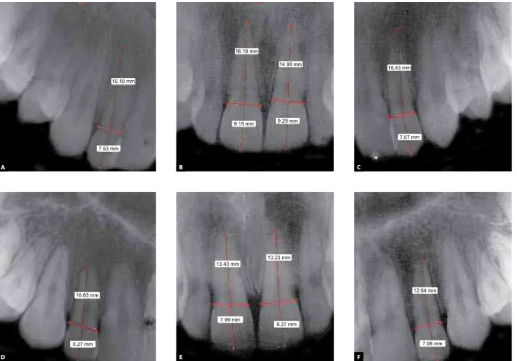
To evaluate the presence or absence of EARR, all periapical radiographs were digitalized using an HP Scanjet scanner 3570c at a resolution of 300 dpm. Images were analyzed directly on the com-puter screen using the software ARARA – An
envi-ronment for image segmentation4 (Fig 1).
Measurements were made according to the
method described by Linge and Linge,23 modified
by Brezniak et al.8 To determine and calculate the
changes in tooth and root lengths between two
radiographs, those authors used, among other landmarks, the midpoint (M) for the cement enam-el junction (CEJ), defined as the midpoint between the mesial and the distal CEJ (Fig 2).
After this landmark has been identified, the fol-lowing distances were measured for all periapical radiographs: from M to root apex, as a measure of root length, and from M to incisal edge as a measure of crown length. These measurements are similar to the r1, r2, c1 and c2 distances described by Linge and Linge23 (Fig 2).
Changes in root length due to treatment were mathematically calculated as follows: during orth-odontic treatment, the length of the crown does not change, unless it is fractured or restored. Therefore, the ratio between initial (c1) and final (c2) length of the crown defines the radiographic change factor.
Figure 1 - Digitalized periapical radiographs before (A,B,C) and after (D,E,F) orthodontic treatment with maxillary incisors measurements of crowns and roots.
16.10 mm
16.16 mm
14.90 mm
9.15 mm 9.29 mm
7.53 mm
16.43 mm
7.67 mm
10.83 mm
13.43 mm
13.23 mm
7.99 mm
8.27 mm
12.64 mm
M B
r1 r2
c1 c2
If there was no change in root length during treat-ment, the ratio between initial and final root length (r1/r2) should be the same as the ratio between ini-tial and final crown length (c1/c2). If the root length shortens during treatment, the amount of resorp-tion is calculated as r1-r2(c1/c2).
Therefore, subjects with less than 2 mm EARR were defined as not affected and included in the con-trol group, and those with only one incisor with EARR
≥ 2 mm were included in the case group (Fig 3).
Examiner calibration
To observe examiner calibration, a single exam-iner repeated measurements of 20 radiographs at an interval of seven days between readings. Results were recorded as presence or absence of readings below 0.5 mm. Kappa statistics was used to evaluate
the reproducibility of radiograph measurements.29
Statistical analysis
Descriptive variables are presented as percent-ages, mean, and standard deviations (SD)
The Student t test, a chi-square test and the Fisch-er exact test wFisch-ere used to analyze variables below 5. The level of significance was set at 5% (p<0.05).
RESULTS
The sample included 72 subjects divided into two groups according to presence (case group, n=32) or absence (control group, n=40) of EARR in maxillary central and lateral incisors after orth-odontic treatment.
Sample characteristics (age, gender, orthodon-tic extractions, Angle classification) and the analy-sis of associations between variables and EARR oc-currence are presented.
Sample characteristics Age
Age ranged from 10 to 50 years, and mean age was 15.7 years (±7.3). In the control group, age ranged from 10 to 34 years, and mean age was 14.6 years; in the EARR group, age ranged from 10 to 50 years, and mean age was 17.2 years. There was a sig-nificant difference in age between groups (Table 1).
Gender
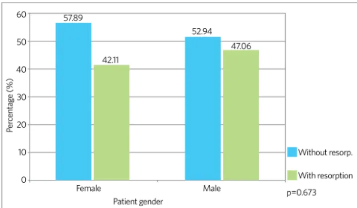
Figure 4 shows the classification of subjects according to gender. EARR was present in 42.1% of the women and 47% of the men. There were no significant differences in EARR regarding gender (p=0.673).
Figure 2 - Identification of midpoint (M) (Brezniak et al8).
Figure 3 - Correction factor for difference in root length: f=c1/c2; external root
resorption: r1-r2(c1/c2) (Linge and Linge23).
Extractions for orthodontic treatment
EARR was found in 43.3% of the subjects in the group that had extractions, and in 45.2% in the group without extractions. There were no signifi-cant differences between groups with or without extractions (p=0.873) (Fig 5).
Angle malocclusion classification
EARR was found in 41.4% of all Class I subjects, 45.4% of the Class II subjects, and 55.5% of Class III malocclusion subjects, but these differences were not statistically significant (P=0.750) (Fig 6).
DISCUSSION
It has been widely accepted that EARR is a frequent iatrogenic outcome of the orthodontic treatment, particularly in maxillary incisors. It is assumed that compression of the periodontal liga-ment, which reduces or interrupts blood supply, leads to aseptic necrosis and during removal of ne-crotic tissue by macrophages and osteoclasts, the
root may be injured.10,16 Meanwhile, the etiology of
EARR remains unclear and subject to the effect of innumerable risk variables. Although several stud-ies have investigated this topic, no single or asso-ciated factor has been identified as responsible for
EARR due to orthodontic treatment.3
Previous studies focused on the magnitude of the force applied,1,25 duration,15,21 and the type of
orthodontic treatment1,6,7 have not established a
causal relation between these factors and EARR. Over the wide range of causal variables related to EARR, this study analyzed the effect of age, gender, ex-tractions for orthodontic treatment and type of mal-occlusion. Our findings did not reveal any statistically significant differences regarding these variables. The results should be interpreted carefully because asso-ciations might have been affected by the study sample.
Difficulties in obtaining a standardized and ho-mogeneous sample are often found in similar stud-ies. In addition, final records from patients that have completed orthodontic treatment are not al-ways available. When available, radiographs are not standardized and, thus, considered useless because they do not show the anatomic landmarks neces-sary for measurements. Therefore, limitations due to sample size are frequent.
Descriptive statistics
Group Min. Max. Mean s.d. p
Global 10 50 15.78 7.32
No EARR 10 34 14.60 4.13 0.162
With EARR 10 50 17.25 9.85
Table 1 - EARR according to age.
Note: Statistical significance according to Student’s t test.
Figure 6 - Classification of subjects according to EARR and Angle malocclusion
classification.
Note: Statistically significant difference according to Fisher exact test.
Figure 4 - Classification of subjects according to EARR and gender.
Note: Statistically significant difference according to chi-square test.
Female 60 50 40 30 20 10 0 P er centag e (%) Male p=0.673 Without resorp. With resorption Patient gender 57.89 42.11 52.94 47.06
Figure 5 - Classification of subjects according to EARR and extractions.
Note: Statistically significant difference according to Fisher exact test.
With no extraction With extraction 60 50 40 30 20 10 0 P er centag e (%) p=0.873 Without resorp. Extraction necessity With resorption 54.76 45.24 56.67 43.33 60 50 40 30 20 10 0 P er centag e (%)
Class I Class II Class III
This study showed that age does not seem to be a significant factor in EARR, a finding similar to those reported by several authors in different stud-ies that investigated the association of this variable with EARR.5,14,17
In accordance to our study, several investiga-tions have demonstrated that there is an
associa-tion between gender and EARR.6,7,26,27 According
Kjaer20 (cited by Hartsfield Jr et al,16) a greater
prevalence of EARR among women was found. Although we did not find an association between type of malocclusion and EARR, several types of malocclusion, both dental and skeletal, have been classified as risk factors for EARR. Several factors are involved in the treatment of each type of mal-occlusion, which may potentialize risk variables in EARR, and the existing literature about this topic is still controversial.5,17,27,30
The severity of malocclusion may be associated with the extent of orthodontic tooth movement, and the duration of orthodontic treatment may be
positively associated with the extent of EARR.26
According to Baumrind et al,5 Horiuchi et al,18 and
Linge and Linge,23 orthodontic tooth movement
has been responsible for up to one third of all
EARR variation, whereas Parker and Harris26
as-signed 90% of all EARR variation to the extent of tooth movement. In addition, in cases of more se-vere overjet, a greater amount of retraction should be achieved during orthodontic treatment, and, therefore, there is, in fact, a chance of greater inci-sor root reinci-sorption.6,14
This study did not find any association between extractions for orthodontic treatment and EARR. Extractions might affect the degree of EARR be-cause they are associated with the amount of tooth movement required to close remaining extraction
sites, differently from non-extraction cases.7,24
Therefore, greater EARR may be expected in cases of extraction of four premolars than in cases with-out extractions or with the extraction of only two maxillary premolars.
CONCLUSION
1. Alexander SA. Levels of root resorption associated with continuous arch and sectional archmechanics. Am J Orthod Dentofacial Orthop. 1996;110(3):321-4. 2. Al-Qawasmi RA, Hartsfield JK Jr, Everett ET, Flury L, Liu L, Foroud TM, et al. Genetic
predisposition to external apical root resorption. Am J Orthod Dentofacial Orthop. 2003;123(3):242-52.
3. Al-Qawasmi RA, Hartsfield JK Jr, Everett ET, Weaver MR, Foroud TM, Faust DM, et al. Root resorption associated with orthodontic force in inbred mice: genetic contributions. Eur J Orthod. 2006;28(1):13-9. Epub 2005 Dec 22.
4. Andrade MC. An interactive algorithm for image smoothing and segmentation. Eletr Letters Comp Vis Image Analysis. 2004;4(1):32-48.
5. Baumrid S, Korn EL, Boyd RL. Apical root resorption in orthodontically treated adults. Am J Orthod Dentofacial Orthop. 1996;110(3):311-20.
6. Beck BW, Harris EF. Apical root resorption in orthodontically treated subjects: Analysis of edgewise and light wire mechanics. Am J Orthod Dentofacial Orthop. 1994;105(4):350-61.
7. Blake M, Woodside DG, Pharoah MJ. A radiographic comparison of apical root resorption after orthodontic treatment with the edgewise and Speed appliances. Am J Orthod Dentofacial Orthop. 1995;108(1):76-84.
8. Brezniak N, Goren S, Zoizner R, Dinbar A, Arad A, Wasserstein A, et al. A comparison of three methods to accurately measure root length. Angle Orthod. 2004;74(6):786-91.
9. Brezniak N, Wasserstein A. Root resorption after orthodontic treatment. Part 1. Literature review. Am J Orthod Dentofacial Orthop. 1993;103(1):62-6.
10. Brezniak N, Wasserstein A. Orthodontically induced inflammatory root resorption. Part I: The basic science aspects. Angle Orthod. 2002;72(2):175-9.
11. Brezniak N, Wasserstein A. Orthodontically induced inflammatory root resorption. Part II: The clinical aspects. Angle Orthod. 2002;72(2):180-4.
12. Capelozza Filho L, Silva Filho O. Reabsorção radicular na clínica ortodôntica: atitudes para uma conduta preventiva. Rev Dental Press Ortod Ortop Facial. 1998;3(1):104-26.
13. Consolaro A. Movimentação dentária induzida: biologia aplicada à prática clínica. In: Consolaro A. Reabsorções dentárias nas especialidades clínicas. 2ª ed. Maringá: Dental Press; 2005. p. 304-51.
14. Harris EF, Kineret SE, Tolley EA. A heritable component for external apical root resorption in patients treated orthodontically. Am J Orthop Dentofacial Orthop. 1997;111(3):301-9.
15. Harry MR, Sims MR. Root resorption in bicuspid intrusion. A scanning electron microscope study. Angle Orthod. 1982;52(3):235-58.
REFERENCES
16. Hartsfield JK Jr, Everett ET, Al-Qawasmi RA. Genetic factors in external apical root resorption and orthodontic treatment. Crit Rev Oral Biol Med. 2004;15(2):115-22. 17. Hendrix I, Carels C, Kuijpers-Jagtman AM, Van ‘T Hof M. A radiographic study of posterior apical root resorption in orthodontic patients. Am J Orthod Dentofacial Orthop. 1994;105(4):345-9.
18. Horiuchi A, Hotokezaka H, Kobayashi K. Correlation between cortical plate proximity and apical root resorption. Am J Orthod Dentofacial Orthop. 1998;114(3):311-8.
19. Kanzaki H, Chiba M, Shimizu Y, Mitani H. Dual regulation of osteoclast differentiation by periodontal ligament cells through RANKL stimulation and OPG inhibition. J Dent Res. 2001;80(3):887-91.
20. Kjaer I. Morphological characteristics of dentitions developing excessive root resorption during orthodontic treatment. Eur J Orthod. 1995;17(1):25-34. 21. Kurol J, Owman-Moll P, Lugren D. Time-related root resorption after application
of a controlled continuous orthodontic force. Am J Orthod Dentofacial Orthop. 1996;110(3):303-10.
22. Levander E, Malmgren O. Long-term follow-up of maxillary incisors with severe apical root resorption. Eur J Orthod. 2000;22(1):85-92.
23. Linge L, Linge BO. Patient characteristics and treatment variables associated with apical root resorption during orthodontic treatment. Am J Orthod Dentofacial Orthop. 1991;99(1):35-43.
24. McNab S, Battistutta D, Taverne A. External root resorption following orthodontic treatment. Angle Orthod. 2000;70(3):227-32.
25. Owman-Moll P, Kurol J, Lundgren D. Repair of orthodontically induced root resorption in adolescents. Angle Orthod. 1995;65(6):403-8.
26. Parker RJ, Harris EF. Directions of orthodontic tooth movements associated with external apical root resorption of the maxillary central incisor. Am J Orthod Dentofacial Orthop. 1998;114(6):677-83.
27. Sameshima GT, Sinclair PM. Predicting and preventing root resorption: Part I. Diagnostic factors. Am J Orthod Dentofacial Orthop. 2001;119(5):505-10. 28. Sameshima GT, Sinclair PM. Predicting and preventing root resorption: Part II.
Treatment factors. Am J Orthod Dentofacial Orthop. 2001;119(5):511-5. 29. Siegel S, Castellan NJ. Nonparametric statistics for the behavioral science. 2a ed.
New York: McGraw-Hill Book; 1988.