w w w . r b o . o r g . b r
Original
article
Effect
of
hyaluronic
acids
as
chondroprotective
in
experimental
model
of
osteoarthrosis
夽
,
夽夽
Marcello
Zaia
Oliveira
∗,
Mauro
Batista
Albano,
Mario
Massatomo
Namba,
Luiz
Antônio
Munhoz
da
Cunha,
Renan
Rodrigues
de
Lima
Gonc¸alves,
Edvaldo
Silva
Trindade,
Lucas
Ferrari
Andrade,
Leandro
Vidigal
UniversidadeFederaldoParaná,Curitiba,PR,Brazil
a
r
t
i
c
l
e
i
n
f
o
Articlehistory:
Received3February2013 Accepted9April2013
Keywords:
Osteoarthritis Hyaluronicacid
Anteriorcruciateligament Knee
Rabbits
a
b
s
t
r
a
c
t
Objective:to analyzetheeffectsofhyaluronicacidofdifferentmolecular weightsinan experimentalmodelofosteoarthritisinrabbits.
Methods:forty-fourmaleCaliforniarabbitsweredividedrandomlyintothreegroupsand underwentresectionoftheanteriorcruciateligamentinhisrightknee.Afterthreeweeks ofthesurgicalprocedurebeganthreeweeklyintra-articularinjectionsofhyaluronicacid native(Polireumin®)-PR,hyaluronicacidbranchedchain(Synvisc®)-Sand0.9%saline-P.All
animalsweresacrificedaftertwelveweeksofsurgeryandtibialplateauinfiltratedtheknees weredissected.Histologicalcartilageofthesupportareasofthetibialplateauswerestained withAlcianBluepH1.0,AlcianBluepH=2.5andtoluidineblueforresearchontheamount ofproteoglycans.TheintensityofstainingwasquantifiedonaZeissmicroscopeapparatus ImagerZ2MetaSystemsandanalyzedbysoftwareMetaferMsearch.
Results:theeffectofchondroprotetorhyaluronicacidsusedinthestudywasconfirmed whencomparedtothecontrolgroup,butthecomparisonmadebetweenthem,therewas nostatisticallysignificantdifferenceregardingchondroprotetion.
Conclusion:thehyaluronicacidstestedhadchondroprotectiveeffect,withnostatistical dif-ferencewithregardtothedifferentmolecularweights.
©2014SociedadeBrasileiradeOrtopediaeTraumatologia.PublishedbyElsevierEditora Ltda.Allrightsreserved.
Efeito
dos
ácidos
hialurônicos
como
condroprotetores
em
modelo
experimental
de
osteoartrose
Palavraschave:
Osteoartrose Ácidohialurônico
Ligamentocruzadoanterior
r
e
s
u
m
o
Objetivo:analisarosefeitosdoácidohialurônicodediferentespesosmolecularesemmodelo experimentaldeosteoartroseemcoelhos.
Métodos:foramalojadosdemodoaleatório44coelhosdarac¸aCalifornia,machos,emtrês grupos(PR,SeP)esubmetidosaressecc¸ãodoligamentocruzadoanteriordojoelhodireito.
夽
Pleasecitethisarticleas:OliveiraMZ,AlbanoMB,NambaMM,daCunhaLAM,deLimaGonc¸alvesRR,TrindadeES,AndradeLF,Vidigal L.Efeitodosácidoshialurônicoscomocondroprotetoresemmodeloexperimentaldeosteoartrose.RevBrasOrtop.2014;49:62–68. 夽夽
StudyconductedatDepartmentofOrthopedyandTraumatology,UniversidadeFederaldoParaná,Curitiba,PR,Brazil. ∗ Correspondingauthor.
E-mail:[email protected](M.Z.Oliveira).
2255-4971/$–seefrontmatter©2014SociedadeBrasileiradeOrtopediaeTraumatologia.PublishedbyElsevierEditoraLtda.Allrightsreserved.
Joelho Coelhos
Decorridas trêssemanasdoprocedimentocirúrgico iniciaram-seastrêsinjec¸ões intra-articularessemanaisdeácidohialurôniconativo(Polireumin®)-PR,ácido hialurônicode
cadeia ramificada(Synvisc®)-Sesorofisiológico 0,9%-P.Todososanimaisforam
sacrifi-cadosapós12semanasdoatocirúrgicoeosplatôstibiaisdosjoelhosinfiltradosforam dissecados.Corteshistológicosdacartilagemdasáreasdeapoiocommaiorespessurados platôstibiaisforamcoradoscomAlcianBluepH=1,0,AlcianBluePh=2,5eAzuldeToluidina parapesquisadaquantidadedeproteoglicanos.Aintensidadedecolorac¸ãofoiquantificada emumaparelhodemicroscopiaZeissImagerZ2Metasystemseanalisadapelosoftware MetaferMsearch.A análiseestatísticaconsistiunousodostestes Kolmorogov–Smirnov, análisedevariância(Anova),tdeStudentequi-quadrado.Oníveldesignificânciausado foide5%.
Resultado:oefeitocondroprotetordosácidoshialurônicosusadosnoestudofoidemonstrado quandocomparadoaodogrupocontrole,porém,feitaacomparac¸ãoentresi,nãohouve diferenc¸aestatísticaquantoàcondroprotec¸ão.
Conclusão: osácidoshialurônicostestadosobtiveramefeitocondroprotetor,semdiferenc¸a estatísticacomrelac¸ãoaosdiferentespesosmoleculares.
©2014SociedadeBrasileiradeOrtopediaeTraumatologia.PublicadoporElsevier EditoraLtda.Todososdireitosreservados.
Introduction
Osteoarthrosis(OA)isthemostcommonjointdisease world-wide,withprevalencegreater than10%aftertheageof50. Thisconditionexhibitscartilaginoushistologicalchanges,and can result in significant functional limitation.1,2 OA is the resultofseveralfactorsinjoint dysfunctionand is charac-terizedbycartilaginousdegenerationandsimultaneousbone, cartilageandconnectivetissueproliferation.3Amongvarious treatmentmodalitiescurrentlyavailable,thetreatmentwith intra-articularinjectionsofhyaluronicacid(HA)hasshown beneficialeffectsincontrollingthesymptomsofkneeOA.4
HA, a polysaccharide of the glycosaminoglycan family, contributes tothe homeostasis of the normal articulation, showinglowerconcentrationanddecreasedmolecularweight inthesynovialfluidinjointswithosteoarthrosis.5,6HA admin-isteredintheformofintra-articularinjectionsmayenhance theregenerativeeffectsofendogenousHAonjointcartilage, restoretheviscoelasticityofthesynovialfluid,contributingto thesynthesisofHAbysynoviocytes,andpreventthe degra-dation ofproteoglycans and collagen fibers present in the extracellularmatrix.HAstimulatesthemetabolism,prevents apoptosisofchondrocytes,andinhibitschondraldegradation andarticularinflammatoryresponses.6Theseeffectsof ther-apywiththeuseofHAareattributednotonlytoitsabilityto alleviatethesymptomsrelatedtoosteoarthrosis,butalsoto itsinterferenceintheprogressionofjointdegeneration.5–7
Consideringthescope andimplications oftheknee OA, nowadaysweunderstand the importanceofdiagnosis and treatmentin its early stages, so thatits consequencesare minimized.8 So far, there is no interventions capable of inhibitingitsevolution;hence,areessentialoptionsthatallow reducingitsprogression.Intra-articularinjectionsofdifferent typesofHAcouldbeusedforthispurpose.
Toevaluatetheeffectsofthesesubstancesingonarthrosis, inthisresearchweproposedtheuseofanexperimentalOA modelthatresemblesthatmimicstheconditioninhumans. Thesectionoftheanteriorcruciateligament(ACL)ofthe rab-bitknee,the“stiflejoint”(termusedinveterinaryanatomy
forthejointsimilartohumankneeinsmallanimals,suchas rabbitsanddogs),mimicsthemorphologicalandbiochemical changesobservedinhumanosteoarthrosis,whichallowsthe accuratereproductionoftheresults.9,10
Theaimofthisstudywastoevaluatetheeffectof intra-articularinjectionsofnativeHA(Polireumin®,TRBPharma, SãoPaulo,Brazil)andofbranched-chainHA(Synvisc®, Novar-tis,SãoPaulo,Brazil),separatelyandcomparativelybetween themselves,inOAinducedbyACLsectionofrabbitknees.
Materials
and
methods
Thisexperimentwasconductedatthebioteriumofthe post-graduatecourseofFederalUniversityofParaná(UFPR).The ResearchEthicsCommitteeoftheDepartmentofHealth Sci-ences,UFPR,evaluatedandapprovedtheresearchprotocolof thisstudy(registryCEP/SD:001.004SI06-06).
Forty-fourmaleCaliforniarabbits,whichwerekeptbefore andduringtheproceduresincages(twoanimalspercage)in theunitbioterium,wereused.Therationwasstandardized andtheanimalsreceivedwateradlibitum.Therabbitswere keptundercontrolledlight(light–darkcycleof12h),with tem-perature(22±1◦C),humidityandnoiselevelkeptstable,with
anaverageweightof3.5kg.Initially,all animalsunderwent resectionoftheACL.Therightkneehasbeenchosenjustto standardizetheexperiment.
Thesurgicalprocedureconsistedofapre-operative anes-thesiawith10mg/kgofketaminehydrochloride(Dopalen®) and50mg/kgofxylazinehydrochloride(Anasedan®), admin-isteredinthesamesyringebyintramuscular(IM)injectionin thesemimembranosusandsemitendinosusmusclebelliesof therighthindlimb.Onthesameoccasion,therabbitswere treatedwithaninjectionofpenicillin14,400IUand strepto-mycin6mg(PentabioticVeterinaryReinforced®–Eurofarma) asantibioticprophylaxis,andFlunamine® (Bayer)2.2mg/kg IM,aspost-operativeanalgesia.
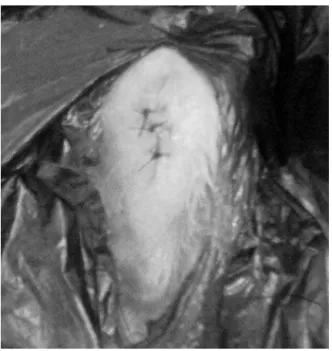
Fig.1–PhotooftheskinincisionandACLcapsulotomy.
skinandsubcutaneoustissue,followedbycapsulotomyand lateraldislocationofthepatella(Fig.1).Then,thekneewas placedinmaximumflexion,allowingvisualizationofthe ante-riorcruciateligament.Thisstructurewassectionedwithan scalpelbladenr.15(Fig.2),thejointwasirrigatedwith nor-malsaline;and acapsulorrhaphy and skinsuturewith4.0 monofilamentnyloncompletedtheoperation(Fig.3).
OnerabbitinGroup2andoneinGroup3developed infec-tion ofthe surgical site with articularextension and were excludedfromthestudy.
Aftersurgery,the rabbitswere keptintheircages with-outrestrictingthesupporttooperatedmembers.Theanimals wererandomizedintothreegroups,with14animalsineach group.Afterthreeweeksofthesurgicalprocedure,theanimals wereinitiatedintheirintra-articularinjections.Theamount ofhyaluronicacidwas0.3ml,similartothevolumeusedin smalljointsofhumans.GroupP:controlgroup,three injec-tions(atweeklyintervals)of0.9%isotonicsaline;GroupPR: threeinjections(atweeklyintervals)ofnativehyaluronicacid (Polireumin®);GroupS:threeinjectionsatweeklyintervalsof branched-chainhyaluronicacid(Synvisc®).
Fig.2–PhotoofACLexposure.
Fig.3– Photoofskinsuture.
The rabbits were euthanized after 12 weeksof surgery; the animals were anesthetized as described previously and subjected tointracardiac injection ofthiopental(5mL) and potassium chloride (10mL). The tibial plateaus were resectedasepticallyandimmersedinaflaskcontaining10% formaldehyde. The vials were labeled for identification of groupsandsenttotheClinicalPathologyService,Hospitalde Clínicas.
Thetibialplateausweredecalcified.Themedialsinthearea ofgreatercartilagethicknessweresubjectedtomicrotomyin thesagittalplane;then,threeslideswerepreparedbytibial plateau,withtheuseofstainsAlcianBluepH1.0,AlcianBlue pH2.5,andToluidineBlueandincludedinparaffin.
ThestainedslidesweresenttothePolytechnicCenterof theLifeSciencesSector,FederalUniversityofParaná.
Thehistologicalslideswereautomaticallyscannedintoa ZeissImagerMetasystemsZ2microscope,usingthesoftware MetaferMSearchwithpost-assemblywithVSlide.Then,the cartilaginousregionswereselectedwiththeMetaViewer soft-ware snapshottool. Thepictures were analyzed byImageJ software, using the RGB Stack toolfor Alcian BluepH 1.0, AlcianBluepH2.5andtheColorDeconvolution“RGB”for Tolu-idineBluestains.Subsequently,thepercentageofmarkedarea wascalculatedafterathreshold,followedbyquantification.11 Allstudysubstanceswereacquiredwithownresourcesof researchers,withoutexternalfinancialsupport.
Table1–NumbersofquantificationofToluidineBlue.
Slides Treatmentgroups
P PR S
1 66.81098 72.71096 96.52204
2 15.6544 50.25438 97.83475
3 48.30333 93.63707 73.56667
4 72.81427 98.97833 64.13119
5 76.35281 90.30573 49.14048
6 83.84236 73.03511 56.87234
7 30.08729 96.62571 66.2721
8 37.47977 73.87755 46.77349
Results
Thedecalcifiedslides,stainedwithAlcianBluepH1.0,Alcian Blue pH 2.5, and Toluidine Blue, showed the presence of glycosaminoglycans in larger quantities in the groups PR (Polireumin®)andS(Synvisc®)versuscontrolgroupP(placebo).
Thesignificancelevelforallanalyzeswas5%.
Theevaluationofthestainingintensityofhistological sec-tionsstainedwithAlcianBluepH1.0,AlcianBluepH2.5and ToluidineBluewithZeissImagerZ2Metasystemsmicroscopy generatedresultsthatwereshownintablesandgraphs.Below are graphs provided bythe program, representative ofthe actualstainingintensitymeasuredineachgroupofthe above-mentioneddyes.
Throughthe analysisconducted, eachslide generateda datumin the set of datafor each group: “P”, “PR”,or “S”, totaling24slides/analyzedindividuals(eightforeachgroup). For the statistical analysis, the null hypothesis (H0) was:
there is no difference between the independent variables, andthealternativehypothesis(H1)was:Thereisdifference
betweentheindependentvariables.Analphavalueof0.05was used.
ThevaluesforeachcolorationarepresentedinTables1–4. TheKolmorogov–Smirnov testfound a normaldistribution ofdata (not shown). Thus, parametric testswere used: an analysisofvariance(ANOVA)forsinglefactorandunpaired Studentt-testassumedequalvariances.IntheF-test,itwas foundthatthevariancesofthegroupsdidnotdifferwithinthe samestain.InToluidineBlueandAlcianBluepH2.5(butnot forpH1.0)stains,ANOVAindicatedastatisticallysignificant
Table3–NumbersofquantificationofAlcianBluepH 1.0.
Slides Treatmentgroups
P PR S
1 34.34797 68.02167 95.38368
2 89.68194 29.72821 97.26896
3 50.03834 81.88278 41.70293
4 41.21177 35.19409 68.65967
5 30.09164 34.54938 99.88211
6 38.7721 55.28347 44.2001
7 10.06779 43.21785 36.21376
8 32.54817 40.86236 29.08637
Table4–NumbersofquantificationofAlcianBluepH 2.5.
Slides Treatmentgroups
P PR S
1 49.86103 61.96043 36.31462
2 37.0048 99.59628 96.2119
3 90.00677 89.87066 94.47586
4 39.21933 38.10635 63.63671
5 41.19301 99.1222 64.21882
6 35.90056 62.46105 60.2876
7 12.16039 72.56529 64.85414
8 28.92869 53.91113 29.84288
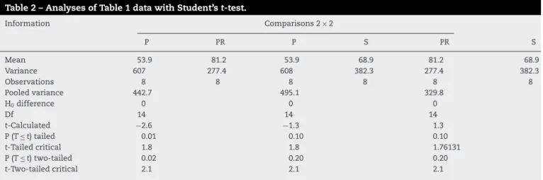
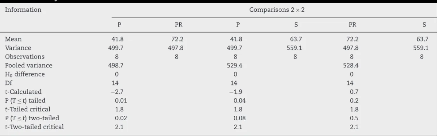
difference (p<0.05) (data not shown). Thus, for these two dyeswith differencein ANOVA,Student’st-tests were per-formed.For ToluidineBluestaining,adifferencewasnoted onlybetweenPversusPRgroups(Table2).ForAlcianBluepH 2.5,adifferencewasobservedbetweenPversusPRandPversus
Sgroups,butthelatterdivergencewasperceivedonlyinthe one-tailedanalysis(Table5).Inordertoverifywhetherthere wereadifferencebetweenthevaluesofPandSgroups, non-parametricanalysisforcategoricalvariableswasperformed. For this purpose, instead ofanalyzing the slidesusing the ImageJsoftware,scoresfrom1to3forstainingintensitywere attributed,thatis,thehigherthescore,themoreintensethe stainingwithAlcianBluepH2.5,asdescribedabove.12
Thestatisticaltestusedwasthecontingencychi-square (2)test.Forthistest,H0is:PandSgroupsdidnotdifferin thepatternofstainingintensity,whileinH1′ PandSgroups
Table2–AnalysesofTable1datawithStudent’st-test.
Information Comparisons2× 2
P PR P S PR S
Mean 53.9 81.2 53.9 68.9 81.2 68.9
Variance 607 277.4 608 382.3 277.4 382.3
Observations 8 8 8 8 8 8
Pooledvariance 442.7 495.1 329.8
H0difference 0 0 0
Df 14 14 14
t-Calculated −2.6 −1.3 1.3
P(T≤ t)tailed 0.01 0.10 0.10
t-Tailedcritical 1.8 1.8 1.76131
P(T≤ t)two-tailed 0.02 0.20 0.20
Table5–AnalysesofTable4datawithStudentttest.
Information Comparisons2× 2
P PR P S PR S
Mean 41.8 72.2 41.8 63.7 72.2 63.7
Variance 499.7 497.8 499.7 559.1 497.8 559.1
Observations 8 8 8 8 8 8
Pooledvariance 498.7 529.4 528.4
H0difference 0 0 0
Df 14 14 14
t-Calculated −2.7 −1.9 0.7
P(T≤ t)tailed 0.01 0.04 0.2
t-Tailedcritical 1.8 1.8 1.8
P(T≤ t)two-tailed 0.02 0.08 0.5
t-Two-tailedcritical 2.1 2.1 2.1
Table6–AnalysisofcategoricaldataofAlcianBluepH 2.5.
Treatmentgroups Notes
1 2 3
P(n=8) 62.5%(5) 12.5%(1) 25%(2) S(n=8) 25%(2) 12.5%(1) 62.5%(5)
Note:2calculated=32.14286.
differinthepatternofstainingintensity.N=16(eightforeach treatmentgroup),alpha=0.05,degreeoffreedom=2,and tab-ulated2=5.991.2calculatedwas=32.14286(Table6).Asthe calculatedvalueof2isgreaterthanthetabulatedvalue,we rejectH0andacceptthealternativehypothesis.
Discussion
In recent decades, studies comparing the effectiveness of hyaluronicacids ofdifferent molecular weights have been published. The data are discrepant because of its results andmethodsofevaluation.13 Inclinicalpractice,the ortho-pedicsurgeons have preferred the hyaluronicacid ofhigh molecularweightforthetreatmentofosteoarthrosis,based onstudies suchasAtamazet al.14 andWobiget al.15 who used the model of osteoarthrosis in humans, compared hyaluronicacidsofdifferentmolecularweightswithsaline intra-articularlyinfiltrated,andobtainedbetterresultswith theuseofhyaluronicacidofhighermolecularweightby clin-icalandnon-histologicalcriteria.
However, according to Karlsson et al.16 who studied hyaluronicacidsofdifferent molecularweightsinjected by intra-articular infiltrations in humans with osteoarthrosis, therewasnosignificant differenceamonghyaluronicacids ofdifferentmolecularweights,concerningclinicaland non-histologicalcriteria.
These controversies have led to the realization of this study,thatcomparedthe effectofahighmolecular weight (Synvisc®)withalowweight(Polireumin®)hyaluronate.For itsrealization,weusedanexperimentalmodelof osteoarthro-sis.
The experimental model in small animals which most closelyresemblestheosteoarthrosispresentinhumanbeings isthetransectionoftheanteriorcruciateligamentinrabbit knee. Thismodelmimicsthe morphologicaland biochemi-cal changesobserved inhumans withosteoarthrosis.5 The rabbitwasthe animalchosenbecauseofitseasyhandling, lower cost, and the vast literature that confirms its use for the purposes of obtaining induced osteoarthrosis. The study period of 12 weeks is relatively short for obtaining AO, but this averagetime isused inmost published stud-iesintheliterature.Theanatomyoftherabbitkneeiseasy to dissect and allows visualization of the anterior cruci-ate ligament,which facilitatesthe surgicalprocedureofits transection.9,10,17,18
Inthepresentstudy,thehandlingoftheanimalsandthe surgicaldissectionoftherabbitkneeswereeasytoperform. Theskinincision,themedialcapsulotomywithvisualization oftheanteriorcruciateligament,anditstransectionhadbeen donequicklyandinapracticalway.Thekneeplateaus under-going this procedure showedsigns ofobvious macroscopic lesions,especiallythoseinthecontrolgroupP(placebo–0.9% saline).19
Specific histological colorations for glycosaminoglycans were chosentoassess jointcartilage,becauseoftheir high sensitivityfordetectionofproteoglycans.AlcianBluepH1.0, AlcianBluepH2.5andToluidineBluehavegreatabilitytostain glycosaminoglycans and proteoglycans, having been used successfully in several histologicalstudies ofcartilage.20–23 TheAlcianBluepH1.0stainsgenerallyglycosaminoglycans indifferenttissues,whereasAlcianBluepH2.5andToluidine Bluespecificallystainproteoglycans.
Theuse ofthe ZeissImagerZ2 Metasystems microscope and of theMetaferMsearch softwareallowedthe measure-ment ofthestainingintensityinthe slidesofcartilaginous tissue.
ofproteoglycansinthecartilaginoustissueoftheknees infil-tratedwithhyaluronates.
There were no statisticaldifferences in the intensity of stainingofslidesofkneestreatedwithinfiltrationwithhigh molecularweightversuslowmolecularweighthyaluronicacid. Thetranspositionofthedatafoundinrabbitsinthisstudymay notreflectthesamefindingsinhumans.
Atthesametime,anevaluationofthestainingintensityby directvisionoftheslides,thatis,dependentontheexaminer, wasdone.In thissense,weasked abiologisttoassessthe slides,ascribingscores.Thisprofessionalratedwith1,2,and 3pointstheslidesstainedwithAlcian BluepH2.5aspoor, intermediateandexcellent,respectively.Theresultsbydirect visionconfirmedtheresultsconferredbytheinstrument,i.e., therewasnostatisticaldifferencebetweenthegroupstreated withhyaluronateswhencomparedwiththeplacebogroup, andtherewas nostatisticaldifference betweenthegroups infiltratedwithhyaluronates,regardlessofmolecularweight. Subjectivedatawereevaluatedbychi-squaretest.
The literature that compares the different molecular weightsofhyaluronatesinthisexperimentalmodelisscarce. Shimizuetal.24inastudyinrabbits,concludedthathyaluronic acidsoflowmolecularweightweresuperiorversusthoseof higher molecular weight. Their study did notmention the dose used in the infiltration; hence we could not make a moreappropriatecomparisonwithourstudy.Inourstudy,the rabbitsweretreatedwithintra-articularapplicationsforthree weeks(asisusuallydoneinhumans)andthevolumeused was0.3mLforthethreesubstancestested(nativehyaluronic acid,branched-chainhyaluronicacid,andnormalsaline).The half-life of native hyaluronic acid is 13h, while branched-chainhyaluronicacid is36h.Wechoseavolumeof0.3mL, becausethisistherecommendeddoseforuseinsmalljoints ofhumans.25
GhoshandGuidolin26usinganexperimentalmodelof tran-section ofthe anterior cruciateligament in dogs,obtained betterresultswithhyaluronicacidsoflowermolecularweight. Thesame authors,inaninvitrostudy,foundbetterresults withtheuseofhyaluronicacidofhighermolecularweight, whichcontradictedtheirstudiesinanimals.Thissubstance wouldbeabetterstimulatoroftheproductionofextracellular matrixcomponents.Thiscouldbepartlyexplained,because thehyaluronicacidoflowermolecularweightwouldpenetrate the extracellularmatrix more easily,maximize its concen-tration,andpromoteits interactionwithtarget cellsinthe synovium.Furthermore,thereisevidencethatthebindingof moleculesofhyaluronicacidwithcellularreceptorsis depend-entonthemolecularweight.12,13
Thereareseveralhypothesestryingtoexplainthe mech-anism of action of hyaluronic acids in non-pathological articulations of humans. In one of them, hyaluronic acid would act as modulator, through the interaction with CD44 receptors present in synoviocytes, and would act biochemically in the joint and decrease the production of cytokines, prostaglandins and metalloproteinases.27–30 Arguably, hyaluronic acid also retrieves the physiological properties of synovial fluid, decreases the pressure of the strength/weight,andimprovesthedistributionoftheweight incident on the joint. Therefore, hyaluronic acids play an importantroleinthemechanicaleffectsofthejoints.31
Insummary,inthisstudy thedataconfirmthe findings ofKarlssonetal.16 whostudiedtheeffectsofhyaluronates ofdifferentmolecularweightsinintra-articularinfiltrations inhumans withosteoarthrosis, and suggestno differences inthemolecularweightofhyaluronicacids,withrespectto chondroprotection.
Conclusion
The analysis of the effects of native and branched-chain hyaluronicacidsinanexperimentalmodelofosteoarthrosis inrabbitsdemonstratedchondroprotectiveproprieties,when compared to the control group (0.9% saline). When native chainandbranched-chain hyaluronicacidswerecompared, nostatisticallysignificantdifferencewasperceived.
Conflicts
of
interest
Theauthorsdeclarethattherewerenoconflictsofinterest.
r
e
f
e
r
e
n
c
e
s
1.WangCT,LinYT,ChiangBL,LinYH,HouSM.Highmolecular
weighthyaluronicaciddown-regulatesthegeneexpression
ofosteoarthritis-associatedcytokinesandenzymesin
fibroblast-likesynoviocytesfrompatientswithearly
osteoarthritis.OsteoarthrCartil.2006;14(12):1237–47.
2.BedsonJ,JordanK,CroftP.Theprevalenceandhistoryofknee
osteoarthritisingeneralpractice:acase–controlstudy.Fam
Pract.2005;22(1):103–8.
3.LotzM.Osteoarthritisyear2011inreview:biology.Osteoarthr
Cartil.2012;20(3):192–6.
4.AltmanRD,MoskowitzR.Intraarticularsodiumhyaluronate
(Hyalgan)inthereatmentofpatientswithosteoarthritisof
theknee:arandomizedclinicaltrial.HyalganStudyGroup.J
Rheumatol.1998;25(11):2203–12.
5.YoshimiT,KikuchiT,ObaraT,YamaguchiT,SakakibaraY,
ItohH,etal.Effectsofhigh-molecular-weightsodium
hyaluronateonexperimentalosteoarthrosisinducedbythe
resectionofrabbitanteriorcruciateligament.ClinOrthop
RelatRes.1994;(298):296–304.
6.SchiavinatoA,FinessoM,CortivoR,AbatangeloG.
Comparisonoftheeffectsofintra-articularinjectionsof
Hyaluronananditschemicallycross-linkedderivative(Hylan
G-F20)innormalrabbitkneejoints.ClinExpRheumatol.
2002;20(4):445–54.
7.HulmesDJ,MarsdenME,StrachanRK,HarveyRE,McInnesN,
GardnerDL.Intra-articularhyaluronateinexperimental
rabbitosteoarthritiscanpreventchangesincartilage
proteoglycancontent.OsteoarthrCartil.2004;12(3):232–8.
8.CooperC,SnowS,McAlindonTE,KellingrayS,StuartB,
CoggonD,etal.Riskfactorsfortheincidenceandprogression
ofradiographickneeosteoarthritis.ArthritisRheum.
2000;43(5):995–1000.
9.MoskowitzRW.Experimentalmodelsofosteoarthritis.In:
MoskovitzRW,AltmanRD,BuckwalterJA,GoldbergVM,
HochbergMC,editors.Osteoarthritis:diagnosisand
medical/surgicalmanagement.2nded.Philadelphia:
Saunders;1992.p.213–52.
10.SahRL,YangAS,ChenAC,HantJJ,HaliliRB,YoshiokaM,etal.
Physicalpropertiesofrabbitarticularcartilageafter
transectionoftheanteriorcruciateligament.JOrthopRes.
11.AndradeLF.Avaliac¸ãodaac¸ãodomedicamentoM1sobreo
melanomamurino“invivo”.Curitiba:UniversidadeFederal
doParaná;2011[dissertac¸ão].
12.ChowMT,TschoppJ,MöllerA,SmythMJ.NLRP3promotes
inflammation-inducedskincancerbutisdispensablefor
asbestos-inducedmesothelioma.ImmunolCellBiol.
2012;90(10):983–6.
13.JüniP,ReichenbachS,TrelleS,TschannenB,WandelS,Jordi
B,etal.,ViscosupplementationTrialGroup.Efficacyand
safetyofintraarticularhylanorhyaluronicacidsfor
osteoarthritisoftheknee:arandomizedcontrolledtrial.
ArthritisRheum.2007;56(11):3610–9.
14.AtamazF,KirazliY,AkkocY.Acomparisonoftwodifferent
intra-articularhyaluronandrugsandphysicaltherapyinthe
managementofkneeosteoarthritis.RheumatolInt.
2006;26(10):873–8.
15.WobigM,BachG,BeksP,DickhutA,RunzheimerJ,
SchwiegerG,etal.Theroleofelastoviscosityinthe
efficacyofviscosupplementationforosteoarthritisofthe
knee:acomparisonofhylanG-F20anda
lower-molecular-weighthyaluronan.ClinTher.
1999;21(9):1549–62.
16.KarlssonJ,SjögrenLS,LohmanderLS.Comparisonoftwo
hyaluronandrugsandplaceboinpatientswithknee
osteoarthritis.Acontrolled,randomized,double-blind,
parallel-designmulticentrestudy.Rheumatology(Oxford).
2002;41(11):1240–8.
17.YoshiokaM,CouttsRD,AmielD,HackerSA.Characterization
ofamodelofosteoarthritisintherabbitknee.Osteoarthr
Cartil.1996;4(2):87–98.
18.LavertyS,GirardCA,WilliamsJM,HunzikerEB,PritzkerKP.
TheOARSIhistopathologyinitiative–recommendationsfor
histologicalassessmentsofosteoarthritisintherabbit.
OsteoarthrCartil.2010;18Suppl.3:S53–65.
19.AlbanoMB,VidigalL,OliveiraMZ,NambaN,SilvaJL,Pereira
FilhoFA,etal.Análisemacroscópicadosefeitosdos
hialuronatosedocorticoesteroidenotratamentoda
osteoartroseinduzidaemcoelhosdecoelhos.RevBrasOrtop.
2010;45(3):273–8.
20.NaitoK,WatariT,FuruhataA,YomogidaS,SakamotoK,
KurosawaH,etal.Evaluationoftheeffectofglucosamineon
anexperimentalratosteoarthritismodel.LifeSci.
2010;86(13–14):538–43.
21.SchmitzN,LavertyS,KrausVB,AignerT.Basicmethodsin
histopathologyofjointtissues.OsteoarthrCartil.2010;18
Suppl.3:S113–6.
22.KurodaT,MatsumotoT,MifuneY,FukuiT,KuboS,Matsushita
T,etal.Therapeuticstrategyofthird-generationautologous
chondrocyteimplantationforosteoarthritis.UpsJMedSci.
2011;116(2):107–14.
23.LinW,ShusterS,MaibachHI,SternR.Patternsofhyaluronan
stainingaremodifiedbyfixationtechniques.JHistochem
Cytochem.1997;45(8):1157–63.
24.ShimizuC,KuboT,HirasawaY,CouttsRD,AmielD.
Histomorphometricandbiochemicaleffectofvarious
hyaluronansonearlyosteoarthritis.JRheumatol.
1998;25(9):1813–9.
25.FurtadoR,NatourJ.Infiltrac¸õesnoaparelholocomotor:
técnicaspararealizac¸ãocomesemoauxíliodeimagem.
Artmed:PortoAlegre;2011.
26.GhoshP,GuidolinD.Potentialmechanismofactionof
intra-articularhyaluronantherapyinosteoarthritis:arethe
effectsmolecularweightdependent?SeminArthritisRheum.
2002;32(1):10–37.
27.PrietoJG,PulidoMM,ZapicoJ,MolinaAJ,GimenoM,Coronel
P,etal.Comparativestudyofhyaluronicderivatives:
rheologicalbehaviour,mechanicalandchemicaldegradation.
IntJBiolMacromol.2005;35(1–2):63–9.
28.TakeshitaS,MizunoS,KikuchiT,YamadaH,NakimiO,
KumagaiK.TheinvitroeffectofhyaluronicacidonIl-1
productioninculturedrheumatoidsynovialcells.Biomed
Res.1997;18(3):187–94.
29.TanakaS,HamanishiC,KikuchiH,FukudaK.Factorsrelated
todegradationofarticularcartilageinosteoarthritis:areview.
SeminArthritisRheum.1998;27(6):392–9.
30.PelletierJP,Martel-PelletierJ,MalemudCJ.Canine
osteoarthritis:effectsofendogenousneutral
metalloproteoglycanasesonarticularcartilageproteoglycans.
JOrthopRes.1988;6(3):379–88.
31.PeyronJG.Intraarticularhyaluronaninjectionsinthe
treatmentofosteoarthritis:state-of-the-artreview.J