782 Brazilian Journal of Physics, vol. 35, no. 3B, September, 2005
Study of the Doppler Broadening of Positron Annihilation Radiation in Silicon
E. do Nascimento, O. Helene, V. R. Vanin,
Instituto de F´ısica, Universidade de S˜ao Paulo, C.P. 66318, 05315-970, S˜ao Paulo, SP, Brazil
and M. Moralles
Instituto de Pesquisas Energ´eticas e Nucleares - IPEN, C.P. 11049, 05422-970, S ˜ao Paulo, SP, Brazil
Received on 15 July, 2005
We report the measurement of Doppler broadening annihilation radiation in silicon, using22Na as a positron source, and two Ge detectors arrangement. The two-dimensional coincidence energy spectrum was fitted using a model function. The model function included at rest positron annihilation with valence band, 2p, 2s, and 1s electrons. In-flight positron annihilation was also fitted. The detectors response functions included backscatte-ring, and a combination of Compton effects, pileup, ballistic deficit, and pulse shaping problems. The obtained results agree well with the literature.
I. INTRODUCTION
Doppler broadening study of positron-electron annihilation radiation is an important tool in the field of materials science [1, 2]. Usually, the results of Doppler broadening experiments have been analyzed through the comparison of the calculated annihilation probability density with the experimental data. In this work we went in the inverse direction: a model func-tion was fitted to the experimental data in order to obtain the distribution of electron momenta, similar to the analysis ac-complished for the aluminum [3, 4]. In these works the coin-cidence energy spectrum was fitted using a model function, accounting for both Doppler broadening and detector system response. Intensities of the thermalized positron annihilation with band, 2p, 2s, and 1s electrons, and in-flight positron an-nihilation were fitted. The binding energies of the 1s, 2s, and 2p electrons and the Fermi cutoff parameters of the band elec-trons were also fitted. This procedure allows the experimen-tal determination of the annihilation parameters and response function parameters with their uncertainties and theχ2-test of the obtained results.
II. EXPERIMENTAL SETUP
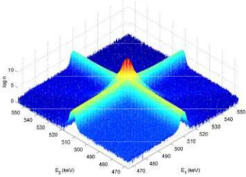
The two annihilation gamma-rays energiesE1andE2were measured using two Ge detectors forming an angle of 180o with each other and separated by 5 cm. A 3.7×10⁵Bq (10 µCi)22Na source was placed between two 2 mm thick silicon mono-crystal sheets (Czochralski). Previous lifetime measu-rements results obtained were 219.0 ps with 97.7% of inten-sity, 473 ps with 2.0% of inteninten-sity, and 2800 ps with 0.3% of intensity. An192Ir source was simultaneously measured in order to calibrate the detectors and to follow any energy ca-libration drift during the experiment. 60Co and137Cs sources were also measured. A two dimensional spectrum was taken for about 730 h. The two dimensional histogram in theE1,E2 plane in the region of interest is presented in (Figure 1).
This spectrum of Figure 1 can be interpreted as follows. The crest along the lineE1+E2=1022 keV is mainly due to at rest positron annihilation with core and band electrons. The
ridges parallel to the axes are due to the coincidence between an annihilation ray and a Compton scattered gamma-ray (either the other annihilation radiation or the 1274.5 keV gamma-ray from22Na decay). When in-flight positron an-nihilates with a low momentum electron, two gamma rays are emitted. Near the 511 keV-511 keV peak in the two dimensio-nal spectrum this annihilation can be approximated by a crest along a circular arc function centered atE1=E2=3/2mc2 [4]. This curve can be barely seen in (Figure 1).
III. FITTING - FUNCTION MODEL
To describe quantitatively the measured spectrum, a two di-mensional function was fitted to the experimental histogram in an 87 keV x 87 keV region around the two photon annihilation peak. Positron annihilation with valence band was represen-ted by
fv= 2
∑
i=1
Ci(E1−E2−αi)(E1−E2+αi) + Ave
−(E1−E2)2 2σ2v
√ 2πσv
along the lineE1+E2+Bv=1022 keV, whereBvis the gap energy of the silicon [5],E1andE2are energies in detectors 1 and 2 respectively,αiare the cutoff parameters (Ci=0 when |E1−E2|>αi). The parabolas were used in the fitting be-cause they fitted better to the experimental data. Positron an-nihilation with 2p electrons was fitted by
f2p= A2pe
−(E1−E2)2 2σ22p
√ 2πσ2p
along the lineE1+E2+B2p=1022 keV, whereB2p is the 2p electron binding energy [6]. Positron annihilation with 2s electrons was fitted by
f2s= A2se
−(E1−E2)2 2σ2
2s
E. do Nascimento et al. 783
along the line E1+E2+B2s=1022 keV, whereB2s is the 2s electron binding energy [6]. Positron annihilation with 1s electrons was fitted by
f1s= A1se
−(E1−E2)2 2σ21s
√ 2πσ1s
along the lineE1+E2+B1s=1022 keV, whereB1sis the 1s electron binding energy [6]. Finally, when(E1,E2)is a point inside the circle centered at (3m0c2/2,3m0c2/2), a function given by
ff =Afe−λd,
where
d=m0c 2
2 −
(E1−3m0c2/2)2+ (E2−3m0c2/2)2
is the distance from(3m0c2/2,3m0c2/2)to(E1,E2), and was used to take into account the in-flight positron annihilation. Finally, two internal(E<Eg)and two external(E>Eg) ex-ponential queues were included in order to simulate the non-Gaussian part of the detectors response functions. Two in-ternal and two exin-ternal ridges along the linesE1=511 keV andE2=511 keV, proportional to the peak intensity, were included in the fit. The background in channel (i,j) was em-pirically considered as proportional to the product of the total number of counts (peak excluded) along the lines j=constant by the total number of counts along the line i=constant; the single fitted parameter was the proportionality factor. The backscattering was described by a parabola, along the line E1+E2=1022 keV and centered at 511keV-511keV. Functi-ons f1s,2s,2p,vand the four exponential queues, after summing, were convoluted with the detector response functions given by two Gaussians. The fitted parameters wereA1s,A2s,A2p,Av,
σ1s,σ2s,σ2p,σv,C1,2,α1,2,a1,2, the background parameter, the peak positions in the two energy axes, the angular inclina-tion of theE1+E2=1022 keV line respect to the main axes, the sixteen exponential queues parameters (eight amplitudes and eight attenuation factors), the detector resolutions, and four parameters for the ridges intensities. Differently from ref. [4], the electron binding energies were not fitted. The fit was done by using the least-squeares method. The Gauss-Marquardt method was used to consider the non-linear para-meters [7],[8]. The chi-squared value was calculated by
χ2=
∑
i,j
(ni j−Fi j)2 Fi j
whereni jis the number of observed events in channel(i,j)of the two dimensional spectrum (Figure 2), andFi jis the fitted function (Figure 3).
IV. RESULTS AND CONCLUSION
The obtained results to the core (2p, 2s, and 1s electrons) and valence band annihilation intensities were 2.27(3)% and
FIG. 1: The two-dimensional representation for the electron-positron annihilation peak, in this figure the in-flight annihilation coincidence is represented.
FIG. 2: Experimental two-dimensional spectrum of the annihilation gamma-rays.
97.73(3)%, respectively. The intensities obtained of literature [9] to the core and valence band were 2% and 98%, respec-tively. We have found that a complete analysis of the two-dimensional Doppler annihilation radiation spectrum, in the case of the silicon, is possible. Unlike the usual approach, this procedure allows the determination of the data uncertainties. Thus, hypotheses can be tested and different results can be averaged.
784 Brazilian Journal of Physics, vol. 35, no. 3B, September, 2005
Acknowledgments
We wish to acknowledge the support of Conselho Nacio-nal de Desenvolvimento Cient´ıfico e Tecnol´ogico - CNPq and
Fundac¸˜ao de Amparo `a Pesquisa do Estado de S˜ao Paulo, and the help of Dr. M. C. A. Fantini.
V. REFERENCE
[1] K G Lynn, J R MacDonald, R A Boie, L C Feldman, J D Gabbe, M F Robbins, E Bonderup, and J Golovchenko, Phys Rev Lett
38, 241 (1977).
[2] A W Hunt, D B Cassidy, F A Selim, R Haakenaasen, T E Cowan, R H Howell, K G Lynn, and J A Golovchenko, Nucl Instr and Meth B (2000) 44.
[3] E do Nascimento, O Helene, V R Vanin, and C Takiya, Braz J Phys34, 1017 (2004).
[4] E do Nascimento, O Helene, C Takiya, and V R Vanin, Nucl Instr
Meth A538, 723 (2005).
[5] N W Ashcroft, N D Mernin (1976)Solid State Physics, Saunders College Publishing, USA
[6] R B Firestone and V S Shirley,Table of Isotopes, eighth edition, vol II, John Willey & Son, (1996)
[7] V. R. Vanin, G. Kenchian, M. Moralles, O. A. M. Helene, and P. R. Pascholati, Nucl Instr Meth A391, 338 (1997).
[8] D. W. Marquardt, J Soc Appll Math11, 431 (1963).