Ar
ti
cle
0103 - 5053 $6.00+0.00
*e-mail: [email protected]; [email protected]
Cytotoxic Lipidic
α
-amino Acids from the Zoanthid
Protopalythoa variabilis
from the Northeastern Coast of Brazil
Diego Veras Wilke,a Paula Christine Jimenez,a Claudia Pessoa,a Manoel Odorico de Moraes,a
Renata Mendonça Araújo,b Wildson Max Barbosa da Silva,b Edilberto Rocha Silveira,b Otília Deusdênia
Loiola Pessoa,b Raimundo Braz-Filho,b Norberto Peporine Lopesc and Letícia Veras Costa-Lotufo*,a
aDepartamento de Fisiologia e Farmacologia, Faculdade de Medicina, Universidade Federal do Ceará,
60430-270 Fortaleza-CE, Brazil
bDepartamento de Química Orgânica e Inorgânica, Universidade Federal do Ceará, 60021-940 Fortaleza-CE, Brazil
cDepartamento de Física e Química, Faculdade de Ciências Farmacêuticas de Ribeirão Preto,
Universidade de São Paulo, 14040-903 Ribeirão Preto-SP, Brazil
Dois α-aminoácidos lipídicos 1a e 1b foram isolados do zoantídeo Protopalythoa variabilis através de fracionamento guiado pela atividade citotóxica. As estruturas foram determinadas por diferentes métodos espectroscópicos, tais como, RMN (ressonância magnética nuclear) 1H e 13C, IV
(infravermelho) e espectrometria de massa de alta resolução (modo positivo). A atividade citotóxica dos extratos, das frações e 1a/1b foi avaliada in vitro através do teste do MTT contra quatro linhagens de células tumorais. Este achado tem implicações biológicas e químicas importantes para essa classe de compostos. Este é o primeiro relato de α-aminoácidos lipídicos a partir de uma fonte natural, bem como de sua atividade citotóxica.
Two lipidic α-amino acids 1a and 1b were isolated from the zoanthid Protopalythoa variabilis using a bioguided fractionation for cytotoxic activity. The structures of the metabolites were determined by spectroscopic methods, including NMR (nuclear magnetic resonance) 1H e 13C, IR
(infrared) and high resolution mass spectrometry (positive mode). The cytotoxic activity of the crude extract, as well as of the mixture of 1a and 1b were measured in vitro using the MTT assay for four human tumor cell lines. This inding has important biological and chemical implications for this type of compound. This is the irst report of lipidic α-amino acids from natural sources, as well as of their cytotoxic activity.
Keywords:Protopalythoa variabilis, lipidic α-amino acids, cytotoxic activity
Introduction
Cnidarians produce a wide variety of compounds showing biological activities, such as antineoplastic,1 antioxidant,2
osteoporosis suppression3 and anti-acetylcholinesterase.4
The most proliic ones are soft bodied sessile corals, such as gorgonians and zoanthids.5 Palytoxin, the most potent
non-proteic toxin known, was irst isolated from the zoanthid Palythoa toxica.6 Despite the interest of the chemical
community due to the complexity of palytoxin molecule, it is a useful tool for probing cellular processes involving sodium as a second messenger, since it binds in Na+,
K+-ATPase and transforms the pump into a channel
permeable to monovalent cations.7,8 Moreover, new
steroids9,10 and zoanthamine alkaloids,some of them with
potent biological activity,11-15 were also isolated from species
of the Zoanthidae family. Herein, in the continuing search to discover anticancer agents from marine sources16-19 we
have investigated the zoanthid Protopalythoa variabilis Duerden 1898, an abundant cnidarian at the coast of Ceará State, northeast of Brazil.
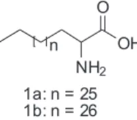
In this paper the isolation and identiication of two new lipidic α-amino acids (1a and 1b, Figure 1) from
the zoanthid Protopalythoa variabilis is described. The literature describes the lipidic α-amino acids (LAAs) as
wide as lubricants,23 cosmetics,24 polishes25 and surface
improvers for ceramics.26 They are also medicinally useful.
LAA amides with saturated or unsaturated long chain amines as well as LAA esters with saturated or unsaturated long chain alcohols, both have been synthesized.27 These
compounds inhibited both porcine pancreatic28 and human
platelet phospholipase A2 (PLA2)27 being potential
antiinlammatory agents. LAAs and their oligomers have been suggested to work as a drug delivery system.29 A
Lipid-Core-Peptide system has been designed and used as a combined adjuvant-carrier-vaccine system.30
Results and Discussion
In preliminary experiments, an aqueous-MeOH extract from Protopalythoa variabilis showed a potent cytotoxic activity. After a liquid-liquid partitioning, the biological activity was distributed among all fractions, but the hexane phase and the aqueous-MeOH residue demonstrated higher cytotoxicity as shown on Table 1. Comparative TLC (thin layer chromatography) showed some common spots in all fractions suggesting the presence of common compounds distributed among all fractions. Due to the higher activity and larger yield, the hexane fraction was chosen for further analytical separation procedures.
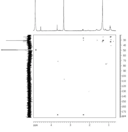
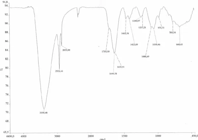
From the hexane fraction, a greasy solid named PV-Hex SII was obtained by successive solubilization/precipitation with CH3CN/H2O at different proportions of solvents using different sample concentrations. Part of this solid was soluble in pyridine-d5, and after the solvent evaporation afforded the most active fraction designated PV-Hex-SIIS. This fraction showed a purple color when reacted with a 0.1% EtOH solution of nynhidrine on TLC plates. Its IR spectrum showed absorptions bands at 1633 and 1413 (-COO-) and 1644 and 1462 (-NH
3
+), characteristic of
amino acids. The very simple 1H NMR spectrum of PV-Hex
SIISexhibited only a signal at d 3.57 (br t, H-2), typical of a nitrogenated methine, an intense and broad signal at
d 1.27, consistent with the long methylene moiety, and a broad triplet at d 0.88 characteristic of the terminal methyl of fatty chains. The 13C NMR (CPD composite pulse
decouplingand and DEPT 135) spectra revealed a signal at d 54.7 (C-2) for the aminated α-carbon, which in the
HMQC (heteronuclear multiple quantum coherence)
spectrum showed correlation with the signal at d 3.57 (H-2), highly suggestive of an α-amino acid moiety, in addition to
multiple carbon signals between d 32.5 to 23.3 (long chain methylenes) and a carbon signal at d 14.6 for the terminal methyl, suggesting the presence of a long alkyl chain. Despite the non observation of the carboxyl carbon in the
13C NMR spectrum, these spectroscopic data suggested
that the compounds of the PV-Hex SIIS were aliphatic amino acids. Indeed, the HR-ESI-TOF (positive ion mode) spectrum of this sample showed some cationizated ions at m/z 490.4566 [M + Na]+ indicating the molecular formula
C30H61NO2 for 1a, while the ion peak at m/z 504.4706 [M+ Na]+ was in agreement with the molecular formula
C31H63NO2 for 1b.
Additionally, the structures were confirmed after preparation of the methyl ester mixture (1a’/1b’) and its correspondent N-acetyl methyl ester derivatives (1a”/1b”). The HMBC (heteronuclear multiple-bond connectivity) spectrum of the methyl ester clearly revealed a correlation for the amino hydrogens (d 2.31) with the carbonyl carbon (d 176.6), as well as with the methylene carbons at d 30.8 (C-3) and 26.2 (C-4). Furthermore, a long-range coupling was also observed between the methyl ester group (d 3.65) with the carbonyl carbon. Further support to the structures of 1a/1b was obtained after acetylation of the methyl ester mixture 1a’/1b’. A signal at d 2.19 in the
1H NMR spectrum of the acetylated methyl esters conirmed
the N-acetylation of both 1a’/1b’. Therefore, the structures of 1a/1b could be unambiguously established.
Conclusions
There are three major points of chemical and biological implications that can be raised from these indings. First, there is no previous report on LAAs isolation from natural sources. Second, the number of carbon atoms for already known synthetic LAAs ranges from eight to twenty four22
what is different for the ones reported here. Third, the cytotoxic activity of LAAs has never been reported in the literature.
Experimental
General Experimental Procedures
IR spectrum (ilm on NaCl discs) was recorded using a Perkin-Elmer FT-IR 1000 spectrometer. NMR spectra were recorded on a Bruker Avance DRX-500 (500 MHz for 1H and
125 MHz for 13C) spectrometer using pyridine-d
5 as solvent. Chemical shifts, given on the d scale, were referenced to the residual undeuterated portion of the deuterated solvent Figure 1. Chemical structures of the lipidic α-amino acids from
for proton (dH 8.74, 7.58, 7.22) and the center peak of the
deuterated pyridine carbons (dC 150.35, 135.91, 123.87).
High resolution mass spectrometry were measured on a quadrupole-time of light instrument (UltrOTOF-Q, Bruker Daltonics, Billerica, MA). The analyses were performed in positive ion ESI mode at a capillary voltage of 3400 V and N2 drying gas temperature of 180 °C. NaTFA 10 mM was used as a standard for internal and external calibration. TLC was performed on precoated silica gel polyester sheets (kieselgel 60 F254, 0.20 mm, Merck). The compounds were detected by spraying with an EtOH solution of 0.1% nynhidrine.
Biological Material Collection and Identiication
Protopalythoa variabilis colonies were manually collected at Paracuru beach (Paracuru, Ceará State, Brazil) at the intertidal zone during low tide. Voucher specimens (Nº 000975), identiied by Dr. Antonio Carlos Marques have been deposited at the Zoological Museum of the University of São Paulo (Museu de Zoologia, Universidade de São Paulo - MZUSP).
Extraction and Bioguided Fractionation
A large amount of colonies of Protopalythoa variabilis after collection was stored in plastic coolers containing crushed ice and immediately transported to the laboratory. The contaminants (little rock pieces and algae specimens) were removed from the colonies. The material was weighted (11.3 kg), blended and soaked with MeOH (2 × 5L) during 48 h at 10 °C. After filtration, the MeOH extract was
evaporated under reduced pressure at 45 °C to approximately 400 mL of an aqueous suspension (named PV-HE) to which an equal amount of MeOH was added and liquid partitionated with hexane, followed by partition with CH2Cl2 and inally with EtOAc. After solvent evaporation, 1.6 g of PV-Hex, 178.2 mg of PV-CH2Cl2,1.2 g of PV-EtOAc and 41.4 mg of PV-MeOH/H2O extracts were obtained. All fractions were biologically assayed showing cytotoxicity, but PV-Hex and PV-MeOH/H2O were the most actives (see data on Table 1). Due to its higher amount PV-Hex was chosen for further puriication through high performance liquid chromatography (HPLC) analysis, but this procedure was not successful due to the poor solubility of this fraction. The treatment of PV-Hex with a mixture of CH3CN/H2O (1:1) yielded an inactive precipitate (548 mg), while the soluble part (PV-Hex S, 680 mg) retained and improved the cytotoxic activity. The PV-Hex S was only partially soluble in CH3CN/H2O (8:2), being the stronger activity observed for the insoluble residue (PV-Hex SII, 90 mg). The solubility of this solid was systematically investigated using deuterated solvents, and only the soluble fraction obtained in pyridine-d5 (PV-Hex SIIS, 26.7 mg) revealed a much stronger cytotoxic activity than all other tested fractions (see Table 1). 1H and 13C NMR analysis of PV-Hex SIIS revealed it to be a mixture
of 1a and 1b, as conirmed by HRESIMS (high resolution electrospray ionization mass spectrometry).
Lipidic α-amino acids (1a/1b): Brown wax, IR ν cm-1
3390, 2924, 2853, 1644, 1633, 1462, 1413; 1H NMR
(pyridine-d5, 500 MHz) 0.88 (br s, H-30/H-31), 1.27 (br s, methylene hidrogens), 3.57 (br s, H-2); 13C NMR
(pyridine-d5, 125 MHz) 54.7 (C-2), 32.4 (C-28), 30.4
Table 1. Cytotoxic activity of PV-HE, its derivative fractions and PV-Hex-SIIS on tumor cell lines evaluated by the MTT, assay for 72 h incubation. Data are presented as IC50 with their CI 95% values by non-linear regression. Experiments were performed in triplicate. N.T. not tested
Samples IC50 (µg/mL) CI 95%
HL-60 HCT-8 SF-295 MDA-MB-435
Doxorubicin 0.02
0.01 – 0.02
0.04 0.03 – 0.05
0.24 0.17 – 0.36
0.48 0.34 – 0.66
PV-HE 0.40
0.27 – 0.59
0.36 0.09 – 1.38
4.24 3.25 – 5.52
0.73 0.48 – 1.09
PV-MeOH/H2O 0.76
0.62 – 0.94
0.40 0.32 – 0.49
0.49 0.40 – 0.59
0.085 0.02 – 0.41
PV-EtOAc 2.94
1.94 – 4.46
2.25 1.26 – 4.02
1.85 1.29 – 2.65
1.42 1.08 – 1.87
PV-CH2Cl2 5.43
4.86 – 6.07
2.67 2.19 – 3.24
2.95 2.68 – 3.25
2.82 2.40 – 3.29
PV-Hex 0.43
0.39 – 0.47
0.24 0.22 – 0.27
0.29 0.25 – 0.33
0.38 0.34 – 0.43 PV-Hex SIIS (1a/1b) 0.13
0.12-0.14
0.05 0.05-0.06
0.07
(C-5/C-27), 29.9 (C-3), 26.8 (C-4), 23.3 (C-29/C-30), 14.6 (C-30/C-31); HR-ESI-TOFMS (1a) m/z 490.4566 [M + Na]+ (calcd. for C
30H61NO2Na, 490.4599); (1b) m/z
504.4706 [M + Na]+ (calcd. for C
31H63NO2Na, 504.4756).
Methylation of 1a/1b: An aliquot (4.0 mg) of the lipidic
α-amino acids (1a/1b) was dissolved in MeOH with
catalytic amounts of HCl, and stirred under relux during 4 h. Usual work-up afforded 1a’/1b’ (4.2 mg). 1H NMR
(pyridine-d5, 500 MHz) 0.93 (t, J = 6.45 Hz, H-30/H-31), 1.32 (br s, methylene hydrogens), 2.31 (NH2), 3.66 (br s, H-2), 3.65 (s, OCH3); 13C NMR (pyridine-d
5, 125 MHz) 176.6 (C-1), 52.1 (C-2), 33.2 (C-28), 30.8-23.8 (C-3/C-29), 14.6 (C-30/C-31).
Acetylation of 1a’/1b’: This material was dissolved in a mixture of pyridine/acetic anhydride 1:2 (1 mL) and stirred for 24 h at room temperature. After this, the reaction mixture was neutralized with a solution of HCl 1mol L-1 (4 drops)
and extracted with CH2Cl2 (4 x 10 mL). The CH2Cl2 layer was evaporated under reduced pressure to yield 1a”/1b”
(4.2 mg). 1H NMR (CDCl
3, 500 MHz) 0.89 (br t), 1.27 (br
s, methylene hydrogens), 2.19 (s), 3.68 (s, OCH3). Cytotoxic assay
The cytotoxic potential of the extract and of all derived fractions was evaluated against four tumor cell lines (National Cancer Institute, Bethesda, MD, USA): HCT-8 (human colon), MDA-MB-435 (melanoma), SF-295 (CNS glioblastoma) and HL-60 (leukemia) using the 3-(4,5-dimethyl-2-thiazolyl)-2,5-diphenyl-2H-tetrazolium (MTT) assay described by Mosmann31 after 72 h incubation.
Doxorubicin was used as positive control.
Acknowledgment
The authors would like to acknowledge the inancial support in the form of grants and/or fellowships from the following Brazilian agencies: CNPq, FINEP and Institute Claude Bernard (InCB). The authors are grateful to Antonio Carlos Marques, from University of São Paulo for taxonomic identiication of the zoanthid. The technical assistance of Silvana França is gratefully acknowledged.
Supporting Information Available
Supplementary information for compounds 1a and 1b is available free of charge as PDF ile at http://jbcs.sbq.org.br.
References
1. Pettit, G.; Fuji, Y.; J. Nat. Prod. 1982, 45, 640.
2. Dunlap, W.C.; Yamamoto, Y.; Comp. Biochem. Physiol. 1995, 112B, 105.
3. Yamaguchi, K.; Yada, M.; Tsuji, T.; Kuramoto, M.; Uemura, D.; Biol. Pharm. Bull. 1999, 22, 920.
4. Turk, T.; Macek, P.; Suput, D.; Toxicon 1995, 33, 133. 5. Harper, M.K.; Bugni, T.S.; Copp, B.R.; James, R.B.; Lindsay,
B.S.; Richardson, A.D.; Marine Chemical Ecology. In the McClintock, JB & Baker BJ eds.; Boca Raton, Florida, 2001. 6. Moore, R.E.; Scheuer, P.J.;Science 1971, 172, 495. 7. Wang, D.Z.; Mar. Drugs 2008, 6, 349.
8. Jha, R.K.; Zi-rong, X.; Mar. Drugs 2004, 2, 123.
9. Guerriero, A.; Traldi, P.; Pietra. F.; J. Chem. Soc., Chem. Commun. 1986, 40.
10. Suksamrarn, A.; Jankam, A.; Tarnchompoo, B.; Putchakarn, S.; J. Nat. Prod. 2002,65, 1194.
11. Behenna, D.C.; Stockdill, J.L.; Stoltz, B.M.; Angew. Chem.
2008, 47, 2365.
12. Daranas, A.H.; Fenandez, J.J.; Gavin, J. A.; Norte, M.; Tetrahedron 1999, 55, 5539.
13. Rao, C.B.; Anjaneyula, A.S.R. Sarma, N.S.;Venkatateswarlu. Y.; J. Am. Chem. Soc. 1984, 106, 7983.
14. Fattorusso, E.; Romano, A.; Taglialatela-Scafati, O.; Achmad, M.J.; Bavestrello, G.; Cerrano, C.; Tetrahedron Lett. 2008, 49, 2189.
15. Villar, R.M.; Gil-Longo, J.; Daranas, A.H.; Souto, M.L.; Fernández, J.J.; Peixinho, S.; Barral, M.A.; Santafé, G.; Rodríguez, J.; Jiménez, C.; Bioorg. Med. Chem. 2003, 11, 2301. 16. Jimenez, P.C.; Fortier, S.C.; Lotufo, T.M.C.; Pessoa, C.; Moraes,
M.E.A.; Moraes, M.O.; Costa-Lotufo, L.V.; J. Exp. Mar. Biol. Ecol. 2003,4040, 1.
17. Takeara, R.; Lopes, J. L. C.; Lopes, N. P.; Jimenez, P. C.; Costa-Lotufo, L. V.; Quim. Nova, 2007,30, 1179.
18. Takeara, R.; Jimenez, P. C.; Costa-Lotufo, L. V. ; Lopes, J. L. C.; Lopes, N. P.; J. Braz. Chem. Soc. 2007, 18, 1054. 19. Jimenez, P.C.; Wilke, D.V.; Takeara, R.; Lotufo, T.M. C.; Pessoa,
C.; Moraes, M.O.; Lopes, N.P.; Costa-Lotufo, L.V.; Comp. Biochem. Physiol. 2008, 151, 391.
20. Roberts, D.C.; Vellaccio, F.; In: Gross E, Meinhofer J. eds.; Academic Press: New York,1983.
21. Williams, R.M.; Synthesis of opticaly active alpha-amino acids. Pergamon Press, Oxford, 1989.
22. Kokotos, G.; Martin, V.; Constantinou-Kokotou, V.; Gibbons, W.A.; Amino Acids 1996, 11, 329.
23. Takino, H.; Sagawa, K.; Kitamura, N.; Kilahara, M.; Jpn. Kokai Tokkyo Koho 62,151,495 1987 (CA 107:220318c)
25. Sagawa, K.; Takehara, M.;Jpn. Kokai Tokkyo Koho 62 30,171
1987a (CA 107:79672e).
26. Sagawa, K.; Takehara, M.;Jpn. Kokai Tokkyo Koho 62 65,964
1987b (CA 107:63607b).
27. Kokotos, G.; Constantinou-Kokotou, V.; Noula, C.; Nicolaou, A.; Gibbons, W.A.; Int. J. Peptide. Protein. Res. Ther. 1996, 48, 160.
28. Nicolaou, A.; Kokotos, G.; Constantinou-Kokotou, V.; York, J.; Gibbons, W.A.; Biochem. Soc. Trans. 1995,23, 614S.
29. Toth, I.; J. Drug. Targeting. 1994, 2, 217.
30. Toth, I.; Danton, M.; Flinn, N.; Gibbons, W.A.; Tetrahedron
1993, 34, 3925.
31. Mosmann, T.; J. Immunol. Met. 1983,65, 55.
Received: August 12, 2008
Web Release Date: August 20, 2009
Su
pp
le
m
enta
ry
Inf
or
m
ati
on
0103 - 5053 $6.00+0.00
*e-mail: [email protected]; [email protected]
Cytotoxic Lipidic
α
-amino Acids from the Zoanthid
Protopalythoa variabilis
from the Northeastern Coast of Brazil
Diego Veras Wilke,a Paula Christine Jimenez,a Claudia Pessoa,a Manoel Odorico de Moraes,a
Renata Mendonça Araújo,b Wildson Max Barbosa da Silva,b Edilberto Rocha Silveira,b Otília Deusdênia
Loiola Pessoa,b Raimundo Braz-Filho,b Norberto Peporine Lopesc and Letícia Veras Costa-Lotufo*,a
aDepartamento de Fisiologia e Farmacologia, Faculdade de Medicina, Universidade Federal do Ceará,
60430-270 Fortaleza-CE, Brazil
bDepartamento de Química Orgânica e Inorgânica, Universidade Federal do Ceará, 60021-940 Fortaleza-CE, Brazil
cDepartamento de Física e Química, Faculdade de Ciências Farmacêuticas de Ribeirão Preto,
Universidade de São Paulo, 14040-903 Ribeirão Preto-SP, Brazil
Figure S3. 13C NMR spectrum (125 MHz, C
5D5N) ofamixture of two lipidic α-amino acids (1a and 1b)
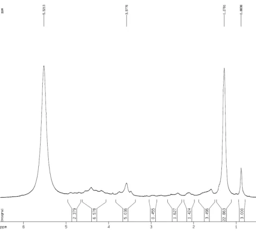
Figure S2. 1H NMR spectrum (500 MHz, C
Figure S5. 1H NMR spectrum (500 MHz, MeOD) of a methyl ester mixture (1a’/1b’)
Figure S4. HRESI-MS spectrum ofamixture of two lipidic α-amino acids (1a and 1b). The major ions: 1am/z 490.4566 [M+Na]+ (calc. for C
30H61NO2Na, 490.4599); 1bm/z 482.4959[M+H]+ (calc. for C
31H64NO2, 482.4936); 1bm/z 504.4706 [M+Na]
+ (calc. for C
Figure S6. HMBC spectrum (500 MHz, MeOD) of methyl ester derivatives (1a’/1b’)
Figure S7. 1H NMR spectrum (500 MHz, CDCl
![Figure S4. HRESI-MS spectrum of a mixture of two lipidic α -amino acids (1a and 1b). The major ions: 1a m/z 490.4566 [M+Na] + (calc](https://thumb-eu.123doks.com/thumbv2/123dok_br/18993076.461333/8.892.187.727.478.968/figure-hresi-spectrum-mixture-lipidic-amino-acids-major.webp)