RevBrasAnestesiol.2017;67(3):314---317
REVISTA
BRASILEIRA
DE
ANESTESIOLOGIA
PublicaçãoOficialdaSociedadeBrasileiradeAnestesiologiawww.sba.com.br
CLINICAL
INFORMATION
Ultrasound-guided
central
venous
catheterization
---‘‘Syringe-Free’’
approach
Francisco
Matias
∗,
Edgar
Semedo,
Cláudia
Carreira,
Paula
Pereira
DepartamentodeAnestesiologia,CentroHospitalareUniversitáriodeCoimbra,Coimbra,Portugal
Received7September2014;accepted29September2014 Availableonline11March2015
KEYWORDS
Ultrasonography; Interventional; Jugularveins; Catheterization; Centralvenous; Internaljugularvein cannulation; Obliqueview
Abstract
Backgroundandobjectives: Centralvenous catheterization of the internaljugular vein isa commonly performed invasive procedure associated with a significant morbidity and even mortality.Ultrasound-guidedmethodshaveshowntoimprovesignificantlythesuccessofthe techniqueandarerecommendedbyvariousscientificsocieties,includingtheAmerican Soci-etyofAnesthesiologists.Theaimofthisreportistodescribeaninnovativeultrasound-guided centrallineplacementoftheinternaljugularvein.
Technique:Theauthorsdescribeaninnovativeultrasound-guidedcentrallineplacementofthe internaljugularveinbasedonanobliqueapproach---the‘‘Syringe-Free’’approach.This tech-niqueallowsimmediateprogressionoftheguidewireinthevenouslumen,whilemaintaining areal-timecontinuousultrasoundimage.
Conclusions:The describedmethodaddstothetraditionalobliquetechniquethepossibility ofachievingacontinuousreal-timeultrasound-guidedvenipunctureandaguidewireinsertion thatdoesnotneedremovingtheprobefromthepuncturefield,whilehavingasingleoperator performingthewholeprocedure.
©2015SociedadeBrasileiradeAnestesiologia.PublishedbyElsevierEditoraLtda.Thisisan openaccessarticleundertheCCBY-NC-NDlicense( http://creativecommons.org/licenses/by-nc-nd/4.0/).
PALAVRAS-CHAVE
Ultrassonografia; Intervenc¸ão; Veiasjugulares; Cateterizac¸ão;
Cateterizac¸ãovenosacentralguiadaporultrassom---abordagem‘‘Syringe-Free’’
Resumo
Justificativaeobjetivos: Acateterizac¸ãovenosacentraldaveiajugularinternaéum procedi-mentoinvasivorealizadofrequentementeeassociadoamorbilidadesignificativaeatémesmo
∗Correspondingauthor.
E-mail:[email protected](F.Matias). http://dx.doi.org/10.1016/j.bjane.2014.09.011
Ultrasound-guidedcentralvenouscatheterization---‘‘Syringe-Free’’approach 315
Veiacentral; Canulac¸ãodaveia jugularinterna; Abordagemoblíqua
mortalidade.Osmétodosguiadosporultrassonografiatêmdemonstradoumamelhoradosucesso desteprocedimentoesãorecomendadosporváriassociedadescientíficas,incluindoaAmerican SocietyofAnesthesiologists.Oobjetivodesteartigoédescreverumaabordageminovadorade cateterizac¸ãovenosacentralguiadaporultrassonografiaaoníveldaveiajugularinterna.
Técnica: Osautoresdescrevemtécnicaecoguiadainovadoradecateterizac¸ãovenosacentral daveiajugularinterna,baseadanumaabordagemoblíqua---aabordagem‘‘Syringe-Free’’.Esta técnicapermiteumaprogressãoimediatadofio-guiaaolongodolúmenvenoso,mantendouma visualizac¸ãoecográficaemtemporealecontínua.
Conclusões: Atécnicadescritaacrescentaàtécnicaoblíquatradicionalapossibilidadede,com umúnicooperador,conseguirumapunc¸ãovenosacentralcomvisualizac¸ãoecográficacontínua eemtemporealassociadoàinserc¸ãodofio-guiasemnecessidadedeafastamentodotransdutor deultrassonografiadocampodepunc¸ão.
©2015SociedadeBrasileiradeAnestesiologia.PublicadoporElsevierEditoraLtda.Este ´eum artigo OpenAccess sobumalicenc¸aCCBY-NC-ND( http://creativecommons.org/licenses/by-nc-nd/4.0/).
Introduction
The centralvenouscatheter(CVC)insertionis an invasive procedurecommonlyperformednotonlywithin anesthesi-ology,butalsoinmultiplespecialtiesrangingfromoncology toemergencymedicine.1
Thetraditionalanatomiclandmarktechniqueforplacing centralvenouslines,howevervaluable,hasbeenlinkedto anumberofprocedure-relatedcomplicationssuchas arte-rialpunctureor cannulation,venousinjury,pneumothorax andhemothorax.2,3Comparatively,ultrasound-guided meth-ods have shown some advantages; namely, a decrease in the number of inadvertent arterial puncture or cannula-tion,lessincidenceofpneumothorax,highersuccessrates and favorablecost---benefit ratios.1,4---6 Itsuse is therefore recommendedbyvariousscientificsocieties,includingthe AmericanSocietyofAnesthesiologists.6---9
Among anesthesiologists,the most frequently selected site for central access is the internal jugular vein (IJV). Techniques described include the traditional ultrasound transverseandlongitudinalapproaches,aswellasan alter-nativeobliqueview.10---13 Inrelationtothelatter,different variantshavebeenillustrated;specifically,medial-oblique, lateral-obliqueandmedial-transversaltechniques.13---15The referredultrasound-guidedmethodssharethefactthatthe operator can confirm the location of the vessel by aspi-rating blood. However, subsequently, the operator has to disconnect the syringe in order to pass the guide wire throughtheneedleorpass itthroughaguidewiresyringe device. Regardless of which methodis used,this require-mentincreasesthepossibilityofneedledislocation(withall theassociatedimplications,suchasarterialpuncture, nee-dle tipexteriorization fromthe venous lumen, and nerve injury). In addition, it requires the presence of a second operatorinordertoachieveacontinuousultrasound imag-ing fromthe skin punctureuntil the complete guidewire insertion.
This paper describes an innovative ultrasound-guided CVCplacementintheIJVthatisbasedontheobliqueview, butonly requiresasingleoperator andprovidesreal-time continuous ultrasound imaging. This procedure is achiev-ablebyusingthelateral-medialvariantapproachwiththe
guidewireadaptedtotheneedlefromthebeginningofthe procedure.
Technique
Thepatientisplacedina15◦Trendelenburgpositionwitha
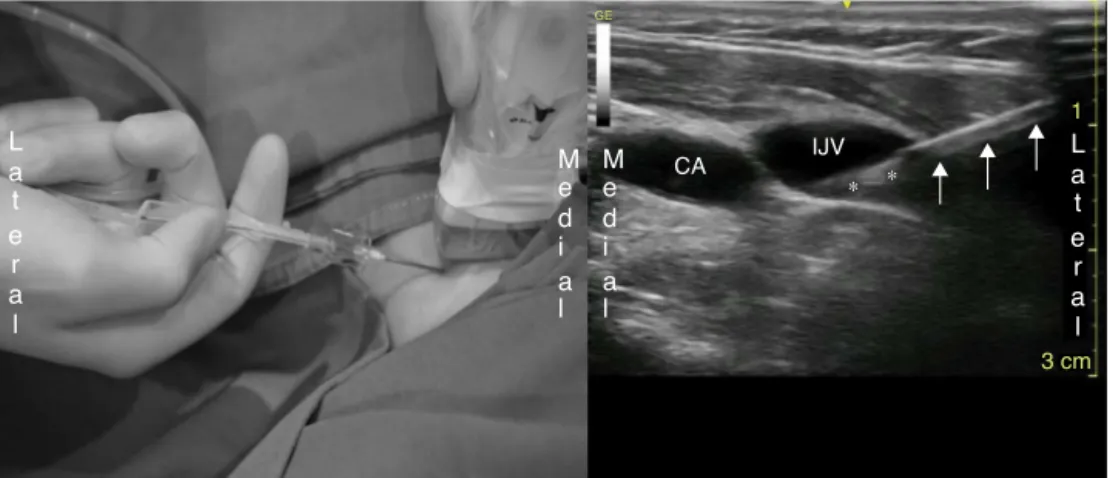
slightheadrotationtothesideoppositetotheoperator.The authorsrecommendplacingthe ultrasoundscreenonthat side,sothattheoperatorcanlookupdirectlyatthescreen withinthe same field of vision used to advancethe nee-dle.Aseptic techniqueforpreparationis mandatory.Also, thehighfrequencylinearprobeandrespectivecableshould beisolated withan appropriate sterile sleeve. The guide wireisadaptedtothepunctureneedlebeforeinitiatingthe procedure(Fig.1).
As describedby PhelanandHagerty,13 inorderto visu-alizethecarotidarteryandtheIJV,theprobeisplacedin aplanetransversaltotheneckat thelevelofthesternal andclavicularheadsofthesternocleidomastoid(SCM) mus-cle.Whencomparedtothecarotidartery,theIJVgenerally appearstobenon-pulsating,islargerandmoresuperficial, hasathinner vesselwall, andis more easilycompressed. Itisimperativetocorrectlypositiontheprobesothatthe structuresontherightsideoftheultrasoundimage corre-spondtothesamesideonthepatient.Oncethedescribed imageisobtained,the probeshouldberotated45◦
316 F.Matiasetal.
Figure1 Guidewireadaptedtothepuncture needlesince thebeginningoftheprocedure.
Advantages
This techniqueincludes the recognizedadvantages of the obliqueapproach(visualizationofboth thearteryandthe vein,aswellasoftheentireneedle),andatthesametime allowsacontinuous visualization of theentire procedure, potentially increasingits safety. Inaddition, this continu-ousvisualization isachievedwithout theneed ofasecond operator,sinceitdoes notrequiresomeone todisconnect thesyringe beforethe guidewire insertionis completed. Alltheotherpreviouslydescribeddifferenttechniques per-formedby asingleoperator requirethiscriticalperiodof interrupting theultrasoundimage, sincethe operatorhas toletgo of theultrasound probein orderto have afree hand for the syringe disconnection and subsequent guide wireinsertion.Withthe‘‘Syringe-Free’’approachthisdoes nothappenbecausetheguidewireisadaptedtothe punc-tureneedlefromthebeginning.Moreover,thephysicianis
incontroloftheentireprocessandmaycorrectinrealtime, asneeded,theneedletipposition,thealignmentandthe progressionofthewire.Complicationsassociatedwiththis stage,suchascephalicprogressionofthewireor perfora-tionoftheposteriorormedialwalloftheIJV,aretherefore reduced.
ThecatheterizationoftheIJVwiththismethodhasbeen successfully employed in the past year. It has been per-formed by the authors, who regularly use ultrasound in regionalanesthesiaandcentralvenouscatheterization.
Limitations
This techniquecomprisessome limitations.In mostcases, thecarotidarteryisfoundtobeinthesametrajectoryas the needle, which could lead to the idea that thereis a greaterrisk ofcarotidarterypuncture.However,the nee-dle is inserted in-plane and at a superficial level, which in theory allows an easy track of the needle. Neverthe-less, it is mandatory that this technique is executed by anexperiencedoperatorwhoisskilledinmanipulatingand interpretingultrasoundimaging,inordertoachievea con-tinuous observation of the needletip. Ifthese conditions aremet, the risk of carotidartery punctureis minimized and theconfirmation by blood aspiration becomes unnec-essary, sincethe operator is able to visualize the needle enteringthelumenoftheIJV.Furthermore,basedontheir experience,theauthorsfoundthattheneedle’slumenfills upwithbloodafterthevenouspuncture.Itshouldbenoted thatinhypovolemicpatientstheexecutionofthetechnique becomesmore challenging, giventhe smallerdiameter of theIJV.Fornoviceultrasoundusersand/ornon-expert sono-graphers,ultrasoundmaygiveafalsesenseofsecurity,and theconstantvisualizationoftheneedleanditstipmightnot beachieved.Therefore,themainlimitationofthedescribed procedureisassociatedwiththeoperator’slevelof experi-enceinultrasoundmanipulation.
Theauthorshavefoundanotherchallengeinthe imple-mentationofthistechniquerelatedtothefactthatnotall central venous catheterization setsprovide a stable con-nection between the needle hub and the dispenser unit of the guide wire. The authors usually use the Certofix®
L a t
e r a l
3 cm
GE
1
M e d i
a l
M CA IJV
∗ ∗ e
d i
a l
L a t
e r a l
Ultrasound-guidedcentralvenouscatheterization---‘‘Syringe-Free’’approach 317
Duo (B-Braun, Melsungen AG, Germany) central venous catheterizationset,whoseguidewiredispensercanbe sta-bly connectedtothe needle.Thus, thetechnique can be performed without undue difficulty. However, ifthis con-nectionisnotsecure, the‘‘Syringe-Free’’approachis not recommended.
Conclusions
The authors suggest this ‘‘Syringe-Free’’ approach for ultrasound-guided CVC placement, which surpasses the alreadydescribedadvantagesoftheobliquetechnique13as it allows real-time continuous ultrasound imaging carried out by a singleoperator. It is nowimperative toprovide evidence through randomized prospective studies on the benefitofthistechnique,mainlywithrespecttoitssafety, successrateandtimeofexecution.
Conflicts
of
interest
Theauthorsdeclarenoconflictsofinterest.
References
1.LennonM,ZawNN,PoppingDM,etal.Proceduralcomplications of central venous catheter insertion. Minerva Anestesiol. 2012;78:1234---40.
2.BowdleA.Vascular complicationsofcentral venouscatheter placement:evidence-basedmethodsforpreventionand treat-ment.JCardiothoracVascAnesth.2014;28:358---68.
3.Wu S, Ling Q, Cao L, Wang J, Xu M, Zeng W. Real-time two-dimensionalultrasoundguidanceforcentral venous can-nulation.Anesthesiology.2013;118:361---75.
4.YoshidaH,KushikataT,KitayamaM,etal.Time-consumption riskofreal-timeultrasound-guidedinternaljugularvein cannu-lationinpediatricpatients:comparisonwithtwoconventional techniques.JAnesth.2010;24:653---5.
5.CalvertN,HindD,McWilliamsRG,etal.Theeffectivenessand cost-effectiveness of ultrasound locating devicesfor central venousaccess:asystematicreviewandeconomicevaluation. HealthTechnolAssess.2003;7:1---84.
6.RuppSM,ApfelbaumJL,BlittC,etal.Practiceguidelinesfor centralvenousaccess:areportbyAmericanSocietyof Anesthe-siologistsTaskForceonCentralVenousAccess.Anesthesiology. 2012;116:539---73.
7.Lamperti M, Bodenham AR, Pittiruti M, et al. International evidence-basedrecommendationsonultrasound-guided vascu-laraccess.IntensiveCareMed.2012;38:1105---17.
8.NationalInstituteforHealthandClinicalExcellence.The clin-icaleffectivenessandcosteffectivenessofultrasoundlocating devices for the placement of central venous lines. Tech-nology appraisalreport49; 2002. Available at: http://www. nice.org.uk/TA49
9.TroianosCA,HartmanGS,GlasKE,etal.Specialarticles: guide-lines for performing ultrasoundguided vascularcannulation: recommendations ofthe American Societyof Echocardiogra-phyandtheSocietyofCardiovascularAnesthesiologists.Anesth Analg.2012;114:46---72.
10.RoseJS,BairAE.Vascularaccess.In:MaOJ,MateerJR,editors. Emergencyultrasound.1sted.NewYork:McGraw-Hill Profes-sional;2002.p.349---60.
11.WeinerMM,GeraldP,MittnachtAJ.Ultrasound-guidedvascular access:acomprehensivereview.JCardiothoracVascAnesth. 2013;27:345---60.
12.Yamauchi M, Sasaki H, Yoshida T, et al. Ultrasound-guided supraclavicularcentralvenouscatheterizationinpatientswith malignanthematologicdiseases.JAnesth.2012;26:775---8. 13.PhelanM,HagertyD.Theobliqueview:analternativeapproach
for ultrasound-guided central line placement.J Emerg Med. 2009;37:403---8.
14.DiLisio R, Mittnacht A. The ‘‘medial-oblique’’ approach to ultrasound-guided central venous cannulation --- maximize the view, minimize the risk. J Cardiothorac Vasc Anesth. 2012;26:982---4.