BrazJOtorhinolaryngol.2016;82(4):487---490
www.bjorl.org
Brazilian
Journal
of
OTORHINOLARYNGOLOGY
CASE
REPORT
Extramedullary
relapse
of
acute
myeloid
leukemia
mimicking
a
necrotizing
external
otitis:
could
mononuclear
leukocyte
scintigraphy
be
the
best
diagnostic
method?
夽
Recidiva
extramedular
de
leucemia
mieloide
aguda
mimetizando
otite
externa
necrotizante:
a
cintigrafia
com
leucócitos
mononucleares
pode
ser
o
melhor
método
diagnóstico?
Roberta
Laurindo
a,
Sergio
Souza
b,∗,
Jaqueline
Moura
a,
Shiro
Tomita
a,
Lea
Barbosa
da
Fonseca
b,
Bianca
Gutfilen
baServiceofOtolaryngology,DepartmentofClinicalMedical,HospitalUniversitárioClementinoFragaFilho,UniversidadeFederal
doRiodeJaneiro(UFRJ),RiodeJaneiro,RJ,Brazil
bDepartmentofRadiology,LaboratóriodeMarcac¸ãodeCélulaseMoléculas,UniversidadeFederaldoRiodeJaneiro(UFRJ),
RiodeJaneiro,RJ,Brazil
Received26January2015;accepted23April2015 Availableonline7September2015
Introduction
Necrotizing external otitis (NEO) is an invasive infec-tious disease involving the cartilaginous and/or bony external auditory canal (EAC) that particularly affects immunocompromised patients.1 Patients usually present
withunrelentingotalgiathatisdisproportionatetothe clin-ical signsand purulent otorrhea. The diagnosis of NEO is based ona combination of clinical, laboratory,and imag-ing findings.2 Current tools used todiagnose NEO include
夽 Pleasecitethisarticleas:LaurindoR,SouzaS,MouraJ,Tomita
S, da Fonseca LB, Gutfilen B. Extramedullary relapse of acute myeloid leukemia mimickinga necrotizing external otitis: could mononuclearleukocytescintigraphybethebestdiagnosticmethod? BrazJOtorhinolaryngol.2016;82:487---90.
∗Correspondingauthor.
E-mail:[email protected](S.Souza).
computed tomography (CT), magnetic resonance imaging (MRI),andscintigraphy.Thecombinationofradiologicaland radionuclideexamsiscrucial inboth, theinitial diagnosis andthefollow-upoftreatmentresponse.
Case
report
A19-year-oldmandiagnosedwithacutemyeloidleukemia (AML)inremissionpresentedwithsevereotalgia,aural full-ness,rightfacialpalsy(scoringVontheHouse-Brackmann scale).Otoscopicfindingsincluded,edemaoftheposterior wallof theexternal earcanal and otorrhea. Diagnosis of NEOwasconsidered,andthepatientwastreatedempirically withimipenemfor12days.Thepatienthadapoorresponse toimipenem, leading the medicalteam toreplace it for cefepime.Evenafterthechangeofthetreatmentregimen thepatient’ssymptomsdidnotshowanyimprovement.
http://dx.doi.org/10.1016/j.bjorl.2015.04.003
488 LaurindoRetal.
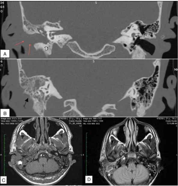
Figure1 High-resolutioncoronalcomputedtomographyimagesofthemastoids(AandB).(A)Erosionoftheposteriorwallofthe externalauditorycanal(EAC;whitearrow)andthickeningandenhancementofthesofttissueintheregionoftheEAC(redarrow). (B)Erosionofthemastoidcortex(blackarrow)anddestructionofthebonyseptae.Magneticresonanceimagingofthemastoids (CandD).(C)Axial,T1-weightedimagedemonstratinginfiltrationofthesofttissuesaroundtheEAC,subcutaneoustissue,and parotidgland.(D)Axial,T2-Flairimageshowingthetympanicandmastoidcavity,withtissuecontrastuptakespreading anteroin-feriorly.
ComplementaryexamsrevealedhighreactiveC-protein levelsandopacificationofmastoidcellsandmiddleearinCT images,whichwasassociatedwitherosionoftheposterior wallofthe EAC(Fig.1AandB)T2-weighted MRIrevealed gadolinium enhancement inside the tympanic cavity and around the carotid canal, extending to the ear pavilion andadjacent soft tissues (Fig.1C andD). The findings of
99mTc-MDPbonescintigraphywereconsistentwithan
inflam-matory/infectiousprocessoccurringinthetemporalbone. However,99mTc-mononuclearleukocytecintigraphydidnot
showanyevidenceofaninflammatoryprocess.
Given these results and the fact that symptoms were present after 6 weeks of antibiotic therapy, the medical team considered the hypothesis that the patient had a neoplasticdisease(Fig.2).Consequently,thepatient under-went a tympanomastoidectomy. Myeloid sarcoma (MS), a typeofextramedullary recurrenceofAML, wasconfirmed
byhistopathological study.There werenosignsof disease in the bone marrow. We initiated a chemotherapy proto-coland observedrapid improvementinterms of painand by otoscopy. The facial palsy regressed to scoring III on the House-Brackmann scale after the treatment. Heterol-ogous transplantation wasrecommended,and thepatient completelyrecovered.
Discussion
MS isa rarecondition characterizedby theoccurrence of oneormoretumorscomposedofimmaturemyeloidcellsin anextramedullarysite.Thisdiseaserarelyaffectsthe tem-poral bone,and itis mostcommonly found in bones,soft tissue,skin,andthecentralnervoussystem.3Symptomsof
Mononuclearleukocytescintigraphyandnecrotizingexternalotitis 489
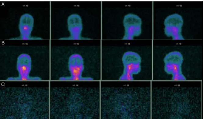
Figure2 99mTc-mononuclearleukocyte scintigraphy(A---C).Planar scintigraphsfrom one,three, and24hafterlabeled
mono-nuclearleukocyteadministrationindicatenouptakealongthemastoidtopographyovertime.
palsy,tinnitus,earfullness,earpain,andretrauricularand externalauditorycanalswellingthatcanmimic otomastoidi-tisor NEO.ImagingfindingsintheearlystagesofNEOare frequentlysubtle,andeveninadvancedcases,theimaging findingsmaynotberecognizedasNEOunlessthediagnosis isalreadyunderconsideration.
Nuclearimagingplaysamajorroleinthediagnosisand follow-up of patients who have NEO. In fact, bone scans using99mTc-MDPprovidebetterinformationabout
inflamma-tionbecausetheyhighlightareaswithosteoblasticactivity. Abonescancanshowpositiveresultsbeforeapparent radio-logicalchangesoccurandmaybeusefulinearlydetection ofthedisease.However,99mTc-MDPisnotspecificfor
infec-tion,asitcanalsobepositiveinmalignantdiseaseanddoes not detect soft tissue spread without bone involvement.4
Bonescansalsoremainpositiveafterthediseaseisresolved and arenot useful for monitoring response totreatment. Gallium-67scintigraphyhasbeenshowntobe70%sensitive and93% specific todiagnose osteomyelitis; thus, it is the investigativetechnique of choice for monitoring response totreatment. CTishighlyeffectiveatdefiningtheextent ofbonydestructionintheearcanalwallorthebaseofskull bydelineatingnormalfatplanesandbonecortices,andthe importanceofCTfordiagnosingNEOiswell-established.MRI isconsideredmoresensitivethanCTfordelineatingsoft tis-sueplanes.Mostskullbaseabnormalitiesexhibitalowsignal on T1-weighted images and a high signal onT2-weighted images.5
99mTc-mononuclearleukocytescintigraphyisusedto
diag-nosisinfection,osteomyelitis,graftrejection,andfeverof unknownorigin.Itis stillconsideredthegold standardfor chronicposttraumaticorpostoperativeosteomyelitis.6,7To
the bestof the authors’ knowledge, this is the first time
that a patient with suspected NEO has undergone 99m
Tc-mononuclearleukocytescintigraphy.Inthiscase,thebone scintigraphywaspositive,CTdemonstratederosionofthe bone inthe earcanal wall, andMRI wascompatible with an inflammatory response. The only finding that did not corroborate an inflammatory/infectious etiology was the
99mTc-mononuclearleukocytescintigraphy,whichwasmore
compatiblewithaneoplasticetiology.
Conclusion
NEO has a variety of clinical presentations and, conse-quently, a broad range of radiological appearances. In the case reported here, the clinical findings and most of the radiological examinations led this group to diag-nose the patient with NEO and to initiate treatment. The99mTc-mononuclearleukocytescintigraphywastheonly
radiologicalexaminationthatmorecloselyalignedwiththe eventual histopathologicalfindings. Additional studies are underwaytodetermineifleukocytescintigraphycouldbea betteroptionfordiagnosingNEOthanothermethodsmore commonlyappliedinclinicalpractice.
Conflicts
of
interest
Theauthorsdeclarenoconflictsofinterest.
References
490 LaurindoRetal.
2.ManiN,SudhoffH,RajapogalS,MoffatD,AxonPR.Cranialnerve involvementinmalignantexternalotitis:implicationsforclinical outcome.Laryngoscope.2007;117:907---10.
3.MurakamiM,UnoT,NakaguchiH,YamadaSM,HoyaK,Yamazaki K, et al. Isolated recurrence of intracranial and temporal bone myeloid sarcoma --- case report. Neurol Med (Tokyo). 2011;51:850---4.
4.Chen CN, Chen YS, Yeh TH, Hsu CJ, Tseng FY. Outcomes of malignant otitis: survival vs mortality. Acta Otolaryngol. 2010;130:89---94.
5.PatmoreH,JebreelA,UppalS,RaineCH,McWhinneyP.Skullbase infectionpresentingwithmultiplelowecranialnervepalsies.Am JOtolaryngol.2010;31:376---80.