r e v b r a s o r t o p . 2017;52(2):220–223
SOCIEDADE BRASILEIRA DE ORTOPEDIA E TRAUMATOLOGIA
w w w . r b o . o r g . b r
Case
report
Snapping
scapula.
Arthroscopic
resection
of
osteochondroma
of
the
subscapularis
superomedial
angle.
Case
report
and
literature
review
夽
Alexandre
Tadeu
do
Nascimento
∗,
Gustavo
Kogake
Claudio
HospitalOrthoservice,SãoJosédosCampos,SP,Brazil
a
r
t
i
c
l
e
i
n
f
o
Articlehistory:
Received29March2016
Accepted10May2016
Availableonline5February2017
Keywords:
Arthroscopy
Boneneoplasms
Humerus Osteochondroma
a
b
s
t
r
a
c
t
Snappingscapulasyndromehasseveraletiologies,includingsubscapularosteochondroma.
Whenthistumorneedstoberemoved,thiscanbedonearthroscopically,aprocedurethat
hasrestrictedindications.Theauthorspresentacaseofapatientwithsuperomedial
sub-scapularosteochondromawhounderwentascapulothoracicarthroscopyforitsremoval.
©2016SociedadeBrasileiradeOrtopediaeTraumatologia.PublishedbyElsevierEditora
Ltda.ThisisanopenaccessarticleundertheCCBY-NC-NDlicense(http://
creativecommons.org/licenses/by-nc-nd/4.0/).
Ressalto
de
escápula.
Ressecc¸ão
artroscópica
de
osteocondroma
subescapular
da
região
superomedial.
Relato
de
caso
e
revisão
da
literatura
Palavras-chave:
Artroscopia
Neoplasiasósseas
Úmero Osteocondroma
r
e
s
u
m
o
Aescápulaem ressaltoéumasíndromecomdiversasetiologias,entreelaso
osteocon-dromasubescapular.Quandoessetumornecessitaserretirado,épossívelfazê-loporvia
artroscópica,umprocedimentoqueapresentaindicac¸õesrestritas.Osautoresapresentam
nesteartigoocasodeumapacientecomosteocondromadaregiãosuperomedialdaface
ventraldaescápula,submetidaaprocedimentocirúrgicoporartroscopiaescápulotorácica
parasuaretirada.
©2016SociedadeBrasileiradeOrtopediaeTraumatologia.PublicadoporElsevierEditora
Ltda.Este ´eumartigoOpenAccesssobumalicenc¸aCCBY-NC-ND(http://
creativecommons.org/licenses/by-nc-nd/4.0/).
夽
StudyconductedattheHospitalOrthoservice,GrupodeOmbroeCotovelo,SãoJosédosCampos,SP,Brazil.
∗ Correspondingauthor.
E-mail:[email protected](A.T.Nascimento).
http://dx.doi.org/10.1016/j.rboe.2017.01.007
2255-4971/©2016SociedadeBrasileiradeOrtopediaeTraumatologia.PublishedbyElsevierEditoraLtda.Thisisanopenaccessarticle
rev bras ortop.2017;52(2):220–223
221
Introduction
Snapping scapula is a disorder that varies in its
clin-ical manifestations from a mild to a limiting disorder,
characterized byscapulothoracic movements that produce
an audible and/or palpable crackling, pain, and snapping
sensation.1
Manycauseshavebeensuggestedforthissyndrome.One
of them is repeated shoulder movement, which produce
microtraumasand alocalbursitisthatcangenerateabony
spuratthelevelofthemuscularinsertioninthescapulaand
subsequentcrepitus.2
Occasionally, there is no identifiable cause. Structural
abnormalitiesthatmayleadtothissyndromeinclude
scoli-osis,thoracickyphosis,bonyprominences(suchasLuschka’s
tubercle),abnormalcurvatureofthesuperiorscapularangle,
Sprengeldeformity,vertebralborderbulging,subscapularrib
irregularities,subscapularribexostosis,osteogenicsarcoma,
andosteochondroma.3
Osteochondromais the mostcommon benigntumor of
thebone,accountingforapproximately35%ofbenignbone
tumors and 9% of all bone tumors. This tumor is often
diagnosed incidentally, as most are asymptomatic, but it
maycausemechanicalsymptomsdependingonlocationand
size.4
In an extensive reviewof the literature, Carlson et al.3
identified89 casesofsnapping scapulasyndromereported
between1867and 1996.Scapularosteochondroma wasthe
causeof16%(14cases).
Scapulothoracicarthroscopyisa procedurewithlimited
indications. Few articles have been published on the
sub-ject, and they refer to case reports and series with a
reduced number of patients. The current indications for
thisprocedurearesnappingscapulasyndrome,
scapulotho-racicbursitis,foreignbodyresection,benigntumorresection,
and treatment of chronic pain refractory to conservative
treatment.5
Thearthroscopicanatomywasdescribedinthestudyby
Rulandetal.,6stipulatingsafeportalsandavoidinginjuryto
neurovascularstructures.Thescapulothoracicjointhastwo
triangular spaces,the serratusanterior spaceand the
sub-scapularspace,whichare dividedobliquelybytheanterior
serratusmuscle.Thelimitsofserratusanteriorspaceinclude
theanteriorserratusmuscleposteriorly,therhomboidmuscle
medially,andthethoracicwallanteriorly.Inthesubscapular
space,theanteriorserratusmuscleislocatedanteriorly,the
subscapularismuscleposteriorly,andtheaxillalaterally.
Inthearthroscopicprocedure,patientisplacedinprone
position,withthearminfullinternalrotationandshoulder
extension(chickenwingposition)toincreasethe
scapulotho-racicspace.Theinitialentranceportalislocatedmedialtothe
scapularangleimmediatelybelowthelevelofscapulothoracic
spine,3cmfromthemedialborderofthescapula;asecond
portalispositionedapproximately4cmbelowthefirstportal
topreventdamagetothenerve,thedorsalscapularartery,and
thespinalaccessorynerve,aswellastoallowaperpendicular
orientationrelativetothechestwall.Toaccessthesuperior
angleofthescapula,itmaybenecessarytocreateaportal
superiorlytothescapula.6
Case
report
A female patient, 21 years of age, university student and
horseracingpractitioner,complainedofpainandcrepitation
ofthescapulaforthreeyears.Shewasattendedtoatseveral
services,withadiagnosisofparaescapulardyskinesia;during
that time, sheunderwent physiotherapywithout
improve-mentofthecondition.
Atphysicalexamination,shepresentednormalrangeof
motion,butwithaudibleandpalpablecrepitus,evenin
pas-sivemovements.
Aradiographofthescapularregionshowedabonetumor
attheanteriorborderofthescapula(Fig.1).Theinvestigation
wascomplementedwithcomputedtomographyandmagnetic
resonanceimaging(MRI;Fig.2),andthediagnostichypothesis
ofosteochondromawasreached.
Then,patientunderwentasurgicalprocedurefor
arthro-scopicremovalofthetumor(Fig.3),accordingtothetechnique
describedbyRuland.6Oneportalwascreatedatthelevelof
the scapular spineinthe spine ofthelevel ofthe scapula
and the other portal at 4cm below the first portal,
main-taining adistanceofat least3cm from the medialborder
ofthescapula.Therewasnoneedtousethesuperior
por-tal, whichis anoption when it is necessary toaccess the
superomedial regionofthescapula.Thetissuesamplewas
sentforanatomopathologicalanalysis,whichconfirmedthe
diagnosisofosteochondroma.Postoperativeradiographyand
tomographywereperformed(Fig.4),whichshowed
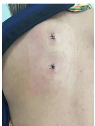
success-fulremovalofthetumor,withexcellentestheticappearance
(Fig. 5). Eight months postoperatively, patient presented a
Fig.1–Radiographinscapularprofileshowingabony
222
rev bras ortop.2017;52(2):220–223Fig.2–Tomographyshowingbonetumorinthesuperomedialregionofthescapula,inclosecontactwiththecoastal
arches,andmagneticresonanceimagingshowingosteochondroma“perforating”thesubscapularismuscle(whitearrow).
Fig.3–Intraoperativeimagesbeforeandafterthetumorremovalprocedure.
Fig.4–Postoperativeradiographandtomographyshowingsuccessfulremovaloftheentiretumor.
significantimprovementintheappliedscores.The
Disabili-tiesofArm,ShoulderandHand(Dash)scoredecreasedfrom
43.3preoperativelyto0.83postoperatively.TheUniversityof
CaliforniaatLosAngeles(UCLA)scoreincreasedfrom22
pre-operativelyto35postoperatively.Thevisualanalogscale(VAS)
decreasedfrom6inthepreoperativeperiodto0inthe
post-operativeperiod.
Discussion
Snappingscapulaisadisorderthatvariesinitsclinical
man-ifestations from a mild to limiting disorder. Many causes
havebeensuggestedforthissyndrome,andosteochondroma
accountsforapproximately15%ofcases.7Osteochondromais
themostcommonbenignbonetumor,accountingfor
approx-imately35%ofthebenigntumorsand9%ofalltumors.This
tumorisoften diagnosedincidentally, asmostare
asymp-tomatic,butmaycausemechanicalsymptoms,dependingon
location andsize.4 Despitebeingthemostcommonbenign
tumorthat affectsthescapula, it israrelyobservedinthis
location.8Itiscommonlyfoundinyoungpatients,generally
agedbelow30years,withamale:femaleratioof1.5:1.9
When reviewing the cases described in the literature,
there was only one case retrieved in which the
loca-tion of the osteochondroma occurred in the superomedial
region ofthe scapula.10 All other cases were located ator
belowthe equatorofthescapula. Thus,thiscasebecomes
of special presentation.11 Tumors of the inferior scapular
region usually reach larger sizes, due to the space they
have to develop; depending on the size, they can
pre-cludearthroscopicresection.12Althoughtechnicallycomplex,
arthroscopic surgery forsnappingscapula syndromeoffers
several theoretical advantages over open surgery. These
includeminimizingthedissection,preservingmuscle
attach-ments,andtherebyeliminatingtheneedforimmobilization
rev bras ortop.2017;52(2):220–223
223
Fig.5–Estheticaspectofthearthroscopicscapulothoracic
surgery.
Thepresentpatienthadanosteochondromawith
partic-ularcharacteristics,duetoitssuperomedialpositionandits
closecontactwiththesecondcostalarch.Asitispossibleto
observeinthetomography,itsgrowthtooktheformofahook,
probablyduetothemechanicaleffectofwearonitssurface,
whichwasinconstantfrictionwiththecostalarch.Another
peculiaritythatwas observedatthe MRIisthatthe tumor
advancedthroughthesubscapularismuscle,notrepellingit,
but insteadpuncturing it,which increasedthe difficulty of
localizationandresectionofthelesion.
Scapulothoracicarthroscopyisaprocedurethathasbeen
increasinglyperformedinorthopedicpractice,allowingthe
treatmentofpathologiesthataffectthescapulainaneffective
andminimallyinvasiveapproach.12Similarlytothepresent
report, many studies have demonstrated excellent results
inosteochondroma resections of the ventral region ofthe
scapulathroughscapulothoracicarthroscopy.14,15
Basedonthe reviewoftheliteratureand observationof
the present case,it can beconcluded that scapulothoracic
arthroscopyisanewprocedurewithlimitedindications,but
withgoodeffectivenessandgoodfinalestheticappearance.
Conflicts
of
interest
Theauthorsdeclarenoconflictsofinterest.
r
e
f
e
r
e
n
c
e
s
1.ManskeRC,ReimanMP,StovakML.Nonoperativeand operativemanagementofsnappingscapula.AmJSports Med.2004;32(6):1554–65.
2.KuhneM,BoniquitN,GhodadraN,RomeoAA,Provencher MT.Thesnappingscapula:diagnosisandtreatment. Arthroscopy.2009;25(11):1298–311.
3.CarlsonHL,HaigAJ,StewartDC.Snappingscapulasyndrome: threecasereportsandananalysisoftheliterature.ArchPhys MedRehabil.1997;78(5):506–11.
4.Ermis¸MN,AykutUS,Durakbas¸aMO,OzelMS,Bozkus¸FS, Karakas¸ES.Snappingscapulasyndromecausedby subscapularosteochondroma.EklemHastalikCerrahisi. 2012;23(1):40–3.
5.PavlikA,AngK,CoghlanJ,BellS.Arthroscopictreatmentof painfulsnappingofthescapulabyusinganewsuperior portal.Arthroscopy.2003;19(6):608–12.
6.RulandLJ3rd,RulandCM,MatthewsLS.Scapulothoracic anatomyforthearthroscopist.Arthroscopy.1995;11(1):52–6.
7.KumarN,RamakrishnanV,JohnsonGV,SouthernS. Endoscopically-assistedexcisionofscapular
osteochondroma.ActaOrthopScand.1999;70(4):394–6.
8.GalateJF,BlueJM,GainesRW.Osteochondromaofthe scapula.MoMed.1995;92(2):95–7.
9.SivanandaP,RaoBK,KumarPV,RamGS.Osteochondromaof theventralscapulacausingscapularstaticwingingand secondaryriberosion.JClinDiagnRes.2014;8(5).LD03-5.
10.SureshSS.Superomedialangleosteochondromaofthe scapulaasacauseofsnappingscapula.KeralaJOrthop. 2011;25(1):37–42.
11.FukunagaS,FutaniH,YoshiyaS.Endoscopicallyassisted resectionofascapularosteochondromacausingsnapping scapulasyndrome.WorldJSurgOncol.2007;5:37.
12.AndreoliCV,EjnismanB,PochiniAC,MonteiroHC,CohenM, FaloppaF.Artroscopiadaarticulac¸ãoescapulotorácica:relato decasos.RevBrasOrtop.2009;44(4):351–6.
13.HarperGD,McIlroyS,BayleyJI,CalvertPT.Arthroscopic partialresectionofthescapulaforsnappingscapula:anew technique.JShoulderElbowSurg.1999;8(1):53–7.
14.FrecheS,JuchF,NusseltT,DelankKS,HagelA.Arthroscopic treatmentofbilateralsnappingscapulasyndrome:acase reportandreviewoftheliterature.ActaOrthopTraumatol Turc.2015;49(1):91–6.
15.SarikayaB,SuluovaF,CetinBV,SarikayaZB.Endoscopically assistedresectionofararemass:intra-articular